Preparation of a UCST type of thermos-responsive sulfobetaine polymer modified poly(ethylene terephthalate) films by Ar plasma-post polymerization
method and recovery of attached HeLa cells on the film by heating with laser irradiation
Akinori IMAJO†2, Taiji ITO †2, Tatsuki NOUSOU †2 and Kohei SHIRAISHI †1,†2
Abstract
For high-throughput cell-diagnosis and/or non-invasive target cell recovery by laser irradiated fluorescence detection and heating to microarray(µAy) using immobilized with single cell or several number of cells, the surface-modified poly(ethylene terephthalate) films(PET) as a component of µAy substrate with poly{2-[(methacryloyloxy)ethyl]dimethyl(3-sulfopropyl)ammonium hydroxide} [poly(SBMA)] and SBMA/butyl methacrylate(BMA) copolymer [poly(SBMA-co-BMA)] as a biocompatible and upper critical solution temperature (UCST) polymer was prepared by Ar plasma-post polymerization method. After a PET film was irradiated with Ar plasma, a known amount of SBMA and SBMA/BMA were post-graft polymerized in the presence of water/ethanol mixed solution at 80°C for 20 h.From the SPM measurements, poly(SBMA) and poly(SBMA-co-BMA) brushes were observed on the PET substrate. From the contact angle(θ) measurements to water at 26°C, the θ values of poly(SBMA)-g-PET and poly(SBMA- co-BMA)-g-PET decreased from θ=75° (the untreated PET) to θ=49°, θ=29°, respectively. On the other hand, the θ values of poly(SBMA)-g-PET and poly(SBMA-co-BMA)-g-PET decreased from θ=49° to θ=46°, θ=29° to θ=27° on heating at 40°C, respectively. The evaluation of HeLa cells attachment and mechanical/thermal stimuli-exfoliation of attached HeLa cells on the g-PETs by laser heating were also examined.
Keywords: 2-[(Methacryloyloxy)ethyl]dimethyl-(3-sulfopropyl)ammonium hydroxide, Upper critical solution temperature (UCST), Ar plasma-post polymerization, Poly(ethylene terephthalate) film
1. INTRODUCTION
A multipotent stem cell such as an embryonic stem cell(ES) or an induced pluripotent stem(iPS) cell have attracted much attention as a potential cell source for regenerative medicine or drug assessment for personalized medicine. In the next decade, the related technology is expected to expand the industrial application in medical, agricultural, and food science fields1). Especially, one of key technology in bio-medical application is focused the diagnosis or the selective recovery of induced differentiated cells2), 3). Flow cytometry is a powerful technique on their cell
operations, but a required number of cells is not comparatively small and is limited for the enabling the induced rare cells to be isolated and collected. Therefore, the development of novel equipment is required for small amount of cells with safe and high-throughput cell sorting and recovery.
Microarray(µAy)s are recognized as one of major tools in the assessment of gene expression via cDNA or
RNA4), 5)and are now accepted as a powerful
experimental tool for high-throughput screening of a large number of samples. Besides, µAy can be performed drug screening6)~8) and assessment of pharmaceuticals and chemicals9)~11). Single cell or several cells attached µAys also have a potential function for high-throughput screening for specific multipotent cells differentiation. The authors have been
†1 Department of Biotechnology and chemistry, Faculty of Engineering, Kindai University
†2 Graduate School of Systems Engineering, Kindai University
Kindai University No.50 2016, pp.1-6
developing precise cell diagnosis and non-invasive target cell recovery technique by using µAy12). For this purpose, surface treatments of substrate as the µAy components are necessary with a control of cell attachment and exfoliation in a manner with non-cell damage.
A polymer with lower critical solution temperature (LCST) has a phase transition to random coil from globule in the LCST below temperature. The hydrophobic-hydrophilic change of the surface immobilized LCST polymers on a substrate such as plastics, metal, or ceramics is induced by thermal stimulation based on phase transition of LCST polymer and used as a method for non-invasively cell recovery lowering the culture temperature of the adherent cells13). Currently, we have been developing the application of the selective recovery of targeting cells using µAy fabricated with patterned glass spots at several µm- regular intervals around gold substrate. The glass spots of µAy is modified with precise controlled chain density and chain lengths of poly(N-isopropylacrylamide)14). On using the µAy, the selective recovery of targeting cells is required after removing unnecessary cells by using highly precise positioning laser light irradiation to cell attached the local μAy spots14). On the other hand, upper critical solution temperature (UCST) types polymer has a phase transition to random coil from globule in the UCST above temperature15). Therefore, it is considered that the immobilization of UCST polymer on the glass spots of μAy enables one path selective recovery of targeting cells by local heating with highly precision positioning continuous wave laser irradiation.
The grafting of zwitterionic biocompatible polymers has promised an effective improvement of the biocompatibility of medical devices16)~18). One of zwitterionic poly{2-[(methacryloyloxy)ethyl]- dimethyl(3-sulfopropyl)ammonium hydroxide}
[poly(SBMA)] has biocompatibility and UCST property.
In this report, to fabricate novel µAy with spots of poly(ethylene terephthalate) (PET) as cell attachment sites around gold substrate, the surface-modified PET films with poly(SBMA) and SBMA/butyl methacrylate(BMA) copolymer [poly(SBMA-co-
BMA)] as a biocompatible and UCST polymer was attempted to prepare by Ar plasma-post polymerization method19). We were also evaluating the surface characterization and test for cell attachment and thermal cell-exfoliation test on the UCST polymer immobilized PET(g-PET).
2. MATERIALS and METHODS
2.1. Materials
2-[(Methacryloyloxy)ethyl]dimethyl-(3-sulfopropyl) ammonium hydroxide (SBMA) was purchased from Sigma-Aldrich (St. Louis, MO). Ethanol was purchased from Wako Pure Chemical Co.(Japan). n-Butyl methacrylate (BMA) was purchased from Wako Pure Chemical Co.(Japan) and used as distillation under reduced pressure[60°C/30 mmHg]. PET film (φ 12 mm, thickness = 50 µm) was kindly supplied by Mitsubishi Plastics, Inc.(Japan) and washed with ethanol, dried in a vacuum at room temperature. Minimum essential medium(MEM), fetal bovine serum (FBS), non-
essential amino acids (NEAA) were purchased from GIBCO. Phosphate buffered saline(PBS)[pH 7.4] and 0.25%Trypsin-EDTA solution were prepared according in the usual manner.Sterile water was autoclaved using distilled water. Distilled water was used for all experiments.
2.2. Preparation of poly(SBMA) and poly(SBMA- co-BMA) grafted PET
Plasma irradiation was carried out according to the previous report20). A PET film (φ 12 mm, thickness = 50 µm) was washed with ethanol, dried in a vacuum at room temperature. After the film was irradiated with Ar plasma (initial pressure = 0.1 Pa, RF power = 20 W, Ar flow rate = 10 sccm, irradiation time = 120 sec; Jeol EH-MN0005A). After plasma treatment, it was exposed to the air for 1 min. After plasma treatment PET film and SBMA 0.528 g(1.89 mmol) in water(0.674 mL)/ethanol(1.326 mL) mixed solution(2 mL) in a glass tube was degassed by the freeze-thaw technique using a liquid nitrogen bath, and then sealed under reduced pressure. After post-polymerization at 80°C for 20 h,the grafted PET film[poly(SBMA)-g-PET] washed for 30 min shaking at 80°C and ultrasonic cleaning using distilled water. Then, poly(SBMA)-g-PET was drying in vacuo at room temperature. poly(SBMA-co-BMA) copolymer grafted PET film [poly(SBMA-co-BMA)-g- PET] was prepared using a similar technique.
poly(SBMA-co-BMA)-g-PET was copolymer consisting of SBMA and BMA [poly(SBMA-co-BMA):
SBMA/BMA=99/1(molar ration)]. The amount of SBMA and SBMA/BMA grafted onto the PET film was calculated from the weight of poly(SBMA)-g-PET and poly(SBMA-co-BMA)-g-PET. The number average molecular weight(Mn) of polymerization of polymers grafted onto PET surface was calculated from the amount of grafted polymer and the amount of free radical21).
2.3. Characterization of grafted PET surface
g-PET surface was evaluated using the following.
Static contact angles (θ) of water were measured at ambient temperature using the sessile drop method with an Erma contact angle meter (Japan), Model G-I20). Scanning probe microscope(SPM) measurements of a Shimadzu SPM-9500J3 with Budget Sensors Tap 300AI-G(Innovative Solutions Bulgaria Ltd., USA) by phase imaging mode.
2.4. Adhesion and growth of HeLa cells on the grafted PET film
g-PET was immobilized on a multidish with 4 wells cell culture multidish(MULTIDISH 4 WELLS NUNCLON DELTA SI:Nunc) using double-sided tape for optical(HJ-3160W:Nitto Denko Corporation). The immobilized g-PET surface was washed with sterile water and 70% ethanol in a clean bench. Then, g-PET surface was sterilized for 1 h by ultraviolet irradiation.
After sterilization, g-PET was washed with PBS. HeLa cells originated from RIKEN cell bank (Japan) were cultured in MEM medium supplemented with 10% FBS and 1% NEAA in a 5%CO2/95% air atmosphere at 37°C.
HeLa cells were detached by the addition of 0.25%
trypsin. HeLa cells(3×10⁴ cells/mL) were added dropwise on each of the modified PET film in multidish with 4 wells each with a diameter of 15 mm and kept for 24 h in a humidified incubator (APC-50D:ASTEC) condition, to 5%CO2/95% air atmosphere at 30°C.
2.5. Thermal stimuli-exfoliation of HeLa cells on g-PET
In accordance with Section 2.4., we cultured HeLa cells. The unattached HeLa cells were removed from the PET film surface by rinsing with PBS. Then, it was observed with an inverted research microscope (IX71:OLYMPUS) and photographed using a microscope digital camera(DP72-BSW:OLYMPUS).
For thermal stimuli-exfoliation, the multidish was placed at 37°C for 2 h to thermoplates(FTP-28190:AS ONE). After 2 h, Exfoliated HeLa cells were removed from the PET film surface rinsing with PBS. The cells remaining on the PET film was observed with an inverted research microscope(IX71:OLYMPUS) and photographed with a microscope digital camera(DP72-BSW:OLYMPUS). Number of adherent cells was estimated by measuring the image taken with a digital camera. Cell exfoliation rate(%) was calculated from the difference between the cell adhesion number of the before thermal stimuli-exfoliation and after thermal stimuli-exfoliation.
2.6. Thermal stimuli-exfoliation using laser irradiation
The laser irradiation was carried out using a new laser system under development (Stec. Co., Ltd, Japan). The laser irradiation system consisted an inverted research microscope(ECLIPSE Ti-S:Nicon) and a Q-switched solid-state pulse laser(Spectra physics Explore349) was patented in Japan[Japan patent JP 5765763B]. The laser irradiation was adjusted through an optical system and focused on the outermost surface from the backside of g-PET attached the culture multidish described with Section 2.4.. The laser irradiation conditions were as follows; wavelength 349 nm, pulse energy; 6 μJ at 1 kHz, irradiation time; 100 msec, reputation; 5 times, and beam radius; 50 μm. The number of attached cells and exfoliated cells were determined in the same manner as in Section 2.5..
3. RESULTS and DISCUSSION
3.1. Preparation and characterization of UCST type of polymer grafted PET
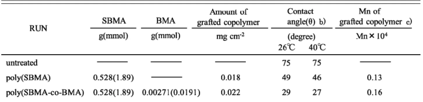
For high-throughput cell-diagnosis and/or non-invasive target cell recovery by laser irradiated fluorescence detection and heating to µAy using immobilized with single cell or several number of cells, the surface-modified PET films as a component of µAy substrate with poly(SBMA) and SBMA and BMA copolymer [poly(SBMA-co-BMA)] as a biocompatible and UCST polymer was prepared by Ar plasma-post polymerization method. The number average molecular weight(Mn) of polymerization of polymers grafted onto PET surface was calculated from the amount of grafted polymer and the amount of free radical(Table 1). The amount of free radical on the PET surface after plasma irradiation have reported in the previous report21). Free radicals on the PET after Ar plasma-post polymerization conditions were detected by using DPPH radical scavenge technique. Free radical amount of PET film surface per unit area(cm2) was reported to be 1.38×10⁻⁵ mmol cm-2. The number average molecular weight(Mn) of poly(SBMA)-g-PET and poly(SBMA-co-BMA)-g- PET were Mn=0.13×10⁴ , 0.16×10⁴ , respectively.
SPM measurements (Fig. 1.) and contact angle(θ) measurements were performed to confirm the modifications of the polymers of the g-PET surface.
From the SPM measurements(30 µm × 30 µm), poly(SBMA) or poly(SBMA-co-BMA) brushes were observed on the PET substrate comparing with untreated PET. From the contact angle(θ) measurements to water at 26°C, the θ values of poly(SBMA)-g-PET
and poly(SBMA-co-BMA)-g-PET decreased from θ=75° (the untreated PET) to θ=49°, θ=29°, respectively.
The surface of g-PETs shows an improved wettability due to grafting of the hydrophilic polymer (Table 1).
It is considered that the hydrophilicity of poly(SBMA-co-BMA) was higher than that of poly(SBMA) due to the number average molecular weight of grafting poly(SBMA-co-BMA) was longer than that of grafting poly(SBMA). The detail is now under consideration. On the other hand, the θ values of poly(SBMA)-g-PET and poly(SBMA-co-BMA)-g-PET decreased from θ=49° to θ=46°, θ=29° to θ=27° on heating from 26°C to 40°C, respectively. The surfaces of g-PETs showed that the hydrophilicity increases on heating. Therefore, the UCSTs of grafting polymer on the g-PET surface was considered to be between 40°C and 26°C based on changing the polymer chains from globule to random coil.
3.2. Cell culture and thermal stimuli-exfoliation of grafted PET surface
To know the behavior of cell recovery attached on the g-PET, the exfoliation of thermal stimulation or laser irradiated heating after cell culture at 30°C below the UCST was examined using HeLa cells on the g-PET surface. g-PET(φ 12 mm) was immobilized on a multidish with 4 wells using double-sided tape for
optical. After UV sterilization, HeLa cells (3×10⁴ cells/mL) were seeded onto g-PET. Fig. 2. exhibits the photomicrographs of HeLa cells after being cultivated for 24 h at 30°C. The unattached HeLa cells were removed from the PET film surface by rinsing with PBS.
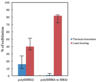
For thermal stimuli-exfoliation, the multidish was placed at 37°C for 2 h on a thermoplate. Exfoliated HeLa cells were removed from the PET film surface by rinsing three times with PBS. The % of thermal stimuli-exfoliation on poly(SBMA)-g-PET and poly(SBMA-co-BMA)-g-PET was approximately 16%
and ~0%, respectively(Fig. 3.). It is considered that the low thermal stimuli-exfoliation on the poly(SBMA)-g- PET and the poly(SBMA-co-BMA)-g-PET was attributed to a restriction of conformational change of the zwitterionic polymer chains with salts in the culture medium. The thermoresponsive polymer immobilized surface is greatly affected by the density of the polymer chains and molecular weight21). Therefore, it is necessary to adjust an optimum surface properties for cell attachment and thermal stimuli-exfoliation by controlling the surface structure constructed on the grafting UCST polymers. From the results of contact angle measurements and thermal stimuli-exfoliation of attached HeLa cells, the properties of poly(SBMA) grafted PET surface such as hydrophilicity, UCST, or cell attachment are able to be effectively modified by introducing the hydrophobic BMA segments in the copolymer. The thermal stimuli-exfoliation using laser irradiated heating were carried out by using a novel microscope equipped with laser irradiated system on the g-PET. In Fig. 3., % of cell exfoliation on the poly(SBMA)-g-PET and poly(SBMA-co-BMA)-g-PET after laser irradiation was approximately 40% and 82%, respectively. The % of exfoliation of laser irradiated heating on the g-PETs was much higher than that of thermal stimuli-exfoliation. The effective local heating a) see Fig.1., b) to water, c)Calculated from amount of grafted (co)polymer and amount of free radicals on the PET after Ar-plasma irradiation / post-polymerization conditions by using DPPH(diphenyl picryl hydrazyl) radical scavenge method; [free radicals] = 1.38×10⁻⁵ mmol/cm2 on the PET, Mn; number average of molecular weight.
Fig. 1. SPM image of g-PET(a) Untreated PET, (b) poly(SBMA)-g-PET, and (c) poly(SBMA-co-BMA)-g- PET.
of outermost surface of UCST polymer grafted PET was achieved by focusing precisely the laser beam to exfoliate the attached HeLa cells thermally induced the change of aggregation of the UCST polymer chains.
The proliferation of recovery HeLa cells also showed similar to that of HeLa cells as seeded. The more detailed laser irradiation conditions for the improvement of thermal stimuli-exfoliation is proceeded now.
4. CONCLUSION
1)The biocompatible UCST types of poly(SBMA) and poly(SBMA-co-BMA) were immobilized on a PET by low-temperature plasma and post-polymerization technique.
2)The resulting g-PET showed UCST properties on the surface, and the UCST was able to be controlled by changing with introduced BMA content in poly(SBMA) segment.
3)HeLa cells were attached on the g-PET with a globule chain under UCST with grafting poly(SBMA) or
poly(SBMA-co-BMA).
4)The low % of exfoliation of attached HeLa cells were observed on the g-PETs by thermal stimulation (Heating 30°C 37°C), on the other hand, the effective laser ⇒ induced thermal stimuli-exfoliation was also observed on the g-PETs.
ACKNOWLEDGEMENT
The authors gratefully acknowledgement support for this research by a grant the Ministry of Economy, Trade and Industry, Strategic Core Technology Advancement Program (Supporting Industry Program; 24163212008).
REFERENCES
1) Eric R. Deutsch and Robert E. Guldberg, Journal of Materials Chemistry, 20, 8942-8951 (2010).
2) H. Yamazoe and H. Iwata, Journal of Bioscience and Bioengineering, 100(3), 292-296 (2005).
3) T. Fukuda, S. Shiraga, M. Kato, S. Yamamura, Y.
Morita, E. Tamiya, T. Hori, S. Suye, M. Ueda, Nanobiotechnology, 1(1), 105-111 (2005).
4) J. Ziauddin and David M. Sabatini, Nature Biotechnology, 411, 107-110 (2001).
5) T. Yoshikawa, E. Uchimura, Daniel P. Funeriu, M.
Miyake, J. Miyake, Journal of Controlled Release, 96, 227-232 (2004).
6) Florence J. Wu, Julie R, Friend, C. C. Hsiao, Michael J. Zilliox, Wen-Je Ko, Frank B. Cerra, and Wei-Shou Hu, Biotechnology and Bioengineering, 50, 404-415 (1996).
7) Steve N. Bailey, Randy Z.Wu and David M. Sabatini, Drug Discovery Today, 7(18), 113-118 (2002).
8) Steve N Bailey, David M. Sabatini , and Brent R.
Stockwell, Proceedings of the National Academy of Sciences of the United States of America, 101(46), 16144-16149 (2004).
9) Maria Jose Gomez-Lechon, Jose Vicente Castell, Maria Teresa Donato, Chemico-Biological Interactions, 168, 30-50 (2007).
10) Camatini M, Bonfanti P, Colombo A, Urani C, Crippa S, Alternatives to Laboratory Animals, 27(3), 325-337 (1999).
11) K. Schlotmann, M. Kaeten, A. F. Black, O.
Damour, M. Waldmann-Laue and T. Forester, International Journal of Cosmetic Science, 23, 309-318 (2001).
12) R. Hamawaki, T. Ishihara, A. Tominaga,
K. Shiraishi, K. Sugiyama, Y. Nitta, T. Nakatani, and K. Okamoto, Journal of Photopolymer Science and Technology, 24(4), 447-452 (2011).
13) M. Yamato, C. Konno, A. Kushida, M. Hirose, M.
poly(SBMA)
poly(SBMA-co-BMA)
Fig. 2. Phase-contrast microscope observation of HeLa cells attached(A), thermal stimuli-
exfoliation(B), and after laser irradiation(C) on g-PET.
Fig. 3. % of exfoliation of attached HeLa cells on the g-PET by thermal stimulation from 30°C to 37°C and laser heating.
Utsumi, A. Kikuchi, T. Okano, Biomaterials, 21, 981-986 (2000).
14) T. Nousou, A. Imajo, K. Shiraishi,
Kobunshi ronbunshu, 72(6), 354-360 (2015).
15) Jan Seuring, Seema Agarwal, Macromolecular Rapid Communications, 33, 1898-1920 (2012).
16) K. Sugiyama, N. Tanigawa, K. Shiraishi, Journal of Biomaterials and Nanobiotechnology, 2, 337-346 (2011).
17) T. Moro, Y. Takatori, M. Kyomoto, K. Ishihara, K.
Saiga, K. Nakamura, H. Kawaguchi, Osteoarthrits and Cartilage, 18, 1174-1182 (2010).
18) Ai T. Nguyen, Jacob Baggerman, Jos M. J. Paulusse, Han Zuilhof, and Cees J. M. van Rijn, Langmuir, 28, 604-610 (2012).
19) Yu-Ju Shih and Yung Chang, Langmuir, 26(22), 17286-17294 (2010).
20) K. Sugiyama, T. Matsumoto, Y. Yamazaki, Macromolecular Materials and Engineering, 282, 5-12 (2000).
21) K. Shiraishi, M. Ohdan, K. Maeda, K. Sugiyama, K.
Suzuki and H. Hosoya, Kobunshi Ronbunshu, 63(9), 613-620 (2006).