九州大学学術情報リポジトリ
Kyushu University Institutional Repository
産業利用を目指した海洋性真核微生物の高度不飽和 脂肪酸生合成経路に関する研究
松田, 高宜
九州大学大学院生物資源環境科学府
https://doi.org/10.15017/21692
出版情報:Kyushu University, 2011, 博士(農学), 課程博士 バージョン:
権利関係:
Study on the Biosynthetic Pathway of Polyunsaturated Fatty Acids in Unicellular Marine Eukaryotes
Aiming for Industrial Applications
Takanori Matsuda
2012
i
CONTENTS
GENERAL INTRODUCTION 1
CHAPTER 1. The development of a transformation system for thraustochytrids 1-1. INTRODUCTION 22
1-2. MATERIALS AND METHODS 24
1-3. RESULTS 29
1-4. DISCUSSION 31
1-5. SUMMARY 33
FIGURES AND TABLES 34
CHAPTER 2. Molecular cloning of a Pinguiochrysis pyriformis oleate-specific microsomal ∆12-fatty acid desaturase and functional analysis in yeasts and thraustochytrids 2-1. INTRODUCTION 38
2-2. MATERIALS AND METHODS 40
2-3. RESULTS 46
2-4. DISCUSSION 51
2-5. SUMMARY 54
FIGURES AND TABLES 55
CHAPTER 3. The analysis of ∆12-fatty acid desaturase function revealed that two distinct pathways are active for the synthesis of polyunsaturated fatty acids in Thraustochytrium aureum ATCC 34304 3-1. INTRODUCTION 65
3-2. MATERIALS AND METHODS 68
3-3. RESULTS 75
3-4. DISCUSSION 82
3-5. SUMMARY 86
FIGURES AND TABLES 87
GENERAL DISCUSSION 106
REFERENCES 112
ACKNOWLEDGEMENTS 130
ii
Abbreviations
ACP: acyl carrier protein
ARA: arachidonic acid (C20:4 n-6) ATCC: American Type Culture Collection Blar: blasticidin resistance
cDNA: complementary DNA CoA: coenzyme A
DHA: docosahexaenoic acid (C22:6 n-3) EF-1α: elongation factor-1α
EPA: eicosapentaenoic acid (C20:5 n-3) FAME: fatty acid methyl ester
FAS: fatty acid synthse GC: gas chromatography
GC-MS: gas chromatography mass spectrometry GFP: green fluorescence protein
GL: glycolipid
GLA: γ-linolenic (C18:3 n-6)
Hygr: hygromycin resistance LA: linoleic acid (C18:2 n-6) Neor: neomycin resistance NL: neutral lipid
OA: oleic acid (C18:1 n-9) ORF: open reading frame
iii
PCR: polymerase chain reaction PKS: polyketide synthase PL: phospholipid
PUFA: polyunsaturated fatty acid
RACE: rapid amplification of cDNA ends SFA: saturated fatty acid
SDS-PAGE: sodium dodecyl sulfate polyacrylamide gel electrophoresis TLC: thin-layer chromatography
1
GENERAL INTRODUCTION
In this thesis, the author describes the establishment of a transformation system for thraustochytrids and analysis of pathway for fatty acid production in thraustochytrids for industrial applications. Before the experimental details are discussed, some background and fundamental information are given as part of a general introduction.
Finally, the scope of this research is summarized at the end of this chapter.
What are Thraustochytrids and Pinguiochrysis?
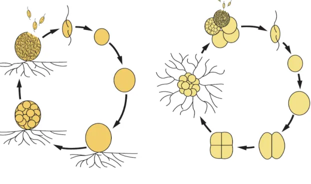
Thraustochytrids, widely distributed in marine and estuarine environments, are heterotrophic eukaryotic microorganisms. They are classified into the kingdom Stramiipila, class Labyrinthulomycetes, family Thraustochytriaceae, and four genera Aurntiochytrium (formerly Schizochytrium), Thraustochytrium, Parientichytrium and Schizochytrium. Thraustochytrids are characterized by the presence of an ectoplasmic network generated by one or more unique cell surface organelles called sagenogenetosomes, a cell wall with noncellulosic scales and a life cycle consisting of vegetative cells, zoosporangia, and zoospores (1, 2). Unlike Thraustochytrium vegetative cells, Schizochytrium vegetative cells divide into halves prior to releasing zoospores from zoosporangia (Fig. gi-1). Interestingly, zoospores of thraustochytrids show a chemotactic response to carbohydrates, amino acids and leaf extracts (3).
Therefore, thraustochytrids can be isolated from environments through the chemotactic response toward pine pollen.
Thraustochytrids play a major role in the degradation and mineralization of detritus in
2
the ocean. They are able to produce extracellular enzymes such as cellulases, proteases, lipases and xylanases, decomposing organic matter as nutrients (4).
Thraustochytrids are also known to be associated with marine invertebrates. Some thraustochytrids are pathogens to juvenile abalone and hard-shell clam (Mercenaria mercenaria) (5, 6). In addition, some species are found on the surface and in the mucus of reef-building coral species such as Favia sp. and Fungia granulose, with which they have mutualistic associations (7, 8).
Particular attention has been given to the thraustochytrids as potential sources of valuable bioactive compounds, such as fatty acids, carotenoids and squalene (9-11).
Thraustochytrids are also expected to be a material for biofuel. Thraustochytrids possess prominent lipid bodies (lipid droplets) and accumulate a great amount of polyunsaturated fatty acids (PUFAs). Therefore, they are expected to be an alternative source of PUFAs, especially docosahexaenoic acid (DHA). Indeed, several products including DHA are now distributed in the USA and European markets.
Thraustochytrids are also used commercially as feed for rotifers (Brachionus sp.) and brine shrimp (Artemia sp.), enriching them with PUFAs.
Many efforts to enhance the production of PUFAs in thraustochytrids have been conducted by changing growth conditions, such as temperature and concentrations of dissolved oxygen (12-16). However, the molecular breeding of thraustochytrids has yet to be fully conducted for the production of PUFAs. There are several basic demands for engineering thraustochytrids as potential sources of beneficial PUFAs: (1) uncovering the full scope of the biosynthetic pathway and, therefore, of the enzymes/genes involved; (2) cloning of the genes necessary for the selective production of specific PUFAs; (3) development of a transformation system for molecular breeding
3
of thraustochytrids.
Studies have proposed two distinct pathways involved in PUFA synthesis in thraustochytrids. One is the desaturase/elongase (standard) pathway, found widely in eukaryotes, in which PUFAs are produced by a series of alternating desaturation and elongation steps from saturated fatty acids. Several genes encoding desaturases and elongases have been cloned from thraustochytrids and functionally characterized in yeasts and plants (17-23). The other is the polyketide synthase-like (PUFA synthase) pathway, which is found in specific marine bacteria (24-26). This pathway, catalyzed by a complex enzyme machinery composed of multi-subunit enzymes homologous to enzymes in the polyketide synthase (PKS) pathway, has been found in Schizochytrium sp. (24-26). However, some enzymes essential for the standard pathway have not been identified in thraustochytrids. Therefore, further investigation is needed to elucidate the full scope of the pathways for PUFA production in thraustochytrids.
Pinguiochrysis pyriformis is an unicellular marine microalga classified into the kingdom Stramiipila, phylum Ochrophyta, and class Pinguiophyceae (27).
Pinguiophyceae is a new class including five genera, Pinguiochrysis, Glossomastix, Phaeomonas, Pinguiococcus and Polypodochrysis (28). All five genera produce large amounts of PUFAs, especially EPA. P. pyriformis cells are typically pear-shaped and frequently change to a subspherical shape. The name of P. pyriformis is based on the large amounts of PUFAs (pingue = fat, grease) and the cell shape (pyriform = pear-shaped). Extracellular structures such as a cell wall and polysaccharide layer are not found around the cells of P. pyriformis. P. pyriformis has an ovoid chloroplast surrounded by a chloroplast endoplasmic reticulum and contains more than three thylakoids. It is suggested that EPA is mainly incorporated into glycolipids, structural
4
components of both envelop membranes and thylakoids in the chloroplast, in Glossomastix chrysoplasta (29).
5
Fig. gi-1. Life cycles of Thraustochytrium and Aurantiochytrium.
6
Polyunsaturated fatty acids (PUFAs)
PUFAs are fatty acids that contain more than one double bond. Fatty acids are often abbreviated as CX:Y∆z, where X stands for the number of carbon atoms of the acyl-chain, Y for the number of double bonds, and Z for the position of the double bond counted from the carboxyl end (Fig. gi-2A). For example, γ-linolenic acid (GLA), abbreviated as C18:3∆6, 9, 12, is an 18-carbon fatty acid with cis double bonds between carbons 6 and 7, carbons 9 and 10 and carbons 12 and 13. PUFAs are also classified into the omega-X form, depending on the position of the first double bond counted from the methyl terminus of the fatty acids. In this way, eicosapentaenoic acid (EPA, C20:5∆5, 8, 11, 14, 17) is abbreviated as C20:5ω3 or C20:5 n-3.
In most eukaryotic and prokaryotic organisms, PUFAs are biosynthesized by the desaturase/elongase (standard) pathway. On the other hand, some marine bacteria produce PUFAs by the polyketide synthase-like (PUFA synthase) pathway (30-34). In mammals, several PUFAs are recognized as essential fatty acids, because they lack the
∆12- and ∆15-fatty acid desaturases necessary to insert a double bond at the ω6 or ω3
position. PUFAs play important roles in many cellular functions as structural membrane components, gene regulatory elements and precursors of eicosanoids such as prostaglandins, thromboxanes, and leukotrienes (Fig. gi-2B) (35-39). For example, EPA and DHA have anti-inflammatory effects through their metabolic products or the stimulation of G protein-coupled receptors, thereby preventing chronic diseases such as coronary heart disease, diabetes, arthritis and cancer (40-44). DHA is also essential for neurological growth and development in infants and for the maintenance of adequate cellular function in the adult brain (45, 46). The eicosanoids from arachidonic acid
7
(ARA, C20:4 n-6) are mediators of acute inflammation and several diseases such as allergic disorders, asthma and cancer.
8
Fig. gi-2. Structure of several PUFAs and their metabolic pathways.
(A) Structures of ARA, EPA and DHA. (B) Outline of the pathways of production of eicosanoids and docosanoids. COX, cyclooxygenase; HETE, hydroxyl-eicosatetraenoic acid; HPETE, hydroperoxy-eicosatetraenoic acid; HPDHA, hydroperoxy-docosahexaenoic acid; HPEPE, hydroperoxy-eicosapentaenoic acid; LOX, lipoxygenase; LT, leukotriene; PG, prostaglandin; TX, thromboxane; Rv, resolvin.
9
Desaturase/elongase (standard) pathway
This pathway consists of alternating desaturation and elongation steps, with molecular oxygen required for the desaturation (Fig. gi-3). The initial step of this pathway occurs with the production of palmitic acid (C16:0) or stearic acid (C18:0) by fatty acid synthase (FAS). The palmitic acid is elongated to form stearic acid and then desaturated to produce oleic acid (OA, C18:1∆9) by a ∆9 -fatty acid desaturase. Next, a
∆12-fatty acid desturase converts the oleic acid into linoleic acid (LA, C18:2∆9, 12), which is further desaturated into α-linolenic acid (ALA, C18:3∆9, 12, 15) by a ∆15-fatty acid desaturase. Subsequently, the LA and ALA are desaturated by a ∆6-fatty acid desaturase. The products of the ∆6-fatty acid desaturation, γ-linolenic (GLA, C18:3∆6,
9, 12
) and stearidonic acid (C18:4∆6, 9, 12, 15), are elongated to form dihomo-γ-linolenic acid (DGLA, C20:3∆8, 11, 14) and eicosatetraenoic acid (ETA, C20:4∆8, 11, 14, 17
), respectively. This elongation is followed by ∆5-fatty acid desaturation to produce arachidonic acid (ARA, C20:4∆5, 8, 11, 14
) and eicosapentaenoic acid (EPA, C20:5∆5, 8, 11, 14, 17). In some fungi, ARA is desaturated to produce EPA by a ω3-fatty acid desaturase (∆17-fatty acid desaturase). Then, ARA and EPA are elongated to form docosatetraenoic acid (DTA, C22:4∆7, 10, 13, 16) and ω3 docosapentaenoic acid (ω3 DPA, C22:5∆7, 10, 13, 16, 19
), respectively. Finally, these elongation products are subjected to further desaturation at the ∆4 position to produce ω6 docosapentaenoic acid (ω6 DPA, C22:5∆4, 7, 10, 13, 16
) and docosahexaenoic acid (DHA, C22:6∆4, 7, 10, 13, 16, 19
).
With regard to the biosynthesis of DHA, a ∆4-fatty acid desaturase-independent pathway has been proposed in mammals (47-49). This “Sprecher pathway” involves two consecutive elongation steps, ∆6-fatty acid desaturation and C2 unit shortening via
10
β-oxidation in the peroxisome. In addition, an alternate pathway for C20 PUFA production has been demonstrated in some organisms, which is independent of ∆6-fatty acid desaturation (50, 51). In the first step of this pathway, LA and ALA are elongated to form eicosadienoic acid (EDA, C20:2∆11, 14) and eicosatrienoic acid (ETrA, C20:3∆11,
14, 17
). In turn, these elongation products are further desaturated to produce DGLA and ETA, respectively, by ∆8-fatty acid desaturase. The DGLA and ETA are then subjected to desaturation and elongation to produce final products in the conventional pathway. A schematic diagram of the desaturase/elongase pathway is shown in Fig.
gi-3.
11
Fig. gi-3. Schematic diagram of desaturase/elongase (standard) pathway.
Dashed arrows indicate the “Sprecher pathway” found in mammals but not thraustochytrids.
12
Fatty Acid Desaturase
Fatty acid desaturases, iron-containing enzymes, introduce a double bond at a specific position in fatty acids (Fig. gi-4) (52). These enzymes require molecular oxygen and an electron transport system (usually ferredoxin-NADPH reductase and ferredoxin, or cytochrome b5 reductase and cytochrome b5) for their reactions. Fatty acid desaturases use activated molecular oxygen to extract hydrogens from the substrate creating a double bond in a fatty acid. Except for the soluble acyl-acyl carrier protein (ACP) desaturase in plant plastids (53), all of these enzymes have several transmembrane domains and contain three histidine boxes, which act as di-iron coordinating centers for catalytic activity (54).
In terms of their regioselectivity or acyl substrate specificity, desaturases are categorized into several groups. Four regioselective classes have been proposed for these fatty acid desaturases (Fig. gi-5). ∆x-fatty acid desaturases introduce a double bond at position x from the carboxyl end of a fatty acid (17, 51). ωx-fatty acid desaturases introduce a double bond between the x and x + 1 carbons from the methyl end (55). The ν + x fatty acid desaturases introduce a double bond at x carbons from the pre-existing double bond (ν) toward the methyl end, while ν – x fatty acid desaturases introduce a double bond at x carbons from the pre-existing double bond ν toward the carboxyl end (56, 57). ∆4, ∆5, ∆6 and ∆8-fatty acid desaturases are also classified as ‘front-end’ desaturases (58). These desaturases introduce a new double bond between the pre-existing double bond and the carboxyl end of fatty acids.
Front-end desaturases are characterized by the presence of cytochrome b5-domain in their amino acid sequences, which is predicted to serve as an electron donor during
13
desaturation.
With regard to the acyl substrate specificity, fatty acid desaturases are separated into three types. The acyl-Coenzyme A (CoA) desaturases use fatty acids esterified to CoA as a substrate, and this type of desaturase is found in animals, insects and microalgae (57, 59, 60). The acyl-ACP desaturases are soluble enzymes, and desaturate fatty acids linked to ACP. The acyl-lipid desaturases introduce a double bond into lipid-bound fatty acids such as glycerophospholipids and sphingolipids (61, 62).
14
Fig. gi-4. Proposed trans-membrane form and mechanism of the ∆9-fatty acid desaturase.
The transfer of 2 electrons and 2 protons from the hydrocarbon chain to water results in the formation of the cis-double bond in the substrate fatty acid. Reducing equivalents were transferred from NADH to the diiron-oxo raction center (gray arrow) through NADH cytochrome b5 dehydrogenase and cytochrome b5. Reducing equivalents reduce the iron atoms to the ferrous state with the release of the oxygen atom as water.
The red boxes indicate the positions of the histidine clusters that form di-iron coordinating centers for catalytic activity.
15
Fig. gi-5. The type of fatty acid desaturase based on their regioselectivity.
16
Elongase
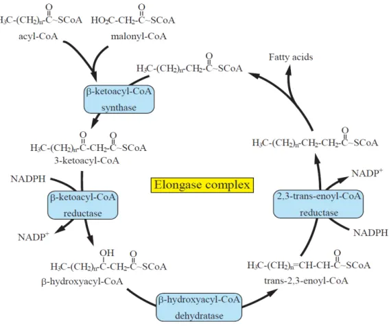
Fatty acid elongation consists of four reactions catalyzed by a multi-enzyme complex composed of four enzymes (Fig. gi-6) (63). The initial step is the condensation of malonyl-CoA with the acyl primer to form β-ketoacyl-CoA catalyzed by β-ketoacyl-CoA synthase (called elongase, ELOVL, KCS or FAE), which is the rate-limiting step in the elongation process. Next, the β-ketoacyl-CoA is reduced to β-hydroxyacyl-CoA by β-ketoacyl-CoA reductase in the presence of NAD(P)H.
Subsequently, dehydraion of the β-hydroxyacyl-CoA to trans-2-enoyl-CoA is mediated by β-hydroxyacyl-CoA dehydratase. Then, reduction of the enoyl-CoA by enoyl-CoA reductase in the presence of NAD(P)H generates a fatty acyl-CoA that is two carbons longer. The substrate specificity in the elongation reaction is determined by the β-ketoacyl-CoA synthase, and predicted elongation activity is restored by the expression of only this enzyme in heterologous hosts (64). The three other enzymes are ubiquitously expressed and shared by diverse elongation systems.
17
Fig. gi-6. Schematic diagram of the fatty acid elongation system.
18
Polyketide synthase-like (PUFA synthase) pathway
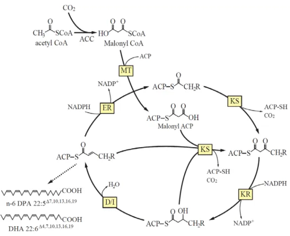
The genes involved in this pathway were cloned from an EPA-producing bacterium, Shewanella sp. strain SCRC-2738, in 1996 (65). After this discovery, homologs were cloned from both prokaryotic and eukaryotic marine organisms (30-34). Among eukaryotes, this pathway has been reported only in thraustochytrids (24-26). Since the pathway does not require molecular oxygen, PUFAs are biosynthesized under both anaerobic and aerobic conditions. This process involves a large multi-domain enzyme complex. Several domains closely resemble homologs of polyketide synthase (PKS) and fatty acid synthase (FAS). The mechanism of action of the PUFA synthase has been predicted by several researchers (66, 67). As shown in Fig. gi-7, the initial step of this pathway is the condensation of an acyl-ACP and a malonyl-ACP to produce a ketoacyl-ACP by ketosynthase. Next, the ketoacyl-ACP is reduced to hydroxyacyl-ACP by ketoreductase, followed by dehydration/isomeration to produce an enoyl-ACP by dehydratase/isomerase. Then, the enoyl-ACP is subjected to reduction to saturated fatty acid by enoyl reductase. Unlike FAS, the PUFA biosynthetic pathway often omits processes such as dehydration/isomeration and reduction.
19
Fig. gi-7. Predicted PUFA biosynthesis in the PUFA synthase pathway.
ACC, Acetyl-CoA carboxylase; MT, Malonyl transferase; ACP, Acyl carrier protein; KR, Ketoreductase; ER, Enoyl reductase; KS, Ketosynthase; D/I, Dehydratase/Isomerase
20
The scope of this study
As described above in this CHAPTER, the development of a transformation system and elucidation of the PUFA biosynthesis pathway are necessary for the selective production of PUFAs using thraustochytrids. This thesis describes the development of a transformation system for thraustochytrids and evaluates the biosynthetic pathway for PUFA production in two typical genera of thraustochytrids, Aurantiochytrium and Thraustochytrium.
CHAPTER 1 describes a transformation system developed for thraustochytrids, which uses the neomycin-resistance (Neor) gene and enhanced-green fluorescence protein (EGFP) as transformation markers. The system is applicable to the genetic manipulation of thraustochytrids as shown in the following chapters.
CHAPTER 2 describes the heterozygous expression of a fatty acid desaturase in Aurantiochytrium limacinum mh0186. The gene encoding a ∆12-fatty acid desaturase was cloned from the marine alga Pinguiochrysis pyriformis, expressed in Saccharomyces cerevisiae, and characterized. This enzyme was expressed functionally in thraustochytrids as well as yeasts. In this chapter, the author discusses the PUFA biosynthesis in A. limacinum mh0186 in terms of ∆12-fatty acid desaturase activity.
Finally, the molecular cloning and characterization of a ∆12-fatty acid desaturase from Thraustochytrium aureum ATCC 34304 are described in CHAPTER 3. This enzyme adopted not only oleic acid (C18:1) but also odd-numbered monounsaturated fatty acids such as C17:1 and C19:1. Disruption of the ∆12-fatty acid desaturase gene in T. aureum resulted in significant changes in fatty acid profiles without decreasing the
21
content of DHA. This result may indicate that the standard pathway functions to produce PUFAs, whereas DHA is mainly produced by the PUFA synthase system.
That is, two distinct pathways are present in T. aureum for production of PUFAs.
The transformation system developed in this study was successfully applied to transgene expression and targeted gene disruption in thraustochytrids. This is the first report describing the heterozygous expression and disruption of a fatty acid desaturase in thraustochytrids. This study also provides evidence that the mode of production of PUFAs differs depending on the genera/species in thraustochytrids. This study could facilitate the molecular breeding of thraustochytrids for the selective production of beneficial PUFAs.
22
CHAPTER 1
The development of a transformation system for thraustochytrids
1-1. INTRODUCTION
Many n-3 PUFAs such as eicosapentaenoic acid (EPA) and docosahexaenoic acid (DHA) function as therapeutic “nutraceuticals” in the prevention and treatment of cardiovascular and inflammatory diseases (41-43). EPA and DHA have historically been obtained from fish oils, but recent decreases in fish stocks make alternative sources desirable. Thraustochytrids are heterotrophic unicellular eukaryotes belonging to the class Labyrinthulomycetes. These organisms have been isolated from a wide range of habitats worldwide including deep-sea and estuarine environments. Because of their ability to accumulate high levels of PUFAs, thraustochytrids have received much attention as a potential commercial source of PUFAs. Considerable effort has been expended in modifying plants to produce desirable PUFAs through the expression of certain elongases and desaturases (68, 69). However, the genetic manipulation of thraustochytrids holds the potential for the accumulation of higher levels of PUFAs than those found in plants because thraustochytrids are capable of storing large amounts of PUFAs in their lipid droplets. Nevertheless, the basic information and tools required for genetic manipulation remain missing for thraustochytrids. Therefore, the development of a transformation system for thraustochytrids will facilitate not only the molecular breeding of thraustochytrids that are highly enriched for desirable PUFAs but also the elucidation of their PUFA biosynthetic pathway.
23
Since its discovery in the jellyfish Aequorea aequorea by Shimomura et al in 1962, green fluorescent protein (GFP) and its derivatives have been applied extensively in the biological sciences, e.g., in promoter assays, the monitoring of protein trafficking, the detection of viral infection and other labeling studies (70, 71). These proteins are also useful tools for evaluating the efficiency of transformation (72) because they are easily detected under a fluorescent microscope and do not require the addition of exogenous substrates.
In this chapter, the author describes the development of a transformation system for thraustochytrids using the neomycin-resistance (Neor) and enhanced-GFP (EGFP) genes as selection markers. These marker genes, driven by promoter/terminator systems derived from elongation factor-1α (ΕF−1α) or ubiquitin, were randomly integrated into the genomes of thraustochytrids and functionally expressed. This transformation system, which is applicable to transgene expression, will make it possible to obtain genetically engineered thraustochytrids for the selective production of beneficial PUFAs.
24
1-2. MATERIALS AND METHODS
Materials
Restriction enzymes and a Ligation-Convenience Kit were purchased from Nippon Gene (Tokyo, Japan). Synthetic oligonucleotides were obtained from Hokkaido System Science (Hokkaido, Japan) and GeneNet (Fukuoka, Japan). D-(+)-Glucose and dry yeast extract were purchased from Nacalai Tesque (Kyoto, Japan). SEA LIFE was purchased from MARINETECH Co., Ltd. (Tokyo, Japan). Neomycin (G418) was purchased from Nacalai Tesque. All other reagents were of the highest purity available.
Strains and culture
Thraustochytrium aureum ATCC 34304 was obtained from the American Type Culture Collection (USA). Aurantiochytrium limacinum mh0186 and Thraustochytrium sp. ATCC 26185, obtained from Dr. M. Hayashi at Miyazaki University, were identified based on the sequences of their 18S rRNA genes (DDJB, accession number: AB362211) (73). All these strains were maintained on potato dextrose agar (PDA) plates (0.8% potato dextrose and 1.2% agar in 50% artificial sea water). The thraustochytrids were cultivated on GY medium, consisting of 3% glucose and 1% yeast extract in 50% artificial sea water (ASW).
Isolation of the promoter and terminator regions of ΕF−1α
T. aureum ATCC 34304 was grown at 25°C in GY liquid medium with shaking at 150 rpm. Cells in the logarithmic growth phase were harvested by centrifugation (3,500 ×
25
g, 4°C, 10 min), and the total RNA was extracted using Sepasol RNA I Super (Nacalai Tesque). The poly(A)+ RNA was purified using an Oligotex-dT Super mRNA
Purification Kit (TaKaRa Bio, Shiga, Japan). First strand cDNA was prepared with a SMART RACE cDNA Amplification Kit (Clontech, CA, USA), and the 3′- and 5′-RACE PCRs were performed according to the manufacturer’s instructions. PCR was then performed using a degenerate primer targeting the conserved region of ΕF−1α, EF-F1 (5′-THG AYG CNC CNG GNC AYM G-3′). The sequence of the 980-bp 3′-RACE product was highly homologous to ΕF−1α, and thus the primer EF-1r
(5′-GTG AAG GCC AGA AGG GCG TG-3′) was designed to perform 5′-RACE PCR.
Consequently, we identified a 1,396-bp region of the ΕF−1α cDNA derived from T.
aureum ATCC 34304 that included a 1,023-bp ORF encoding 341 amino acid residues.
Subsequently, the 5′- and 3′-flanking sequences of the gene, assumed to be the
functional ΕF−1α promoter and terminator regions, were isolated using an LA PCR in vitro Cloning Kit (TaKaRa Bio) according to the manufacturer’s instructions. The PCR primers used were as follows: r3 (5′-CCT CCT TCT CGA ACT TCT CGA TCG TG-3′) for the isolation of the ΕF−1α 5′-flanking sequences; EF-t-F1 (5′-CAT GGT CAA GAT GTA TCC CCT CCA A-3′) and EF-t-F2 (5′-TCA CCA AGG GCG ACA AAT AAA TTC T-3′) for the ΕF−1α 3′-flanking sequences. As a result, 615-bp and
1,414-bp ΕF−1α promoter and terminator regions, respectively, were identified.
Construction of the Neor and Neor/EGFP expression cassettes
The codons of the Neor gene were adjusted according to the codon usage of T.
aureum (http://www.kazusa.or.jp/codon/) to achieve suitable expression in thraustochytrids. The Neor expression cassette (the Neor construct, Fig. 1-1A), driven
26
with an ΕF−1α promoter/terminator system, was prepared by fusion PCR. The Neor construct was subcloned into the Ssp I/Pst I sites of the pUC18 vector (TaKaRa Bio) to generate the plasmid pNeor. A linear DNA fragment of the Neor construct was amplified by PCR using 2F and 5R as the primers and used for the transformation of thraustochytrids. Prior to constructing the Neor/EGFP expression cassette (the EGFP construct, Fig. 1-2A) that is driven by an ΕF−1α or ubiquitin promoter/terminator system, the EGFP expression cassette was generated by fusion PCR. The PCR product was then subcloned into the Kpn I site of pNeor to generate the EGFP construct. The ubiquitin promoter and terminator were obtained from T. aureum ATCC 34304. A linear DNA fragment of the EGFP construct was amplified by PCR with the 2F and pUC18-R primers and used for thraustochytrid transformation. The primers used for the PCR amplification are listed in Table 1-1.
Transformation of Thraustochytrium sp. ATCC 26185
The Neor construct was introduced into Thraustochytrium sp. ATCC 26185 by electroporation. After culturing in GY medium, cells in the logarithmic growth phase were harvested by centrifugation (3,500 × g, 4°C, 10 min) and washed with distilled water plus 1.75% (w/v) SEA LIFE. The cells (5 × 10⁶) were then resuspended in 75 µl of Nucleofector solution L (Amaxa Biosystems, MD, USA), 50 mM sucrose, 100 mM sucrose or 300 mM sorbitol. Subsequently, the suspended cells were mixed with 5 mg of highly purified DNA fragments and then transferred to a 0.1-cm-gap cuvette, which was pulsed twice using a Gene Pulser (Bio-Rad, CA, USA). After the pulses, 1 ml of GY medium was immediately added to the solution, which was incubated at 25°C for 1 day and then spread on a PDA plate (containing G418 at 2 mg/ml).
27
Expression of EGFP in thraustochytrids
The EGFP construct was introduced into Thraustochytrium sp. ATCC 26185 and A.
limacinum mh0186 by electroporation using Nucleofector solution L as the suspending solution. The method for electroporation was described above (pulse conditions: 50 mF, 50 W, 7.5 kV/cm, 2 pulses).
The Biolistic PDS-1000/He system (Bio-Rad) was used for the transformation of T.
aureum ATCC 34304. Gold particles (0.6 mm in diameter) coated with highly purified DNA fragments were prepared according to the manufacturer’s instructions. Cells in the logarithmic growth phase were harvested by centrifugation (3,500 × g, 4°C, 10 min) and spread on a PDA plate (15 × 60 mm) without G418. The bombardment was performed in a Biolistic PDS-1000/He system with DNA-coated gold microcarriers according to the manufacturer’s instructions using the following bombardment conditions: pressure, 7.58 × 10⁶ Pa; target distance, 6 cm; vacuum, 8.80 × 10⁴ Pa.
After the bombardment, the plate was incubated at 25°C for 4 hours, after which the cells were collected and suspended in GY medium followed by respreading on a PDA plate containing G418. The concentrations of G418 added to the PDA plates were 2 mg/ml for Thraustochytrium sp. ATCC 26185 and T. aureum ATCC 34304 and 0.5 mg/ml for A. limacinum mh0186.
Genomic PCR and Southern blot hybridization of thraustochytrid transformants Genomic DNA was prepared from transformants cultured in GY medium containing appropriate amounts of G418. PCR was then performed using the forward primer 2F and the reverse primers 5R or pUC18-R (Table 1-1). For the Southern blot analysis, 3 µg of genomic DNA was digested with Pst I or Hind III and subjected to 0.7% agarose
28
gel electrophoresis. The DNA was then transferred to a nylon membrane (Hybond N+, GE healthcare, Tokyo, Japan). The membrane was hybridized with a probe that was prepared using the PCR DIG Probe Synthesis Kit (Roche Diagnostics K.K., Mannheim, Germany). The 3F and 4R PCR primers were used for the Neor DNA probe synthesis.
The genomic DNA hybridized with the probe was detected with an anti-digoxigenin-AP Fab fragment and an NBT/BCIP stock solution (Roche Diagnostics K.K.).
RT-PCR analysis
Total RNA was prepared from transformants grown in GY medium containing appropriate amounts of G418 using Sepasol RNA I Super, an RNeasy Mini Kit (QIAGEN, Tokyo, Japan) and DNase I (Takara Bio Inc.) and used to produce first-strand cDNA with PrimeScriptTM Reverse Transcriptase (Takara Bio Inc.). PCR was performed using the forward primer 3F and the reverse primer 4R for the amplification of Neor cDNA.
Fluorescence assay
Colonies grown on PDA plates containing appropriate amounts of G418 were observed by fluorescence microscopy (Leica Microsystems, Tokyo, Japan).
EGFP-positive colonies were inoculated on GY medium containing appropriate amounts of G418 and incubated at 25°C with shaking at 150 rpm. Cells in the late logarithmic growth phase were harvested by centrifugation (3,500 × g, 4°C, 10 min) then washed and suspended in sterilized sea water. The harvested cells were then observed under a confocal laser-scanning microscope Digital Eclipse C1 (Nicon, Tokyo, Japan).
29
1-3. RESULTS
Transformation of Thraustochytrium sp. ATCC 26185 with the Neor construct To develop a transformation system for Thraustochytrium sp. ATCC 26185, the author constructed a Neor expression cassette (Neor construct, Fig. 1-1A) driven with an ΕF−1α promoter/terminator system. The ΕF−1α gene with 5′ and 3′ flanking
sequences was isolated from T. aureum ATCC 34304 as described in the MATERIALS AND METHODS. The Neor construct was introduced into Thraustochytrium sp.
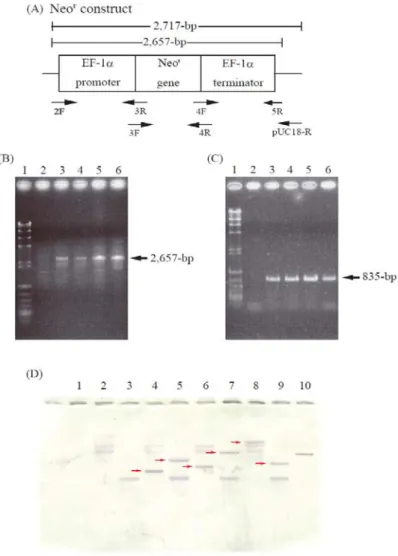
ATCC 26185 by electroporation. The transformants were obtained under several pulse conditions: 0.15 kV/50 Ω/1,000 µF, 0.3 kV/50 Ω/500 µF, 0.3 kV/50 Ω/1,000 µF and 0.75 kV/50 Ω/50 µF with Nucleofector solution L, or 0.15 kV/50 Ω/200 µF and 0.15 kV/50 Ω/1,000 µF with 50 mM sucrose. Transformants grown on GY medium containing G418 were subjected to genomic PCR to examine whether the full-length Neor construct was integrated into the chromosomal DNA. As illustrated in Fig. 1-1B, a 2,657-bp PCR product (corresponding to the Neor construct) was detected in these transformants. Southern blot hybridization using a Neor DNA probe confirmed that the Neor gene was randomly integrated into the genomes of the thraustochytrids (Fig.
1-1D). Furthermore, RT-PCR revealed the presence of 835-bp transcripts of the Neor gene in these transformants (Fig. 1-1C).
Expression of the EGFP gene in the thraustochytrids
To express EGFP in the thraustochytrids, the Neor/EGFP expression cassette (EGFP construct, Fig. 1-2A), driven with an ΕF−1α and ubiquitin promoter/terminator system, was constructed. The EGFP and Neor constructs were separately introduced into
30
Thraustochytrium sp. ATCC 26185, A. limacinum mh0186 and T. aureum ATCC 34304.
Colonies transformed with the EGFP construct, but not the control Neor construct, showed EGFP-derived fluorescence under the fluorescence microscope. The transformants were also subjected to genomic PCR, and single bands corresponding to each full-length construct were detected (Fig. 1-2B, C). Furthermore, cells harboring the EGFP construct, but not the Neor construct, exhibited green fluorescence under the confocal laser-scanning microscope (Fig. 1-3).
31
1-4. DISCUSSION
Thraustochytrids are unicellular eukaryotic microorganisms that are grouped in the class Labyrinthulomycetes based on their 18S rDNA sequences and life cycles.
Thraustochytrid PUFA profiles are used as identifying characteristics in their taxonomy because these profiles differ by strain (74). Because of their ability to synthesize and accumulate PUFAs, thraustochytrids have attracted attention as potential commercial sources of beneficial PUFAs. The development of a transformation system should enable the production of large amounts of desirable PUFAs in thraustochytrids. In addition, the alteration of the PUFA profiles in thraustochytrids by gene manipulation could elucidate the biosynthetic pathways and biological functions of the PUFAs.
However, a transformation system has not yet been established for thraustochytrids.
In this study, the author attempted to develop a transformation system for thraustochytrids. The Neor expression cassette was integrated into the genome when introduced into Thraustochytrium sp. ATCC 26185 (Fig. 1-1B). Southern blot analysis revealed that the integration of the marker gene occurred at a different chromosomal site in each transformant (Fig. 1-1D). Furthermore, the Neor gene was effectively transcribed into Neor mRNA (Fig. 1-1C). To assess the transformation system developed in this study, the Neor/EGFP expression cassette was introduced into Thraustochytrium sp. ATCC 26185, A. limacinum mh0186 and T. aureum ATCC 34304.
EGFP-expressing transformants, which are easily distinguished from wild-type and control mock transformants by fluorescence microscopy, were obtained in the experiments using A. limacinum and T. aureum as host cells. However, the author detected no EGFP-positive transformants when Thraustochytrium sp. ATCC 26185 was
32
used as the host, even when transformed with the Neor construct. The EGFP and Neor genes were confirmed to be integrated into the genomic DNA in A. limacinum and T.
aureum.
The transformation efficiencies of A. limacinum and T. aureum with the EGFP construct were lower than that with the Neor construct (the control). Because the Neor/EGFP construct is longer than the Neor construct, these results suggest that the size of the expression cassette affected the transformation efficiency. Further studies will be required to improve the transformation efficiency. In conclusion, the author developed a transformation system for thraustochytrids by which exogenous genes can be functionally expressed in thraustochytrids. This result indicates that the genetic engineering of thraustochytrids can be performed using this method.
33
1-5. SUMMARY
Thraustochytrids are unicellular eukaryotic organisms with the potential to become an alternative source of useful PUFAs. Although gene manipulations would allow the accumulation of more abundant or specific PUFAs in thraustochytrids, a transformation system has not yet been established for thraustochytrids. In this work, the author reported the development of a transformation system for thraustochytrids. Neor and Neor/EGFP constructs, driven by either an EF-1α or a ubiquitin promoter/terminator system, were introduced into Thraustochytrium sp. ATCC 26185. The Neor gene was integrated into the host genomic DNA at random and then transcribed into Neor mRNA.
However, the EGFP gene was not expressed in Thraustochytrium sp. ATCC 26185. In contrast, the EGFP gene was expressed in T. aureum ATCC 34304 and A. limacinum mh0186 when the Neor/EGFP expression cassette was introduced into these
thraustochytrids. These results indicated that this transformation system is suitable for the expression of functional genes in certain thraustochytrids. This study opens the door to the generation of engineered thraustochytrids with a capacity for the selective production of beneficial PUFAs. Furthermore, the visual screening system using EGFP fluorescence may be useful for developing an optimized transformation system for various thraustochytrids.
34
Fig. 1-1. Molecular characterization of transformants.
(A), thraustochytrid-specific exprerssion construct containing Neor gene (Neor construct, control vector) with sites for primers used. Neor gene was drived with thraustochytorid-derived EF-1α promoter/terminator. (B), Genomic PCR showing Neor construct. 1, λHind III digest/φX174 Hinc II digest; 2, wild type (2F/5R); 3-5, Neor transformants (2F/5R); 6, pNeor (2F/5R). (C), RT-PCR analysis of transformants.
1, λHind III digest/φX174 Hinc II digest; 2, wild type cDNA (3F/4R); 3-5, Neor transformants cDNA (3F/4R); 6, pNeor (3F/4R). (D), Southern blot hybridization using Neor gene-specific probe. 1, λHind III digest/φX174 Hinc II digest; 2, wild type (Hind III); 3, wild type (Pst I); 4, 6, 8, Neor transformants (Hind III); 5, 7, 9, Neor transformants (Pst I); 10, positive control (pNeor, Hind III). The details are shown in MATERIALS AND METHODS.
35
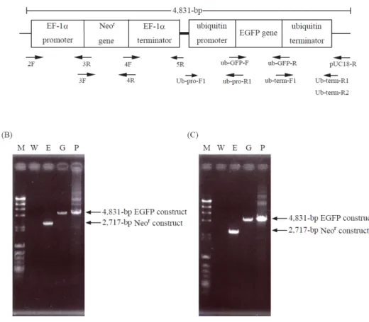
Fig. 1-2. Genomic PCR of transformants harboring EGFP gene.
(A), thraustochytrid-specific exprerssion construct containing Neor and EGFP genes (EGFP construct) with sites for primers used (arrows). Neor and EGFP genes were drived with thraustochytorid-derived EF-1α or ubiquitin promoter/terminator, respectively. (B), Genomic PCR of A. limacinum transformants. (C), Genomic PCR of T. aureum transformants. M, λHind III digest/φX174 Hinc II digest; W, wild type of A. limacinum or T. aureum; E, Neor construct transformants; G, EGFP construct transformants; P, Positive control (EGFP construct). The details are shown in MATERIALS AND METHODS.
36
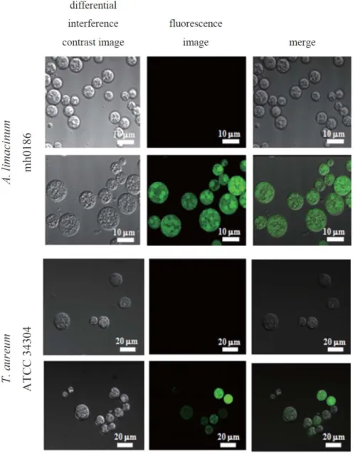
Fig. 1-3. Detection of EGFP-derived fluorescence in transformants.
Images of A. limacinum mh0186 and T. aureum ATCC 34304 transformed with the Neor construct (control, upper) or EGFP construct (lower). The details are shown in MATERIALS AND METHODS.
37
Table 1-1. PCR primers used in this chapter.
5R includes Pst I site (underlined).
Ub-pro-F1 and Ub-term-R2 also have Kpn I site (underlined).
38
CHAPTER 2
Molecular cloning of a Pinguiochrysis pyriformis oleate-specific microsomal
∆12-fatty acid desaturase and functional analysis in yeasts and thraustochytrids
2-1. INTRODUCTION
A body of accumulating evidence shows the cardiovascular benefits of omega-3 polyunsaturated fatty acids (PUFA) such as eicosapentaenoic acid (EPA, C20:5∆5, 8, 11, 14, 17) and docosahexaenoic acid (DHA, C22:6∆4, 7, 10, 13, 16, 19
) (75). Actually, cardiac societies including the American Heart Association and the European Society for Cardiology recommend the intake of 1 g/day of EPA and DHA for the prevention of cardiovascular disease and sudden cardiac death (76). Additionally, DHA, a major fatty acid of phospholipids in the human brain and retina, is thought integral to the growth and development of the brain (77). The major source of EPA and DHA is fish oils such as sardine oil but recent decreases in fish resources require a substitute (78).
This has stimulated plant biotechnology aiming to accumulate beneficial PUFA in seed oils of transgenic plants (79). An alternative approach to the production of omega-3 fatty acids may target thraustochytrids, unicellular eukaryotic marine protists including the genera Thraustochytrium, Ulkenia, and Aurantiochytrium (formerly Schizochytrium) (80). Thraustochytrids are known to accumulate PUFA especially DHA and omega-6 docosapentaenoic acid (DPA, C22:5∆4, 7, 10, 13, 16
), mainly in their lipid droplets.
Compared to plants such as arabidopsis and tobacco, however, basic genetic information is still lacking for thraustochytrids.
39
In the present study, a cDNA encoding a putative fatty acid desaturase (PpDes12) was isolated from the marine microalga Pinguiochrysis pyriformis MBIC 10872 belonging to a new class of Pinguiophyceae, which was found to accumulate EPA in cells (28). The PpDes12 was identified to be a microsomal ∆12-fatty acid desaturase which converts oleic acid (OA, C18:1∆9) to linoleic acid (LA, C18:2∆9, 12). The
∆12-fatty acid desaturase is a key enzyme in the standard (desaturase/elongase) pathway
for production of omega-3 as well as omega-6 fatty acids (Fig. 2-1A). To express the PpDes12 in thraustochytrids, a construct driven by the ubiquitin promoter from Thraustochytrium aureum ATCC 34304 was used. A. limacinum mh0186 transformed with the PpDes12 gene, but not with empty construct, converted exogenously added OA to LA, indicating that the gene product functions as a ∆12-fatty acid desaturase in thraustochytrids. This report, the first to describe the heterozygous expression of a fatty acid desaturase in thraustochytrids, could facilitate a genetic approach to the synthesis of fatty acids in thraustochytrids.
40
2-2. MATERIALS AND METHODS
Materials
TOPO TA Cloning vector was purchased from Invitrogen (California, USA).
Lambda cDNA Library Construction Kits was purchased from Stratagene (California, USA). Synthetic oligonucleotides and all other reagents are the same as described in CHAPTER 1.
Strains and culture
P. pyriformis MBIC 10872 was obtained from the Marine Biotechnology Institute, Kamaishi (Japan) and American Type Culture Collection (USA), respectively. T.
aureum ATCC 34304 and A. limacinum mh0186 were obtained and cultured as described in CHAPTER 1.
Molecular cloning of PpDes 12 from P. pyriformis MBIC 10872
P. pyriformis was grown at 25℃ in ESM medium (81). Cells in a late logarithmic phase of growth were harvested by centrifugation (6,000 x g, 4℃, 15 min), and total RNA was extracted by the phenol-SDS method (82). Poly(A)+RNA was purified and subjected to the first-strand cDNA synthesis. A pair of degenerate primers targeting the conserved region for fatty acid desaturases, F1 (5’-GGI TGG MGI ATH WSI CAY MGN ACI CAY CA-3’; corresponding to the amino acid sequence GWRISHRTHH) and R1 (5’-CCR TAR TCN CKR TCN AYI GT-3’; corresponding to T(V/I)DRDYG).
PCR was then performed using these primers with first-strand cDNA as a template (PCR cycle: 95℃/30 s, 50℃/30 s, 68℃/2 min, 40 cycles). The amplified PCR
41
products were subcloned into the TOPO TA Cloning vector and sequenced. The sequence of an insert showed high identity to known ∆12-fatty acid desaturases, and thus was used as a probe to screen a cDNA library of P. pyriformis MBIC 10872. A cDNA library was constructed using Lambda cDNA Library Construction Kits. Phage was packaged and used to infect Escherichia coli XL1-Blue MRF’. Subsequently, a cDNA library was screened by plaque hybridization with a HRP-labeled probe prepared by ECL Direct Nucleic Labeling. After several rounds of screening, positive clones were excised as a pBluescript SK (-) phargemid by in vivo excision. Finally, a full-length cDNA clone encoding ∆12-fatty acid desaturase, named PpDes12, was obtained. The plasmid containing PpDes12 cDNA was designated pBCN8.
Expression of PpDes12 in yeasts
A cDNA of PpDes12 ORF was amplified by PCR using a 5’ primer containing a Hind III site (P.pyr-F, 5’-TTA AGC TTC AAA ATG TCT CGT GGA GGA AAC CTC TC-3’) and a 3’ primer containing a Xba I site (P.pyr-R, 5’-GTC TAG ATT TAG TCG TGC GCC TTG TAG AAC A-3’), and pBCN8 as a template (94℃/30 s, 61℃/30 s, 72℃/2 min, 30 cycles). The PCR-amplified PpDes12 ORF was digested with Hind III and Xba I, and cloned into the same sites of pYES2/CT (Invitrogen). The resulting PpDes12-expression vector, designated pYp∆12Des, was introduced into the Saccharomyces cerevisiae INVSc1 (Invitrogen) by the lithium-acetate method (83).
The transformants were selected by plating on synthetic agar plates lacking uracil (SC-ura). S. cerevisiae harboring PpDes12 was cultured in uracil-lacking SC medium containing 2% glucose at 25℃ for 3 days, and then cultured for an additional 1 day in uracil-lacking SC medium containing 2% galactose. Cells were collected by
42
centrifugation at 3,500 g for 10 min.
Western blotting of FLAG-tagged PpDes12
The Flag tag sequence was inserted just after the initiation codon of PpDes12 gene by PCR. The PCR was conducted using a forward primer containing the FLAG tag sequences (P.pyr-FLAG-F, 5’-CTA AGC TTC AAA ATG GAT TAC AAG GAT GAC GAT GAC AAG TCT CGT GGA-3’) and reverse primer (P.pyr-R, 5’-GTC TAG ATT TAG TCG TGC GCC TTG TAG AAC A-3’). The underline and italics indicate the Hind III site and FLAG tag sequence, respectively. The PCR fragment was directly subcloned into the yeast expression vector pYES2/CT and subsequencely introduced into S. cerevisiae by the method described above. After incubation of the transformant in SC-ura medium, the cells were harvested and suspended in 0.1 M potassium phosphate buffer, pH 7.2, containing 0.33 M sucrose, 0.1% BSA, 1000 units/ml catalase, and a protease inhibitor coctail (Roche Diagnostics K.K.). Glass beads were added and the resultant slurry was sonicated for 20 sec and centrifuged (3,000 x g for 10 min).
The supernatant (cell lysate) was centrifuged at 100,000 x g for 60 min. The supernatant was used as a cytosolic fraction and the resultant pellet was suspended in 0.1 M potassium phosphate buffer, pH 7.2, containing glycerol (20% by vol.) and used as a microsomal fraction. Ten micrograms of protein was loaded onto a 10%
SDS-PAGE gel and transferred to a PVDF membrane (0.45 µm) using a Bio-Rad Trans-BlotⓇ SD Cell. The membrane was incubated with 5% (w/v) skim milk in TBS buffer containing 0.1% Tween 20 (Tween-TBS) for 1 hour at room temperature, washed with Tween-TBS three times, and incubated at room temperature for 3 hour with an anti-DYKDDDDK tag monoclonal antibody (Wako, Osaka, Japan, 1:5000). It was
43
then washed with Tween-TBS 3 more times and incubated for 3 hours at room temperature with an HRP-conjugated anti-mouse IgG [H+L] goat antibody (Nacalai Tesque; 1:10000). The membrane was again washed with Tween-TBS 3 times.
Protein expression was visualized using a peroxidase staining kit (Nacalai Tesque, Kyoto, Japan; 1:20).
Expression of PpDes12 in thraustochytrids
To express the PpDes12 gene in thraustochytrids, an expression construct (Neor/PpDes12 construct, Fig. 2-6A) was prepared. For control, PpDes 12 gene with ubiquitin promoter/terminator was omitted from the expression construct (Neor construct, Fig. 2-6B). The EF-1α promoter/terminator and ubiquitin promoter/terminator were obtained from T. aureum ATCC 34304. The codons of Neor were adjusted according to the codon usage of T. aureum ATCC 34304 (84). The primers for PCR amplification of these sequences are shown in supplemental Table 2-2.
The expression construct was introduced into A. limacinum mh0186 cells by electrophoration as described in CHAPTER 1. The cells were then immediately re-suspended in 1 ml of GY medium and incubated at 25℃ for 1 day, and spread on potato-dextrose agar plates containing G418 at 0.5 mg/ml. After incubation at 25℃
for 2-5 days, colonies that appeared on the plates were regarded as putative transformants. A. limacinum mh0186 harboring PpDes12 gene was cultured in GY medium at 25℃ for 4 days. Cells were collected by centrifugation at 3,500 g for 10 min.
44
Genomic PCR and southern blot hybridization of thraustochytrid transformants Genomic PCR was performed using the forward primer 2F and reverse primer pUC18-R (Table 2-2) (96℃/2 min, 98℃/20 sec, 60℃/30 sec, 72℃/5 min, 30 cycles).
For Southern blot hybridization, 5 µg of genomic DNA was digested at 37℃ with Xba I overnight. The digested DNA was separated on 1% agarose gel and transferred onto a Hybond-N+. The membrane was hybridized with a probe prepared using the DIG DNA Labeling Kit (Roche Diagnostics K.K.). PCR primers used were PD12d-probe-F (5’-CTG CCC GGC CCG CCG CGA CGA CTA-3’) and PD12d-probe-R (5’-CGG CGT GAA GCT ACG GTC GAT GGT-3’). Genomic DNA hybridized with probe was detected with an anti-Digoxigenin-AP Fab fragment and an NBT/BCIP stock solution (Roche Diagnostics K.K.).
RT-PCR of Neor and PpDes12 in the thraustochytrid transformants
Total RNA was prepared from transformants, grown in GY medium containing appropriate amounts of G418, with a Sepasol RNA I Super (Nacalai Tesque), RNeasy Mini Kit (QIAGEN) and DNase I (Takara Bio Inc.), and used to produce first-strand cDNA with PrimeScriptTM Reverse Transcriptase (Takara Bio Inc.). PCR was performed using the forward primer 3F and reverse primer 4R for amplification of Neor cDNA and forward primer ub pro-D12d-F and reverse primer ub term-D12d-R for amplification of PpDes12 cDNA (96℃/2 min, 98℃/20 sec, 60℃/30 sec, 72℃/90 sec, 30 cycles) (Table 2-2).
Fatty acid analysis
The preparation and extraction of fatty acid methyl esters (FAME) were carried out as
45
described previously (17). The resulting FAMEs were analyzed by gas
chromatography (GC) by the method described in (85). The FAMEs were also subjected to gas chromatography-mass spectrometry (GC-MS) using a Shimadzu GC-MS QP-5000 (SHIMADZU Co., Kyoto, Japan) equipped with a capillary column (DB-1, 0.25 mm i.d. x 30 m, film thickness 0.25 µm, Agilent). The column
temperature was programmed to increase at 4℃/min from 160℃ to 260℃. The injection-port temperature was 250℃. The rate of conversion of substrates to products was calculated as follows; conversion rate (%) = GC peak area of product / (GC peak area of product + GC peak area of substrate) x 100. Furthermore, picolinyl esters were prepared from the FAME as described previously (86) and subjected to GC-MS using the equipment described above. The column temperature was programmed to increase at 2.5℃/min from 240℃ to 260℃ and maintained for 15 min, then increased at
2.5℃/min to 280℃.
46
2-3. RESULTS
Molecular cloning of PpDes12 from P. pyriformis MBIC 10872
P. pyriformis MBIC 10872 has been reported to accumulate omega-3 PUFA, especially EPA, in cells (28). In this study, the author isolated the cDNA fragment (516-bp) of a putative desaturase (PpDes12) from this organism by degenerate PCR as described in MATERIALS AND METHODS. The DNA fragment was used as a probe to isolate a full-length cDNA clone through plaque hybridization with a P. pyriformis MBIC 10872 cDNA library. After the screening of 5.5 x 10⁵ recombinants, a cDNA clone including the putative PpDes12 ORF was isolated and designated pBCN8.
Nucleotide and deduced amino acid sequences of PpDes12
The author sequenced 1,494 nucleotides of pBCN8, and found a 1,314-bp ORF of PpDes12 encoding a putative 437 amino acid residues. As shown in Fig. 2-2, the deduced amino acid sequence of PpDes12 exhibited a high degree of identity with fungal and protozoan ∆12-fatty acid desaturases, such as those from Mortierella alpina (43.4%) (87), Mucor circinelloides (45.3%) (88), Rhizopus oryzae (44.6%) (89), Saprolegnia diclina (48.0%) (90) and Trichoderma brucei (37.2%) (91) (the number in parentheses shows the identity relative to PpDes12). Three histidine boxes, conserved in almost all fatty acid desaturases, were found in the deduced amino acid sequence of PpDes12 (Fig. 2-2, underlined), whereas the cytochrome b5 motif, characteristic of front-end desaturases, was not.
47
Phylogenetic analysis
∆12- and ∆12/∆15-fatty acid desaturases have been classified into the following
groups based on sequence similarity: a fungal & protozoan group, a plant group, a cyanobacterial group, and a chloroplast-localized plant group. The evolutionary relationship between PpDes12 and other ∆12- and ∆12/∆15-fatty acid desaturases was examined in a phylogenetic analysis. PpDes12 was found to be clustered with the fungal & nematode group in which it was most closely related to the S. diclina
∆12-fatty acid desaturase (Fig. 2-3).
Expression of PpDes12 in S. cerevisiae
To clarify the function of PpDes12, a PpDes12-expression construct (pYp∆12Des) and an empty-control construct (pYES2/CT) were separately introduced into the INVSc1 strain of S. cerevisiae and the fatty acid composition of pYp∆12Des and mock transformants was analyzed by GC using fatty acid methyl esters. The peak corresponding to standard LA (18:2∆9, 12) methyl ester was found in pYp∆12Des transformants but not in mock transformants, although OA (18:1∆9), the precursor of LA, was found in both transformants (Fig. 2-4A, B). On the other hand, amounts of endogenous palmitic acid (C16:0), stearic acid (C18:0) and palmitoleic acid (C16:1∆9) were unchanged in pYp∆12Des and mock transformants. GC-MS of the new peak in pYp∆12Des transformants revealed its molecular mass (m/z 294) and fragmentation pattern to be identical to those of the standard LA methyl ester (Fig. 2-4C, D). The rate of conversion of OA to LA was calculated to be 14.3 ± 2.71 % under the conditions used (average from duplicate experiments using 3 different transformants). These results indicate that endogenous OA was converted to LA in pYp∆12Des transformants.
48
However, no double bonds were introduced into myristoleic acid (14:1∆9), palmitoleic acid (16:1∆9), elaidic acid (18:1∆9 trans), LA, γ-linolenic acid (C18:3∆6, 9, 12
), dihomo-γ-linolenic acid (C20:3∆8, 11, 14
), arachidonic acid (C20:4∆5, 8, 11, 14
) and docosatetraenoic acid (C22:4∆7, 10, 13, 16
) when they were added to the culture of pYp∆12Des or mock transformants at 40 µM (data not shown). Taken together, the PpDes12 gene of P. pyriformis MBIC 10872 encodes a ∆12-fatty acid desaturase that catalyzes the conversion of OA to LA by introducing a double bond at the ∆12 position of OA.
Western blotting of FLAG-tagged PpDes12 expressed in the yeasts
The author examined the expression of PpDes12 at the protein level. Yeast cells expressing FLAG-tagged PpDes12 were lysed and fractionated into a microsomal fraction and cytosolic fraction, which were subjected to Western blotting using anti DYKDDDDK-tag antibody. A 51.1-kDa protein band was detected in the cell lysate and microsomal fraction but not cytosolic fraction (Fig. 2-5). The molecular weight (51.1-kDa) was well consistent with that estimated from the deduced amino acid sequence of the desaturase with a FLAG tag. This result indicates that PpDes12 can be classified as a microsomal fatty acid desaturase.
Expression of PpDes12 in A. limacinum
Thraustochytrids are potentially an alternative to fish for the production of omega-3 PUFAs (80). However, the genetic approach to the synthesis of fatty acids in thraustochytrids has not been fully established due to a lack of molecular tools for gene manipulation. In this study, the author designed a thraustochytrid-specific expression
49
construct to express the heterozygous gene in thraustochytrids using a promoter and a terminator of house-keeping genes derived from T. aureum ATCC 34304. To select the transformants, the author used neomycin (G418) and a neomycin-resistance (Neor) gene after adjusting the codons according to the codon usage of T. aureum ATCC 34304.
To confirm whether the PpDes12 is able to function in thraustochytrids, a Neor/PpDes12-expression construct (Fig. 2-6A) and a Neor control construct (Fig. 2-6B) were separately injected into A. limacinum mh0186 by electroporation. Transformants grown on a G418-containing GY agar medium were subjected to genomic PCR to examine whether a full-length Neor/PpDes12 DNA was integrated into the genome of the mh0186 strain. As shown in Fig. 2-6C, a 5,425-bp PCR product (corresponding to Neor/PpDes12 construct, Fig. 2-6A) was detected in the Neor/PpDes12 transformants, whereas a 2,717-bp PCR product (corresponding to Neor construct, Fig. 2-6B) was amplified for control Neor transformants. Southern blot hybridization using a PpDes12 DNA probe confirmed that the PpDes12 gene was integrated into the mh0186 genome (Fig. 2-6D). Furthermore, RT-PCR revealed that transcripts of both Neor gene (835-bp) and PpDes12 gene (1,354-bp) were present in Neor/PpDes12 transformants while the transcript of Neor gene, but not PpDes12, was detected in control Neor transformants (Fig. 2-6E and F). These results clearly indicate that the PpDes12 and Neor genes were integrated into the genome of A. limacinum mh0186 and then translated to the respective mRNA.
Finally, the fatty acid composition of Neor/PpDes12 transformants and control Neor transformants was analyzed by GC using methyl ester derivatives. The peak corresponding to standard LA methyl ester appeared in Neor/PpDes12 transformants (Fig. 2-7B) but not in control Neor transformants (Fig. 2-7A) after adding OA to the
50
culture of both transformants. GC-MS of this new peak revealed its molecular mass (m/z) and fragmentation pattern to be identical to those of the LA picolinyl ester (Fig.
2-7C). The rate of conversion of OA to LA was calculated to be 7.28 ± 1.33 % (average from duplicate experiments using 5 different transformants). No significant change in fatty acid composition except OA and LA was observed in Neor/PpDes12 transformants, compared to control Neor transformants (data not shown).
Additionally, 14C-LA was detected in Neor/PpDes12 transformants but not in control Neor transformants when 14C-oleoyl-CoA was added to the culture of transformants (Fig.
2-8).
Collectively, the Pinguiochrysis gene encoding PpDes12 was integrated into the genome of A. limacinum mh0186 (Fig. 2-6C and D), translated into PpDes12 mRNA (Fig. 2-6F) and functioned as a ∆12-fatty acid desaturase in thraustochytrid cells (Fig.
2-7).
51
2-4. DISCUSSION
In this study, the author cloned a putative fatty acid desaturase (PpDes12) gene from P. pyriformis MBIC 10872 that accumulates omega-3 PUFAs especially EPA (28).
The gene was found to encode an enzyme capable of catalyzing the introduction of a double bond at the ∆12 position of OA but not other fatty acids tested. Western blotting of FLAG-tagged PpDes12 expressed in the yeast revealed that the enzyme was recovered in the microsomal fraction. Furthermore, analysis using TMHMM (http://www.cbs.dtu.dk/services/TMHMM/) suggested that the enzyme has two transmembrane domains. These results indicate that PpDes12 is an oleate-specific microsomal ∆12-fatty acid desaturase. The deduced amino acid sequence of PpDes12 contains three histidine boxes (Fig. 2-2, underlined), commonly conserved in fatty acid desaturases. This region may act as di-iron co-ordinating centers for catalytic activity (54). Meanwhile, PpDes12 possesses no cytochrome b5-like domain which is usually present in front-end desaturases and functions as an electron donor. It has been reported that a T. brucei oleate desaturase did not carry a cytochrome b5-like domain but might use a microsomal cytochrome or the cytochrome b5-like domain of other desaturases as an electron donor (91). PpDes12 could accept electrons in a similar manner to the T. brucei oleate desaturase.
The phylogenetic analysis of ∆12- and bifunctional ∆12/∆15-fatty acid desaturases by the maximum-likelihood method (92) revealed that PpDes12 is a member of a fungal &
nematode ∆12-fatty acid desaturase group. Among the organisms belonging to this group, only P. pyriformis is a “photosynthetic” stramenopile. Although PpDes12 was recovered in the microsomal fraction when expressed in the yeast (Fig. 2-5), its
52
intracellular distribution remains to be clarified. However, PpDes12 could be present in chloroplasts like other ∆12-fatty acid desaturases of higher plants, because the strain MBIC 10872 cells used in this study have one or two typical chloroplasts and accumulate PUFA in the chloroplasts (28, 29). It is worth noting that the activity of
∆12-fatty acid desaturase could not be detected in vitro using the cell lysate or microsomal fraction as an enzyme source possibly because of difficulty with the solubilization of the protein. Thus, reconstitution of the enzyme reaction in vitro remains to be achieved.
Although thraustochytrids accumulate PUFA mainly in lipid droplets, their pathway for production of PUFA has not been well documented. Accumulating evidence, however, suggests that two distinct pathways of fatty acid synthesis are present in thraustochytrids, i.e., polyketide synthase-like (PUFA synthase) and the desaturase/elongase (standard) pathway. The former pathway has been well documented in marine bacteria (93) and thraustochytrids (30), and the latter, in animals from nematodes to mammals (94, 95). It is worth noting that targeted mutagenesis of a PUFA synthase gene of Schizochytrium sp. resulted in auxotrophic mutants that required supplementation with PUFA (26). This result suggests that the regular pathway in Schizochytrium sp. was not capable of synthesizing adequate amount of PUFA under the conditions used, probably due to the absence of a ∆12-fatty acid desaturase (26). The author also found in the present study that A. limacinum mh0186 does not have OA and LA, the former being the substrate of ∆12-fatty acid desaturase, and the latter, the product of ∆12-fatty acid desaturase. Furthermore, exogenously added OA was not converted to LA in mh0186 cells until a Pinguiochrysis ∆12-fatty acid desaturase was expressed in the strain. The author’s observations may indicate that A. limacinum