九州大学学術情報リポジトリ
Kyushu University Institutional Repository
Cardiotonic actions of quercetin and its
metabolite tamarixetin through a digitalis-like enhancement of Ca2+ transients
早水, 憲吾
http://hdl.handle.net/2324/1937170
出版情報:九州大学, 2018, 博士(医学), 課程博士 バージョン:
権利関係:© 2017 Elsevier Inc.
Contents lists available atScienceDirect
Archives of Biochemistry and Biophysics
journal homepage:www.elsevier.com/locate/yabbi
Cardiotonic actions of quercetin and its metabolite tamarixetin through a digitalis-like enhancement of Ca
2+transients
Kengo Hayamizu
a,b, Sachio Morimoto
a,c,∗, Miki Nonaka
a, Sumio Hoka
b, Toshiyuki Sasaguri
aaDepartment of Clinical Pharmacology, Graduate School of Medical Sciences, Kyushu University, Fukuoka, 812-8582, Japan
bDepartment of Anesthesiology and Critical Care Medicine, Graduate School of Medical Sciences, Kyushu University, Fukuoka, 812-8582, Japan
cDepartment of Health Sciences Fukuoka, International University of Health and Welfare, Fukuoka, 831-8501, Japan
A R T I C L E I N F O
Keywords:
Quercetin Tamarixetin Digitalis Inotropic action Cardiomyocyte Ca2+transient
A B S T R A C T
The plant-derivedflavonoid, quercetin (QCT), has many biological actions, including cardioprotective actions, resulting from its antioxidant and anti-inflammatory effects. In this study, effects of QCT and its metabolites on the contraction and Ca2+transients (CaT) of mouse single cardiomyocytes were simultaneously measured and compared with those of isoproterenol and digoxin. Furthermore, cardiac function and plasma concentrations were analyzed after bolus intravenous administration of QCT in mice. QCT and its metabolite, tamarixetin, as well as isoproterenol and digoxin, enhanced the contraction and CaT of cardiomyocytes. The inotropic action of isoproterenol was accompanied by an increase in the velocities of sarcomere shortening and relengthening and CaT decay through activation of cAMP-dependent protein kinase; however, no such lusitropic effects accom- panied the inotropic action of QCT, tamarixetin or digoxin. Intravenous administration of QCT to mice resulted in a sustained increase in cardiac systolic function; QCT was rapidly metabolized to tamarixetin and its plasma concentration was maintained at high levels over a similar time frame as the enhancement of cardiac systolic function. These results suggest that QCT exerts a cardiotonic action in vivo at least, in part, through digitalis-like enhancement of CaT by itself and its metabolite tamarixetin.
1. Introduction
Quercetin (QCT) (3, 3′, 4′, 5, 7-pentahydroxyflavon) is a powerful anti-oxidantflavonoid found in fruits and vegetables[1]. Epidemiolo- gical studies indicated that a higher dietary intake of QCT was asso- ciated with lower cardiovascular disease-related morbidity and mor- tality [2–4]. In vitro studies demonstrated that QCT and its related flavonoids inhibited oxidative modification of LDL by scavenging re- active oxygen species (ROS)[5,6]. In addition to its anti-oxidant action, QCT has been reported to interfere with many biochemical signaling pathways involving physiological and pathological processes[7].
Doxorubicin (Dox), an anthracycline antibiotic, is commonly used for treating a wide range of cancers, but it exerts severe adverse effects on non-tumor tissues, including the heart, through disruption of mi- tochondrial function by ROS generation [8–10]. Several studies in- dicated that QCT protected cardiomyocytes against Dox-induced
toxicity through its antioxidant activity, iron chelation, and inhibition of carbonyl reductase[11]or through up-regulation of Bmi-1, a protein that regulates mitochondrial function and ROS generation[12]. In a previous study, however, we found that Dox treatment of isolated cardiomyocytes directly suppressed their contraction and that QCT not only antagonized the Dox-induced inhibition of cardiomyocyte con- traction, but also enhanced it far beyond pre-treatment levels, which could not be explained by any known actions of QCT, including its anti- oxidant action (unpublished data; Supporting informationFig. 1S).
In this study, we demonstrated that QCT had a strong positive in- otropic action on isolated cardiomyocytes in vitro and mouse hearts in vivo. Further investigation to explore the mechanisms underlying the positive inotropic action of QCT indicated that QCT and its metabolite exerted a positive inotropic action both in vitro and in vivo by en- hancing CaT in cardiomyocytes in a manner similar to the cardiotonic agent, digitalis.
https://doi.org/10.1016/j.abb.2017.11.009
Received 17 June 2017; Received in revised form 11 November 2017; Accepted 15 November 2017
∗Corresponding author. Department of Health Sciences Fukuoka, International University of Health and Welfare, 137-1 Enokizu, Okawa, Fukuoka, 831-8501, Japan.
E-mail address:[email protected](S. Morimoto).
Abbreviations:bmp, beats per minute; CaT, Ca2+transients; COMT, catechol-O-methyl transferase; cTnI, cardiac troponin I; cTnT, cardiac troponin T; DCM, dilated cardiomyopathy;
DIG, digoxin; DMSO, dimethylsulfoxide; EF, ejection fraction; ERK, extracellular signal-regulated kinase; FS, fractional shortening; HR, heart rate; IκB, inhibitory of NF-κB; IL, interleukin;
ISP, isoproterenol; IVST, intraventricular septal wall thickness; JNK, cJun N-terminal kinase; LDL, low-density lipoprotein; LPS, lipopolysaccharide; LV, left ventricle; LVEDD, LV end- diastolic dimension; LVESD, LV end-systolic dimension; LVPWT, LV posterior wall thickness; MLC1, myosin light chain 1; MLC2, myosin light chain 2; MyBP-C, myosin binding protein-C;
MyHC, myosin heavy chain; NF-κB, nuclear factorκ-kinase gene in B cells; PDE, phosphodiesterase; QCT, quercetin; ROS, reactive oxygen species; PKA, protein kinase A; SL, sarcomere length; TAM, tamarixetin; TNF, tumor necrosis factor; VLDL, very low-density lipoprotein
Available online 21 November 2017
0003-9861/ © 2017 Elsevier Inc. All rights reserved.
T
2. Methods 2.1. Animals
All animal care and experimental procedures complied with the Guidelines for Animal Experiments of the Faculty of Medicine, Kyushu University, and the Law (No. 105) and Notification (No. 6) of the Japanese Government and were approved by the Committee of Ethics on Animal Experiments of the Faculty of Medicine, Kyushu University.
Studies involving animals are reported in accordance with the ARRIVE guidelines for reporting experiments involving animals [13,14]. Male C57Bl/6J wild-type mice (20–30 g; 8–12 weeks), were purchased from Charles River Laboratories Japan Inc., Japan. Male knock-in mice on the genetic background of C57Bl/6J (20–30 g; 8–12 weeks), in which three base-pairs coding for K210 in cardiac troponin T (cTnT) was heterozygously deleted from the endogenousTnnt2gene, were used as DCM model[15]. They were housed in polypropylene cages (5–6 mice in each) in a temperature- (23.0 °C) and humidity-controlled (30–50%) room with a 12 h light and dark cycle and were provided with a normal chow diet and water ad libitum.
2.2. Isolation of cardiomyocytes and simultaneous measurements of sarcomere shortening and CaT
Cardiomyocytes were isolated from the left ventricles (LVs) of the wild-type mouse hearts by collagenase treatment as described pre- viously [15]. Isolated cardiomyocytes suspended in Krebs–Henseleit solution were plated onto laminin-coated glass coverslips and incubated for 15 min at 37 °C with 5% CO2. The cardiomyocyte-attached cover- slips were then kept in oxygenated Krebs–Henseleit solution at 37 °C until used for measurements.
The cardiomyocyte-attached coverslip was mounted on a thermo- statically controlled chamber with a capacity of 1 mL located on the stage of an inverted microscope and perfused with oxygenated Krebs−Henseleit solution at 37 °C at a flow rate of 2.8 mL/min.
Cardiomyocytes were loaded with Fura-2 AM (1μM) for 10 min at 37 °C in oxygenated Krebs−Henseleit solution. Contraction was evoked by a bipolar electricalfield stimulation of 5 ms duration with a voltage ap- proximately 30% above the threshold at 1 Hz via platinum wire elec- trodes. CaT and sarcomere length (SL) changes in a single cardiomyo- cyte were simultaneously monitored through a 20 × objective lens using afluorescence and contractility recording system (MyoCam and Photo-Multiplier system with Galvo-Driven HyperSwitch Dual Excitation Light Source, IonOptix, Milton, MA, USA). Ten cycles of CaT and SL transients were averaged for waveform analysis.
The effects of QCT (Wako, Osaka, Japan), isoproterenol (Sigma- Aldrich, St. Louis, MO, USA), digoxin (Sigma-Aldrich), tamarixetin (EXTRASYNTHESE, Genay cedex, France), and isorhamnetin (EXTRASYNTHESE) on isolated cardiomyocytes were determined by dissolving these agents into the perfusates withfinal concentration of DMSO being 0.1% in the cases of QCT, digoxin, tamarixetin and iso- rhamnetin. QCT and its metabolites was found to have fluorescence emission independently of Ca2+concentration when excited at 380 nm but not 340 nm in our detecting system (Supporting information Fig. 2S), therefore, the F380 signal was corrected by subtracting the fluorescence emitted by QCT and its metabolites, which was estimated from the increase in the F380 baseline signal after application of these agents.
2.3. Preparation of skinned cardiac musclefibers and force measurements
Small bundles (0.5 mm in diameter and 7 mm long) of the left ventricular trabeculae of wild-type mice were tied to glass capillary tubes and skinned with relaxing solution containing 0.5% (w/v) Brij 58 for 30 min at 25 °C. The skinned (membrane-disrupted) trabeculae were stored at−20 °C in relaxing solution containing 50% (v/v) glycerol. A
smallfiber (120–300μm in diameter) was dissected from the skinned left ventricular trabeculae muscle and isometric force was measured as described previously[16].
2.4. QCT administration in mice
Induction of anesthesia was performed in an isolation chamber with 2% isoflurane in 100% O2and anesthesia was maintained at 1.5% in 100% O2at aflow rate of 1 L min−1via a small nose cone. QCT dis- solved in DMSO (30 mg mL−1) was intravenously administered to mice through their caudal veins at a dose of 3 mg kg−1(i.e., 100μL/kg) with a 50-μL microsyringe (Hamilton Company, Reno, NV, USA) under in- halation anesthesia with isoflurane.
2.5. Echocardiography
Transthoracic echocardiography (M-mode) was performed under inhalation anesthesia with isoflurane using a 14-MHz linear array probe with a diagnostic ultrasound system (Nemio SSA-550A, Toshiba Medical Systems, Tochigi, Japan) before and after intravenous admin- istration of QCT.
2.6. Blood plasma concentrations of QCT and its metabolites
Blood plasma concentrations of QCT and its metabolites were quantified using HPLC (Alliance 2695 Separations Module, Waters, Milford, MA, USA). Approximately 40μL of blood was taken from the caudal vein under anesthesia with 1.5% isoflurane 5 min to 6 h after intravenous administration of QCT to the same fasted mouse. Plasma was separated by centrifugation at 5000 rpm for 15 min at room tem- perature and stored at −20 °C for 30 min. Three volumes of cold ethanol (−20 °C) and 1/100 volume of 3 M NaOAc (pH 4.9) were added to plasma and stored at−20 °C for 3 h to extract water-insoluble QCT and its metabolites. The samples were then centrifuged at 13,500 rpm for 30 min at−10 °C and the supernatant was weighed and evaporated to dryness for 2 h. Evaporated samples were reconstituted with 40μL of methanol and 40μL of a mobile phase of acetonitrile/
methanol/ammonium acetate (0.6% w/v) (1:3:4 v/v/v). QCT and its metabolites were separated on a reverse-phase column (Inertsil ODS-2, 4.6 × 150 mm, GL science, Tokyo, Japan) at a flow rate of 1.3 mL min−1and monitored at a wavelength of 375 nm (Supporting informationFig. 3S).
2.7. Determination of phosphorylation levels of cardiac troponin I (cTnI) at Ser22/23 and other sarcomeric proteins
Isolated cardiomyocytes suspended in oxygenated Krebs−Henseleit solution were electrically stimulated at 1 Hz for 10 min at 37 °C in a thermostatically controlled chamber in the presence or absence of QCT, isoproterenol (ISP), and digoxin (DIG). Cardiomyocytes were then added into Laemmli's sample buffer and subjected to Western blot analysis as described previously[17], using an anti-cTnI monoclonal antibody (8E10) (HyTest, Turku, Finland) and anti-cTnI phosphoserine- 22/23 monoclonal antibody (ab8291) (Abcam, Tokyo, Japan) or phosphoprotein analysis, using Pro-Q Diamond phosophoprotein gel stain followed by SYPRO Ruby protein gel stain (Thermo Fisher Sci- entific, Yokohama, Japan).
2.8. Statistical analysis
Statistical analysis was performed using GraphPad Prism version 5.00 for Windows (GraphPad Software, San Diego, USA). Data are shown as raw data or means ± SE. Differences between mean values were analyzed by paired t-tests and one-way or repeated measures ANOVA, followed by a post hoc Dunnett's multiple comparison tests. p- values below 0.05 were considered statistically significant.
K. Hayamizu et al. Archives of Biochemistry and Biophysics 637 (2018) 40–47
41
3. Results
3.1. In vitro effects of QCT on sarcomere shortening and CaT of isolated single cardiomyocytes
Perfusion with Krebs solution containing 10μM QCT markedly en- hanced the amplitude of sarcomere shortening of isolated single car- diomyocytes, which were electrically stimulated at 1 Hz (Fig. 1A). Si- multaneous measurements of SL and CaT (F340/F380 ratio of Fura-2) revealed that QCT enhanced the amplitudes of sarcomere shortening of cardiomyocytes, while unexpectedly decreasing the CaT (Fig. 1B). This surprisingfinding suggest that QCT might directly enhance the con- tractility of cardiac myofilament, which overtakes the negative in- otropic effect of CaT attenuation. However, we found that QCT had intrinsic fluorescence emission excited at 380 nm but not at 340 nm, which only increased the F380 signal of Fura-2 independently of Ca2+
concentration in our detecting system (Supporting informationFig. 2S).
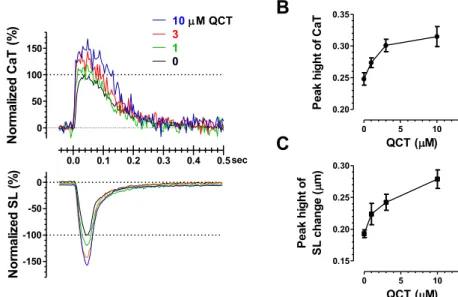
This Ca2+-independent increase in F380 by intrinsic QCTfluorescence must decrease the F340/F380 ratio. Indeed, CaT (F340/F380), in which the F380 signal was corrected by subtracting the intrinsic QCTfluor- escence, was found to be markedly increased by QCT (Fig. 1C). We then found that QCT enhanced the amplitudes of sarcomere shortening and CaT of cardiomyocytes in a similar dose-dependent manner (Fig. 2).
Peak amplitude of F340 transients of Fura-2, on which the intrinsic fluorescence of QCT has no significant effects, was also found to be increased by QCT in a dose-dependent manner, as well as digoxin, an inotropic agent increasing cardiac systolic function through enhancing the CaT in cardiomyocytes (Supporting information Fig. 4S). These results indicated that QCT exerted a positive inotropic effect on isolated cardiomyocytes through enhancement of CaT.
QCT had no effects on the phosphorylation levels of Ser22/23 of cTnI [protein kinase A (PKA) sites] and other sarcomeric proteins in isolated cardiomyocytes (Fig. 3), strongly suggesting that the inotropic
effect of QCT was not caused by mechanisms involving enhancement of SERCA2a activity by activation of the cAMP/PKA pathway through β−adrenergic receptor stimulation or phosphodiesterase (PDE) in- hibition. Consistent with thisfinding, QCT did not change the kinetics of CaT and SL transients in cardiomyocytes (Fig. 4A, D–F), whereasβ- adrenergic receptor stimulation with isoproterenol increased the phosphorylation level of cTnI, decreased the time constant of CaT decay and exerted inotropic and lusitropic effects on cardiomyocytes as manifested by an increase in the maximum amplitudes of sarcomere shortening and velocities of sarcomere shortening and relengthening (Figs. 3 and 4B, D-F). Similar to QCT, digoxin enhanced the amplitudes of CaT and sarcomere shortening without changing their kinetics and phosphorylation levels of sarcomeric proteins (Figs. 3B and 4C, D-F), suggesting that the mechanisms underlying the inotropic action of QCT were common to those of digitalis, which exerts an inotropic effect on cardiomyocytes through inhibition of plasma membrane Na+/K+-AT- Pase.
3.2. In vitro effects of QCT on the Ca2+-activated contraction of skinned cardiac musclefibers
To see whether QCT exerts its inotropic effects on cardiomyocytes through directly activating cardiac myofilaments, we next examined the effects of QCT on membrane-permeabilized (skinned) cardiac muscle. QCT had no significant effects on the Ca2+sensitivity and co- operativity in force-pCa relationships (Fig. 5A) and the maximum force- generating capability (Fig. 5B) of skinned cardiac muscle fibers, in- dicating that QCT did not directly act on the contractile apparatus of cardiomyocytes.
Fig. 1.Effects of QCT on the contraction and CaT of isolated single cardiomyocytes. A. Representative recording of SL changes evoked by electrical stimulation at 1 Hz showing a gradual increase in the amplitude of sarcomere shortening following perfusion with Krebs−Henseleit solution containing 10μM QCT. B, C. CaT and SL transients simultaneously measured in single cardiomyocytes treated with 10μM QCT before and after correcting the increase of F380 by intrinsicfluorescence of QCT, respectively.
3.3. In vivo effects of QCT administration on cardiac systolic function and plasma concentrations of QCT and its metabolites
The intravascular administration of QCT to wild-type mice in- creased cardiac systolic function as evidenced by marked increases in LV ejection fraction (EF) and fractional shortening (FS) determined by echocardiography, which were kept at high levels for at least 3 h after administration without a change in the heart rate (Fig. 6A–C). HPLC analyses of blood plasma revealed that QCT rapidly disappeared (t1/
2 = 0.80 h) and was metabolized to 4′-O-methyl QCT (tamarixetin), which remained at a high concentration for at least 3 h after adminis- tration (t1/2= 2.0 h) (Fig. 6D). 3′-O-methyl QCT (isorhamnetin), an- other water-insoluble QCT metabolite produced by catechol-O-methyl transferase (COMT), was not detected in plasma in the present study.
The intravascular administration of QCT was also found to markedly improve the systolic function of knock-in mouse model of DCM (Table 1).
The QCT metabolite, tamarixetin, had an inotropic effect similar to QCT on isolated cardiomyocytes by enhancing CaT amplitude (Fig. 7A), without changing the time constant of its decay and the maximum velocities of sarcomere shortening and relengthening (Fig. 7B). These data strongly suggested that tamarixetin played a primary role in the
prolonged inotropic effects after intravenous administration of QCT in vivo.
4. Discussion
QCT has a variety of biological actions. Oral intake of QCT inhibited platelet aggregation in humans[18]andflavonoid intake (mostly in- volving QCT) was associated with a reduced risk of death from coronary heart disease[19,20]. The cardioprotective effects of QCT are thought to be associated with its anti-inflammatory and antioxidant effects.
Treatment of macrophages with QCT attenuated the basal expression of inflammatory genes, such as tumor necrosis factor (TNF)-α, interleukin (IL)-6, IL-8, IL-1β, interferon-γ-inducible protein-10, and cycloox- ygenase-2[21]. Moreover, in human adipocytes, QCT attenuated the TNF-α-induced expression and secretion of IL-6, IL-8, and monocyte chemotactic protein-1; TNF-α–mediated phosphorylation of ERK and JNK; degradation of IκB; and TNF-α–induced NF-κB transcriptional activity[22]. In rat myocardial infarction models, QCT reduced the levels of serum LDL and VLDL cholesterol while significantly increasing serum HDL cholesterol through its antioxidant activity and lowered ST- segment elevation on ECG[23]. In addition to the biological actions of QCT based on its anti-inflammatory and antioxidant effects, the present Fig. 2.Dose-dependent effects of QCT on the contraction and CaT of isolated single cardiomyocytes. A. Representative CaT and SL tran- sients simultaneously measured in single cardiomyocytes before and after treatment with 1–10μM QCT. B. Dose dependence of the in- crease in the peak heights (peak-baseline) of CaT and SL transients caused by QCT (n = 25 cardiomyocytes).
Fig. 3.Phosphorylation levels of sarcomeric pro- teins in isolated cardiomyocytes. A. Effects of QCT on the phosphorylation level of cTnI at Ser22/23.
B. Comparison of phosphorylation levels of main sarcomeric proteins in cardiomyocytes stimulated by QCT, isoploterenol (ISP), and digoxin (DIG).
Phosphorylation of sarcomeric proteins in each lane detected with Pro-Q Diamond gel stain were normalized by MLC2 total protein in the same lane detected with SYPRO Ruby stain. Data re- present the means ± SE (n = 5 hearts).
Statistical significance was determined by one- way ANOVA. ns, not significant. MyHC, myosin heavy chain; MyBP-C, myosin binding protein-C;
MLC1, myosin light chain 1; MLC2, myosin light chain 2.
K. Hayamizu et al. Archives of Biochemistry and Biophysics 637 (2018) 40–47
43
study has revealed that QCT is a potent cardiotonic agent with a direct positive inotropic action on cardiomyocytes through augmentation of CaT. To our knowledge, this is the first report that demonstrates the cardiotonic actions of QCT and its metabolite tamarixetin (4′-O-methyl QCT) both in vitro and in vivo. QCT has no effects on the maximum force-generating capability and Ca2+ sensitivity and cooperativity of force generation in skinned cardiac musclefibers, indicating that QCT
does not directly act on the contractile machinery of cardiomyocytes, unlike the Ca2+sensitizer, pimobendan[24], and the myosin activator, omecamtiv mecarbil[25].
QCT was shown to inhibit PDE activity in a cell free system of rat adipocytes (IC50= 32μM)[26], though it conversely potentiated PDE activity in intact rat adipocytes[27]. In the present study, QCT treat- ment of cardiomyocytes did not change the phosphorylation levels of Fig. 4.Effects of QCT, ISP, and DIG on the contraction and CaT of isolated single cardiomyocytes. A. Representative CaT and SL transients simultaneously measured in single cardio- myocytes before and after treatment with 3μM QCT. B. Representative CaT and SL transients simultaneously measured in single cardiomyocytes before and after treatment with 0.1μM ISP. C. Representative CaT and SL transients simultaneously measured in single cardiomyocytes before and after treatment with 1μM DIG. D. Time constants of CaT decay before and after treatment with QCT, ISP, or DIG. E. Maximum velocities of sarcomere shortening before and after treatment with QCT, ISP, or DIG. F. Maximum velocities of sarcomere relengthening before and after treatment with QCT, ISP, or DIG. Data represent the means ± SE for 6 cardiomyocytes. Statistical significance between before and after treatment in each group was determined by pairedt-test. *p < 0.05.
Fig. 5.Effects of QCT on the mechanical properties of skinned cardiac musclefibers. A. Force-pCa relationships independently determined in the presence or absence of QCT. B. Effects of QCT on the maximum force-generating capability. Data represent the means ± SE for 5fibers from different hearts. Statistical sig- nificance was determined by unpairedt-test. pCa =−log10[Ca2+].
the PKA sites of cTnI at Ser22/23 and had no lusitropic effects known to be caused by PDE inhibition[28], strongly suggesting that activation of the cAMP/PKA pathway through PDE inhibition plays no significant role in the inotropic action of QCT on cardiomyocytes.
QCT was also shown to stimulate SERCA2a activity in isolated cardiac sarcoplasmic reticulum (SR) by suppressing the inhibitory regulation of phospholamban (PLB) at low concentrations (< 25μM), while conversely inhibiting it by competition with the nucleotide- binding sites of SERCA2a at higher concentrations[29].β-Adrenergic receptor stimulation of cardiomyocytes increases cytoplasmic levels of cAMP and activates SERCA2a by suppressing the inhibitory regulation of PLB through its phosphorylation with PKA. The present study shows that the selectiveβ-adrenergic receptor agonist, isoproterenol, increases the time constant of CaT decay and the maximum velocities of short- ening and relengthening of sarcomeres in isolated cardiomyocytes. The increase in the shortening velocity of sarcomeres should be caused by an increase in the rate of Ca2+release from the SR resulting from an enhanced accumulation of Ca2+in the SR due to SERCA2a activation.
In addition, the increase in the velocities of CaT decay and sarcomere relengthening should be synergistically caused by an increase in the rate of Ca2+reuptake into the SR due to SERCA2a activation and an increase in the off-rate of Ca2+from cardiac troponin C due to cTnI phosphorylation at Ser22/23. In our study, QCT had no effect on the time constant of CaT decay and the maximum velocities of shortening
and relengthening of sarcomeres in isolated cardiomyocytes, strongly suggesting that modification of SERCA2a activity plays no important role in the inotropic action of QCT on cardiomyocytes.
Digitalis exerts inotropic effects on cardiomyocytes by enhancing CaT though inhibition of sarcolemmal Na+/K+-ATPase, followed by suppression of the extrusion of cytoplasmic Ca2+by sarcolemmal Na+/ Ca2+exchangers owing to a decrease in the Na+gradient across the plasma membrane. QCT has been shown to inhibit purified plasma membrane Na+/K+-ATPase in vitro by inhibiting a different kinetic step in the reaction cycle from that reported for digitalis (IC50= 8μM) [30]. The present study showed that digoxin, a digitalis-derived che- mical compound used for congestive heart failure, enhanced the peak amplitude of CaT in a manner similar to that of QCT without changing the time constant of CaT decay and the maximum velocities of short- ening and relengthening of sarcomeres in isolated cardiomyocytes. This suggests that similar molecular mechanisms may underlie the inotropic actions of QCT and digoxin.
Recently, Santos et al. reported a study similar to our present study, in which QCT enhanced both the sarcomere contraction and CaT in mice isolated cardiomyocytes[31]. Surprisingly, however, they did not refer to the intrinsicfluorescence of QCT and requisite correction of the F380 intensity to reconcile the apparently contradistinctive phenom- enon of enhanced cardiomyocyte contractility with decreased CaT in experiments using Fura-2. It is unbelievable that Santos et al. [31]
noticed this important issue of the interference of Fura-2 signal with intrinsicfluorescence of QCT and intentionally did not mention it at all, making theirfindings completely unreliable. They concluded that QCT activatesβ-adrenergic receptor of cardiomyocytes from the experiments using mice left atrium but not isolated cardiomyocytes. However, our careful experiments using isolated cardiomyocytes clearly indicates that QCT has no“direct”β-adrenergic action on cardiomyocytes as evident from the phosphorylation states of cTnI and the kinetics of sarcomere shortening and relengthening and CaT decay.
Bioavailability of QCT after oral intake is low[20]. Infive human healthy volunteers, median maximum plasma concentration of QCT was reported to be only 0.4μM 6 h after the intake of 150 mg of QCT [32]. In this study, we demonstrated its strong cardiotonic action in vivo by intravenously injecting a very small volume of a high con- centration of QCT to mice to reach plasma concentrations over 1μM, where significant inotropic effect was exerted on isolated Fig. 6.Effects of intravenous administration of QCT in mice on the cardiac function and plasma concentrations of QCT and its metabolites. A-C. Time course of effects on heart rate, EF, and FS after QCT administration (n = 5 mice).
Statistical significance against before administration (time 0) was determined by repeated measures ANOVA followed by a post-hoc Dunnett's multiple comparison test.
*p < 0.05 vs time 0. D. Time course of changes in plasma concentrations of QCT and its metabolite tamarixetin (TAM) after QCT administration (n = 3 mice).
Table 1
Echocardiographic data of DCM mice before and 1 h after single i.v. injection with DMSO (vehicle) and QCT.
DMSO QCT
Before After Before After
No. of mice 5 5 5 5
HR, bpm 418 ± 36 401 ± 15 419 ± 17 436 ± 15
IVST, mm 0.58 ± 0.04 0.60 ± 0.04 0.64 ± 0.02 0.62 ± 0.05 LVESD, mm 3.63 ± 0.35 3.20 ± 0.16 3.60 ± 0.28 2.82 ± 0.23*
LVEDD, mm 4.34 ± 0.16 4.26 ± 0.16 4.32 ± 0.10 4.26 ± 0.10 LVPWT, mm 0.64 ± 0.09 0.68 ± 0.05 0.74 ± 0.05 0.74 ± 0.05
EF, % 44 ± 8 48 ± 6 44 ± 4 68 ± 5*
Data represent the means ± SE. Statistical significance was determined by unpairedt- test. *P < 0.05 vs before injection.
K. Hayamizu et al. Archives of Biochemistry and Biophysics 637 (2018) 40–47
45
cardiomyocytes. We found that a significant proportion of QCT was rapidly metabolized into tamarixetin within 5 min after injection and tamarixetin had a comparable inotropic action on cardiomyocytes. QCT has been shown to be converted to tamarixetin by COMT in platelets [33]. The time course of the decline of tamarixetin concentration in plasma was similar to that of the return of EF and FS to baseline, sug- gesting that tamarixetin plays an important role in the augmentation of cardiac systolic function after QCT injection. QCT metabolites produced by COMT have also been shown to exert a variety of cardioprotective actions. Pretreatment of macrophages with isorhamnetin decreased LPS-induced TNF-αsecretion[34]and tamarixetin inhibited collagen- stimulated platelet aggregation and serotonin secretion [35]. Iso- rhamnetin was not detected in plasma in the present study, but this metabolite also had an inotropic action similar to QCT on isolated cardiomyocytes (Supporting informationFig. 5S).
In conclusion, the present study demonstrated that QCT and its metabolite tamarixetin have a potent cardiotonic action in vivo at least, in part, through digitalis-like enhancement of CaT in cardiomyocytes in addition to antioxidant and anti-inflammatory effects. This strongly suggests that QCT might be more useful than previously thought for the treatment of cardiovascular diseases. Further investigation is warranted to explore the therapeutic efficacy of QCT for cardiovascular diseases through its cardiotonic action.
Funding
This work was supported by Grants-in-Aid for Scientific Research from Japan Society for the Promotion of Science to S. M. (No.
25670130).
Conflict of interest
The authors declare no conflict of interest.
Acknowledgements
We would like to thank Ms Hayakawa and Ms Takami for their technical assistance at the Research Support Centre, Research Centre for Human Disease Modelling, Kyushu University Graduate School of Medical Sciences.
Appendix A. Supplementary data
Supplementary data related to this article can be found athttp://dx.
doi.org/10.1016/j.abb.2017.11.009.
References
[1] J.M. Harnly, R.F. Doherty, G.R. Beecher, J.M. Holden, D.B. Haytowitz, S. Bhagwat, S. Gebhardt, Flavonoid content of U.S. fruits, vegetables, and nuts, J. Agric. Food Chem. 54 (26) (2006) 9966–9977.
[2] M.G. Hertog, D. Kromhout, C. Aravanis, H. Blackburn, R. Buzina, F. Fidanza, S. Giampaoli, A. Jansen, A. Menotti, S. Nedeljkovic, et al., Flavonoid intake and long-term risk of coronary heart disease and cancer in the seven countries study, Arch. Intern Med. 155 (4) (1995) 381–386.
[3] P. Knekt, R. Jarvinen, A. Reunanen, J. Maatela, Flavonoid intake and coronary mortality in Finland: a cohort study, Bmj 312 (7029) (1996) 478–481.
[4] P. Knekt, J. Kumpulainen, R. Jarvinen, H. Rissanen, M. Heliovaara, A. Reunanen, T. Hakulinen, A. Aromaa, Flavonoid intake and risk of chronic diseases, Am. J. Clin.
Nutr. 76 (3) (2002) 560–568.
[5] C.V. de Whalley, S.M. Rankin, J.R. Hoult, W. Jessup, D.S. Leake, Flavonoids inhibit the oxidative modification of low density lipoproteins by macrophages, Biochem.
Pharmacol. 39 (11) (1990) 1743–1750.
[6] A. Negre-Salvayre, R. Salvayre, Quercetin prevents the cytotoxicity of oxidized LDL on lymphoid cell lines, Free Radic. Biol. Med. 12 (2) (1992) 101–106.
[7] M. Sanchez, M. Galisteo, R. Vera, I.C. Villar, A. Zarzuelo, J. Tamargo, F. Perez- Fig. 7.Effects of tamarixetin (TAM) on the contraction and CaT of isolated single cardiomyocytes. A. Representative CaT and SL tran- sients simultaneously measured in single cardiomyocytes before and after treatment with 3μM TAM. B. Time constants of CaT decay and maximum velocities of sarcomere shortening and relengthening before and after treatment with TAM. Data represent the means ± SE for 5 cardiomyocytes. Statistical significance between before and after treatment was determined by paired t-test.
*p < 0.05.
Vizcaino, J. Duarte, Quercetin downregulates NADPH oxidase, increases eNOS ac- tivity and prevents endothelial dysfunction in spontaneously hypertensive rats, J.
Hypertens. 24 (1) (2006) 75–84.
[8] S.E. Lipshultz, S.R. Lipsitz, S.M. Mone, A.M. Goorin, S.E. Sallan, S.P. Sanders, E.J. Orav, R.D. Gelber, S.D. Colan, Female sex and drug dose as risk factors for late cardiotoxic effects of doxorubicin therapy for childhood cancer, N. Engl. J. Med.
332 (26) (1995) 1738–1743.
[9] K.B. Wallace, Doxorubicin-induced cardiac mitochondrionopathy, Pharmacol.
Toxicol. 93 (3) (2003) 105–115.
[10] G.C. Pereira, A.M. Silva, C.V. Diogo, F.S. Carvalho, P. Monteiro, P.J. Oliveira, Drug- induced cardiac mitochondrial toxicity and protection: from doxorubicin to car- vedilol, Curr. Pharm. Des. 17 (20) (2011) 2113–2129.
[11] H. Kaiserova, T. Simunek, W.J. van der Vijgh, A. Bast, E. Kvasnickova, Flavonoids as protectors against doxorubicin cardiotoxicity: role of iron chelation, antioxidant activity and inhibition of carbonyl reductase, Biochim. Biophys. Acta 1772 (9) (2007) 1065–1074.
[12] Q. Dong, L. Chen, Q. Lu, S. Sharma, L. Li, S. Morimoto, G. Wang, Quercetin at- tenuates doxorubicin cardiotoxicity by modulating Bmi-1 expression, Br. J.
Pharmacol. 171 (19) (2014) 4440–4454.
[13] C. Kilkenny, W. Browne, I.C. Cuthill, M. Emerson, D.G. Altman, R. National Centre for the Replacement, R. Reduction of Animals, Animal research: reporting in vivo experiments–the ARRIVE guidelines, J. Cereb. Blood Flow. Metab. 31 (4) (2011) 991–993.
[14] J.C. McGrath, E. Lilley, Implementing guidelines on reporting research using ani- mals (ARRIVE etc.): new requirements for publication in BJP, Br. J. Pharmacol. 172 (13) (2015) 3189–3193.
[15] C.K. Du, S. Morimoto, K. Nishii, R. Minakami, M. Ohta, N. Tadano, Q.W. Lu, Y.Y. Wang, D.Y. Zhan, M. Mochizuki, S. Kita, Y. Miwa, F. Takahashi-Yanaga, T. Iwamoto, I. Ohtsuki, T. Sasaguri, Knock-in mouse model of dilated cardiomyo- pathy caused by troponin mutation, Circ. Res. 101 (2) (2007) 185–194.
[16] S. Morimoto, F. Yanaga, R. Minakami, I. Ohtsuki, Ca2+-sensitizing effects of the mutations at Ile-79 and Arg-92 of troponin T in hypertrophic cardiomyopathy, Am.
J. Physiol. 275 (1 Pt 1) (1998) C200–C207.
[17] H. Nakaura, S. Morimoto, F. Yanaga, M. Nakata, H. Nishi, T. Imaizumi, I. Ohtsuki, Functional changes in troponin T by a splice donor site mutation that causes hy- pertrophic cardiomyopathy, Am. J. Physiol. 277 (2 Pt 1) (1999) C225–C232.
[18] G.P. Hubbard, S. Wolffram, J.A. Lovegrove, J.M. Gibbins, Ingestion of quercetin inhibits platelet aggregation and essential components of the collagen-stimulated platelet activation pathway in humans, J. Thromb. Haemost. 2 (12) (2004) 2138–2145.
[19] M.G. Hertog, E.J. Feskens, P.C. Hollman, M.B. Katan, D. Kromhout, Dietary anti- oxidantflavonoids and risk of coronary heart disease: the Zutphen Elderly Study, Lancet 342 (8878) (1993) 1007–1011.
[20] Y. Guo, R.S. Bruno, Endogenous and exogenous mediators of quercetin bioavail- ability, J. Nutr. Biochem. 26 (3) (2015) 201–210.
[21] A. Overman, C.C. Chuang, M. McIntosh, Quercetin attenuates inflammation in human macrophages and adipocytes exposed to macrophage-conditioned media, Int. J. Obes. (Lond.) 35 (9) (2011) 1165–1172.
[22] C.C. Chuang, K. Martinez, G. Xie, A. Kennedy, A. Bumrungpert, A. Overman, W. Jia, M.K. McIntosh, Quercetin is equally or more effective than resveratrol in
attenuating tumor necrosis factor-{alpha}-mediated inflammation and insulin re- sistance in primary human adipocytes, Am. J. Clin. Nutr. 92 (6) (2010) 1511–1521.
[23] P.S. Prince, B. Sathya, Pretreatment with quercetin ameliorates lipids, lipoproteins and marker enzymes of lipid metabolism in isoproterenol treated cardiotoxic male Wistar rats, Eur. J. Pharmacol. 635 (1–3) (2010) 142–148.
[24] M. Nonaka, S. Morimoto, T. Murayama, N. Kurebayashi, L. Li, Y.Y. Wang, M. Arioka, T. Yoshihara, F. Takahashi-Yanaga, T. Sasaguri, Stage-dependent ben- efits and risks of pimobendan in mice with genetic dilated cardiomyopathy and progressive heart failure, Br. J. Pharmacol. 172 (9) (2015) 2369–2382.
[25] M.S. Utter, D.M. Ryba, B.H. Li, B.M. Wolska, R.J. Solaro, Omecamtiv mecarbil, a cardiac myosin activator, increases Ca2+ sensitivity in myofilaments with a dilated cardiomyopathy mutant tropomyosin E54K, J. Cardiovasc. Pharmacol. 66 (4) (2015) 347–353.
[26] U.R. Kuppusamy, N.P. Das, Effects offlavonoids on cyclic AMP phosphodiesterase and lipid mobilization in rat adipocytes, Biochem. Pharmacol. 44 (7) (1992) 1307–1315.
[27] U.R. Kuppusamy, N.P. Das, Potentiation ofβ-adrenoceptor agonist-mediated lipo- lysis by quercetin andfisetin in isolated rat adipocytes, Biochem. Pharmacol. 47 (3) (1994) 521–529.
[28] F. Afzal, J.M. Aronsen, L.R. Moltzau, I. Sjaastad, F.O. Levy, T. Skomedal, J.B. Osnes, E. Qvigstad, Differential regulation of beta2 -adrenoceptor-mediated inotropic and lusitropic response by PDE3 and PDE4 in failing and non-failing rat cardiac ven- tricle, Br. J. Pharmacol. 162 (1) (2011) 54–71.
[29] E. McKenna, J.S. Smith, K.E. Coll, E.K. Mazack, E.J. Mayer, J. Antanavage, R.T. Wiedmann, R.G. Johnson Jr., Dissociation of phospholamban regulation of cardiac sarcoplasmic reticulum Ca2+ATPase by quercetin, J. Biol. Chem. 271 (40) (1996) 24517–24525.
[30] Y. Kuriki, E. Racker, Inhibition of (Na+, K+)adenosine triphosphatase and its partial reactions by quercetin, Biochemistry 15 (23) (1976) 4951–4956.
[31] M.S. Santos, E.D. Oliveira, A. Santos-Miranda, J.S. Cruz, A.N.S. Gondim, J.E.R. Menezes-Filho, D.S. Souza, L. Pinho-da-Silva, I.C.G. Jesus, D. Roman-Campos, S. Guatimosim, A. Lara, E.A. Conde-Garcia, C.M.L. Vasconcelos, Dissection of the effects of quercetin on mouse myocardium, Basic Clin. Pharmacol. Toxicol. 120 (6) (2017) 550–559.
[32] S. Egert, S. Wolffram, A. Bosy-Westphal, C. Boesch-Saadatmandi, A.E. Wagner, J. Frank, G. Rimbach, M.J. Mueller, Daily quercetin supplementation dose-depen- dently increases plasma quercetin concentrations in healthy humans, J. Nutr. 138 (9) (2008) 1615–1621.
[33] B. Wright, T. Gibson, J. Spencer, J.A. Lovegrove, J.M. Gibbins, Platelet-mediated metabolism of the common dietaryflavonoid, quercetin, PLoS One 5 (3) (2010) e9673.
[34] C. Boesch-Saadatmandi, A. Loboda, A.E. Wagner, A. Stachurska, A. Jozkowicz, J. Dulak, F. Doring, S. Wolffram, G. Rimbach, Effect of quercetin and its metabolites isorhamnetin and quercetin-3-glucuronide on inflammatory gene expression: role of miR-155, J. Nutr. Biochem. 22 (3) (2011) 293–299.
[35] B. Wright, L.A. Moraes, C.F. Kemp, W. Mullen, A. Crozier, J.A. Lovegrove, J.M. Gibbins, A structural basis for the inhibition of collagen-stimulated platelet function by quercetin and structurally relatedflavonoids, Br. J. Pharmacol. 159 (6) (2010) 1312–1325.
K. Hayamizu et al. Archives of Biochemistry and Biophysics 637 (2018) 40–47
47