M
i c e def i c i ent i n t he Shm
t 2 gene have
m
i t oc hondr i al r es pi r at i on def ec t s and ar e
em
br yoni c l et hal
著者
Tani H
ar una, O
hni s hi Saki ko, Shi t ar a H
i r os hi ,
M
i t o Takayuki , Yam
aguc hi M
i dor i , Yonekaw
a
H
i r om
i c hi , H
as hi z um
e O
s am
u, I s hi kaw
a Kaor i ,
N
akada Kaz ut o, H
ayas hi J un- I c hi
j our nal or
publ i c at i on t i t l e
Sc i ent i f i c r epor t s
vol um
e
8
page r ange
425
year
2018- 01
権利
( C) The Aut hor ( s ) 2017
Thi s ar t i c l e i s l i c ens ed under a Cr eat i ve
Com
m
ons At t r i but i on 4. 0 I nt er nat i onal
Li c ens e, w
hi c h per m
i t s us e, s har i ng,
adapt at i on, di s t r i but i on and r epr oduc t i on i n
any m
edi um
or f or m
at , as l ong as you gi ve
appr opr i at e c r edi t t o t he or i gi nal aut hor ( s )
and t he s our c e, pr ovi de a l i nk t o t he Cr
e-at i ve Com
m
ons l i c ens e, and i ndi c at e i f c hanges
w
er e m
ade. The i m
ages or ot her t hi r d par t y
m
at er i al i n t hi s ar t i c l e ar e i nc l uded i n t he
ar t i c l e’
s Cr eat i ve Com
m
ons l i c ens e, unl es s
i ndi c at ed ot her w
i s e i n a c r edi t l i ne t o t he
m
at er i al . I f m
at er i al i s not i nc l uded i n t he
ar t i c l e’
s Cr eat i ve Com
m
ons l i c ens e and your
i nt ended us e i s not per - m
i t t ed by s t at ut or y
r egul at i on or exc eeds t he per m
i t t ed us e, you
w
i l l need t o obt ai n per m
i s s i on di r ec t l y f r om
t he c opyr i ght hol der . To vi ew
a c opy of t hi s
. . .
U
RL
ht t p: / / hdl . handl e. net / 2241/ 00150985
doi: 10.1038/s41598-017-18828-3
have mitochondrial respiration
defects and are embryonic lethal
Haruna Tani
1, Sakiko Ohnishi
1, Hiroshi Shitara
1,2, Takayuki Mito
3, Midori Yamaguchi
2,
Hiromichi Yonekawa
2, Osamu Hashizume
3, Kaori Ishikawa
1,3, Kazuto Nakada
1,3&
Jun-Ichi Hayashi
4Accumulation of somatic mutations in mitochondrial DNA (mtDNA) has been proposed to be responsible for human aging and age-associated mitochondrial respiration defects. However, our previous indings suggested an alternative hypothesis of human aging—that epigenetic changes but not mutations regulate age-associated mitochondrial respiration defects, and that epigenetic downregulation of nuclear-coded genes responsible for mitochondrial translation [e.g., glycine C-acetyltransferase (GCAT), serine hydroxymethyltransferase 2 (SHMT2)] is related to age-associated
respiration defects. To examine our hypothesis, here we generated mice deicient in Gcat or Shmt2 and
investigated whether they have respiration defects and premature aging phenotypes. Gcat-deicient
mice showed no macroscopic abnormalities including premature aging phenotypes for up to 9 months after birth. In contrast, Shmt2-deicient mice showed embryonic lethality after 13.5 days post coitum (dpc), and ibroblasts obtained from 12.5-dpc Shmt2-deicient embryos had respiration defects and retardation of cell growth. Because Shmt2 substantially controls production of N-formylmethionine-tRNA (fMet-N-formylmethionine-tRNA) in mitochondria, its suppression would reduce mitochondrial translation, resulting in expression of the respiration defects in ibroblasts from Shmt2-deicient embryos. These indings support our hypothesis that age-associated respiration defects in ibroblasts of elderly humans are caused not by mtDNA mutations but by epigenetic regulation of nuclear genes including SHMT2.
Because mitochondria produce reactive oxygen species endogenously and preferentially accumulate exogenous chemical carcinogens, mitochondrial DNA (mtDNA) is exposed to these mutagens, resulting in accumulation of somatic mutations with age1–5. Some of these somatic mutations in human mtDNA are pathogenic, because the same mutations are found in patients with mitochondrial diseases caused by mitochondrial respiration defects. herefore, the mitochondrial theory of aging1–5 proposes that accumulation of somatic mutations in mtDNA is responsible for human aging and age-associated mitochondrial respiration defects.
However, it is also possible that abnormalities in nuclear DNA but not in mtDNA induce age-associated mitochondrial respiration defects, because both nuclear DNA and mtDNA encode proteins required for mito-chondrial respiratory function1. To determine which genome, nuclear or mitochondrial, is responsible for the respiration defects in the ibroblasts of elderly humans, we previously carried out intercellular transfer of mtDNA6 or nuclear DNA7 by using mtDNA-less HeLa cells8; the results led us to propose that nuclear recessive mutations induce the age-associated respiration defects. In contrast, the mitochondrial theory of aging has been supported by studies of mtDNA mutator mice9,10, which were generated by introducing a proofreading-deicient mtDNA polymerase gene. hese mice showed accelerated accumulation of somatic mutations in mtDNA, resulting in accelerated expression of respiration defects and premature aging phenotypes9,10.
herefore, it has been controversial whether human aging and age-associated respiration defects are con-trolled by the accumulation of somatic mutations in mtDNA9,10 or by nuclear recessive mutations7. More recently,
Graduate School of Life and Environmental Sciences, University of Tsukuba, - - Tennodai, Tsukuba, Ibaraki, - , Japan. Laboratory for Transgenic Technology, Tokyo Metropolitan Institute of Medical Science, - - Kamikitazawa, Setagaya-ku, Tokyo, - , Japan. Faculty of Life and Environmental Sciences, University of Tsukuba, - - Tennodai, Tsukuba, Ibaraki, - , Japan. University of Tsukuba, - - Tennodai, Tsukuba, Ibaraki, - , Japan. Haruna Tani, Sakiko Ohnishi and Hiroshi Shitara contributed equally to this work. Correspondence and requests for materials should be addressed to J.-I.H. (email: jih @biol.tsukuba.ac.jp) Received: 15 August 2017
Accepted: 12 December 2017
www.nature.com/scientificreports/
epigenetic regulation of cellular senescence has been proposed in human fibroblasts11. Our recent study12 addressed these issues by deep sequencing analysis of mtDNA and showed that mtDNA in ibroblasts from elderly humans does not accumulate somatic mutations. Moreover, reprogramming of these ibroblasts by gen-erating induced pluripotent stem cells (iPSCs) restores normal respiratory function12. his led us to hypothesise that age-associated respiration defects are controlled not by mutations in either nuclear or mtDNA, but by epi-genetic regulation of nuclear genes. Our microarray screening results suggest that epiepi-genetic downregulation of the nuclear genes glycine C-acetyltransferase (GCAT) and serine hydroxymethyltransferase 2 (SHMT2) is involved in age-associated respiration defects of the ibroblasts of elderly humans12. Because the products of both genes are localized in mitochondria and regulate glycine production in mitochondria13,14, their downregulation would induce defects in mitochondrial translation and respiratory function, resulting in the age-associated respiration defects found in the ibroblasts of elderly humans6,7. To examine this possibility, we generated mice deicient in Gcat or Shmt2, and investigated whether these mice would have mitochondrial respiration defects and premature aging phenotypes.
Results
Generation of mice deicient in the
Gcat
or
Shmt2
genes.
We generated knockout mouse strains deicient in the Gcat gene or the Shmt2 gene by using the CRISPR/Cas9 system. Target sequences were designed according to the mouse Gcat and Shmt2 sequences (Supplementary Fig. S1). Cas9 mRNA and single-guide RNAs (sgRNAs) were synthesized as reported previously15, and were microinjected into fertilized eggs (pronuclear stage) from C57BL/6J (hereater referred to as B6J) mice. he microinjected eggs were transferred to the oviducts of pseudo-pregnant females.In the case of Gcat knockout mice, 41 of 70 mice were mutation-positive in the Surveyor assay (see Methods). We analysed the sequence around the target region in the mice with mutations and selected one male mouse with an insertion and a deletion that would disrupt Gcat gene function (Supplementary Fig. S1); we used this mouse as a founder for further breeding to obtain heterozygous (Gcat m/+) females and males. By mating heterozygous females with heterozygous males, we obtained 34 pups. Genotyping showed that 11 pups had no mutation, 19 were heterozygous, and 4 were homozygous (Gcat m/m) (Fig. 1a). We then obtained ofspring (Gcat+/+, m/+, m/m) by in vitro fertilization using heterozygous females and a heterozygous male.
In the case of Shmt2 knockout mice, 20 of 25 mice were mutation-positive in the Surveyor assay. We selected one female mouse with a single-nucleotide insertion (T) resulting in a frame shit that would disrupt Shmt2 gene function (Supplementary Fig. S1) and used this mouse as a founder for further breeding to obtain heterozygous (Shmt2 m/+) females and males. By mating heterozygous females with heterozygous males, we obtained 45 pups. Genotyping by Xcm I digestion of the PCR products showed that 14 mice had no mutation and 31 were heterozy-gous, but no mice had a homozygous mutation (Fig. 1b), indicating the lethality of embryos with a homozygous mutation in Shmt2 (Shmt2 m/m).
Characterization of mice deicient in the
Gcat
gene.
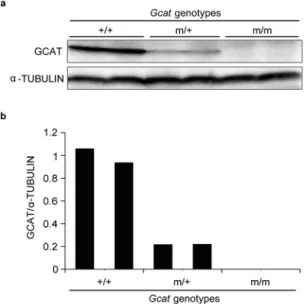
Because the Gcat m/m mice did not show embry-onic lethality, we performed Western blot analysis to conirm the suppression of the Gcat gene in these mice. he GCAT protein was not detectable in livers of 5-month-old Gcat m/m mice, but was detectable in wild-type (+/+) or heterozygous (m/+) mice (Fig. 2). hese observations conirm complete absence of the GCAT protein in Gcatm/m mice.
However, no growth retardation or obvious macroscopic abnormalities including premature aging pheno-types, such as hair greying, alopecia, or kyphosis, were observed in Gcat m/m mice for at least 9 months ater
Figure 1. Genotyping of F1 pups obtained by mating heterozygous females and males. (a) Mutations in Gcat.
PCR products of 266 bp and 234 bp correspond to Gcat without mutations (+/+) and with mutations (m/m; see Supplementary Fig. S1 for sequences), respectively. he presence of both fragments indicates heterozygosity (m/+). Arrowhead shows an additional fragment that may represent heteroduplex molecules. (b) Mutation in
birth, even though they did not have the GCAT protein. Mice with a heterozygous mutation in the Shmt2 gene (Shmt2 m/+ mice) also showed no growth retardation or no obvious macroscopic abnormalities for at least 9 months ater birth. Further investigations would be required to determine whether these mice have any prema-ture aging phenotypes or shorter lifespan than those of mice without the mutations.
Characterization of embryos deicient in the
Shmt2
gene.
he absence of Shmt2 m/m mice among the offspring (Fig. 1b) suggested their embryonic lethality. To investigate the embryonic lethality, we used 12.5-dpc (days post coitum) and 13.5-dpc F1 embryos obtained by mating heterozygous females with heterozy-gous males. We found no macroscopic abnormalities in any of the 12.5-dpc embryos, but detected abnormalities such as small size and anaemia in three of the ten 13.5-dpc embryos (Fig. 3a), indicating that the lethality occurs ater13.5 dpc.hen, we genotyped the embryos by Xcm I digestion of PCR products. Of the ten 13.5-dpc embryos, one had no mutation, six were heterozygous, and three had homozygous mutations (Fig. 3b); the three embryos with homozygous mutations were the same as those showing macroscopic abnormalities. Western blot analysis (Fig. 3c) showed that the amounts of SHMT2 protein in embryos without the mutation were about twice those
Figure 2. Suppression of GCAT protein production in 5-month-old Gcat m/m mice. (a) Western blot analysis
of the GCAT protein in the livers of mice of the indicated genotypes. (b) Quantiication of Western blot data.
Figure 3. Lethality of 13.5-dpc F1 embryos with a homozygous mutation in Shmt2 obtained by mating
www.nature.com/scientificreports/
in heterozygous embryos. In embryos with homozygous mutations, low amounts of the SHMT2 protein were detectable (Fig. 3c), probably due to contaminating maternal cells, such as placenta or blood cells.
Examination of
Gcat
and
Shmt2
expression during embryonic stages.
A question that arises from the absence of embryonic lethality in Gcat m/m mice (Fig. 1a) is whether the expression of the Gcat gene is not required at any embryonic stage. To answer this question, we examined the levels of the GCAT and SHMT2 proteins in the placenta (foetal side), brain, and liver from 11.5-, 12.5- and 13.5-dpc embryos without mutations by Western blot analysis. Both proteins were present in all three tissues at all embryonic stages examined; their levels decreased slightly but signiicantly ater 11.5 dpc, except that the level of SHMT2 in the liver did not change signiicantly (Fig. 4). herefore, the absence of embryonic lethality in Gcat m/m mice cannot be explained by the absence of Gcat expression during embryogenesis.Isolation and characterization of mouse embryonic ibroblast lines from the embryos of
Shmt2
m/m mice.
To investigate the cause of embryonic lethality of Shmt2 m/m mice, we obtained two pregnant mice by mating heterozygous mice, and isolated a mouse embryonic ibroblast (MEF) line from each of the 12.5-dpc embryos. Genotyping showed that three lines had no mutation (Shmt2+/+ MEF), ive were heterozy-gous, and eight had homozygous mutations (Shmt2 m/m MEF). We used three Shmt2+/+ MEF lines and threeShmt2 m/m MEF lines for further investigation (Fig. 5a). he absence of the SHMT2 protein in the Shmt2 m/m MEF lines was conirmed by Western blot analysis (Fig. 5b). Given that MEF lines do not contain maternal cells, the presence of low amounts of SHMT2 in 12.5-dpc Shmt2 m/m embryos (Fig. 3c) was likely due to contamina-tion with maternal cells.
hen, we examined whether the absence of SHMT2 results in mitochondrial respiration defects by using
Shmt2+/+ MEFs and Shmt2 m/m MEFs. First, we performed biochemical analysis of respiratory function, and found the reduced activities of mitochondrial respiratory complexes in Shmt2 m/m MEFs in comparison with Shmt2+/+ MEFs (Fig. 5c). Next, we used Western blot analysis to compare the amounts of nuclear- and mtDNA-coded subunits of respiratory complexes between Shmt2+/+ MEFs and Shmt2 m/m MEFs, and found preferential decrease of mtDNA-coded subunits in Shmt2 m/m MEFs (Fig. 5d). Probably, the absence of SHMT2 (Fig. 5b) induced signiicant respiration defects (Fig. 5c) as a consequence of the reduction of one-carbon metab-olism to produce glycine and N-formylmethionine-tRNA (fMet-tRNA)13,14,16, both of which are required for the mitochondrial translation that produces mtDNA-coded subunits of respiratory complexes.
Finally, we compared the doubling times between Shmt2+/+ MEFs and Shmt2 m/m MEFs, and found sig-niicant growth retardation in Shmt2 m/m MEFs (Fig. 5e). herefore, the embryonic lethality observed in Shmt2
m/m mice is likely due to both the respiration defects and growth retardation caused by the Shmt2 deiciency.
Isolation and characterization of MEF lines from the embryos of
Gcat
m/m mice.
Next, we gen-erated MEF lines from Gcat m/m embryos, and examined their respiratory function and doubling times to inves-tigate why Gcat m/m embryos are not embryonic lethal. We obtained two pregnant mice by mating heterozygous mice, and isolated a MEF line from each of the 12.5-dpc embryos. Genotyping showed that seven lines had no mutation (Gcat+/+ MEF), seven were heterozygous, and three had homozygous mutations (Gcat m/m MEF). We randomly selected three of the seven Gcat+/+ MEF lines and the three Gcat m/m MEF lines for further investi-gation (Fig. 5a). he complete absence of the GCAT protein in Gcat m/m MEF lines was conirmed by Western blot analysis (Fig. 5b).Comparison of respiratory function and doubling time between Gcat+/+ MEFs and Gcat m/m MEFs showed no respiration defects and no growth retardation in Gcat m/m MEFs (Fig. 5c and e). Moreover, the amounts of
Figure 4. Examination of SHMT2 and GCAT protein levels in the placenta (foetal side), brain, and liver of
nuclear- and mtDNA-coded subunits of respiratory complexes did not difer substantially between Gcat+/+ MEF and Gcat m/m MEF lines (Fig. 5d). hese observations suggest that the absence of respiration defects and growth retardation in Gcat m/m MEF lines are related to the absence of embryonic lethality of Gcat m/m mice.
Figure 5. Characterization of MEF lines generated from 12.5-dpc embryos with no mutation (Shmt2+/+;
Gcat+/+) and with homozygous mutations (Shmt2 m/m; Gcat m/m). (a) Genotyping of the mutation. (b) Western blot analysis of SHMT2 protein and GCAT protein. (c) Biochemical analysis of relative enzymatic activities of mitochondrial respiratory complexes. (d) Western blot analysis of the subunits of mitochondrial respiratory complexes encoded by mtDNA (ND1, COX1) and nuclear DNA (NDUFA9, COX4, SDHA). (e) Doubling times in culture. Experiments were performed in triplicate. Data are means ± s.e.m. *P< 0.05,
www.nature.com/scientificreports/
Discussion
In our previous studies12,17, we put forward the hypothesis that age-associated respiration defects in human ibro-blasts are not due to mutations but to the epigenetic regulation, because reprogramming ibroibro-blasts from elderly humans by generating iPSCs restores normal mitochondrial respiratory function. Our hypothesis also proposed that epigenetic downregulation of human GCAT or SHMT2 or both would partly be related to age-associated respiration defects. To test this hypothesis, we generated mice deicient in Gcat or Shmt2, and examined whether suppression of these genes induces respiration defects.
Mice deicient in Shmt2 (Shmt2 m/m) showed embryonic lethality, but mice deicient in Gcat (Gcat m/m) did not (Fig. 1). To further investigate the embryonic lethality of Shmt2 m/m mice, we isolated MEF lines from 12.5-dpc embryos and showed that Shmt2 m/m MEF lines had respiration defects (Fig. 5c). Moreover, the absence of SHMT2 (Fig. 5b) reduced the amounts of the mtDNA-coded subunits of the respiratory complexes (Fig. 5d). herefore, these results are consistent with our hypothesis12,17 that epigenetic downregulation of human SHMT2 is involved in age-associated respiration defects. Probably, age-associated downregulation of human SHMT212 suppresses one-carbon metabolism to produce glycine and fMet-tRNA13,14, both of which are required for trans-lation in mitochondria, and thereby decreases the production of the mtDNA-encoded subunits of the respiratory complexes, resulting in expression of age-associated respiration defects.
Questions that then arise are why Gcat m/m mice are not embryonic lethal (Fig. 1), and why Gcat m/m MEF lines do not show respiration defects (Fig. 5c), even though both Gcat and Shmt2 genes are involved in glycine production in mitochondria13,14. his discrepancy could be resolved by assuming that embryonic lethality and the respiration defects induced by Shmt2 disruption result from fMet-tRNA depletion but not glycine depletion. he SHMT2 pathway generates fMet-tRNA via two processes; one is conversion of serine and tetrahydrofolate (THF) to glycine and 5,10-methylene-THF, and the other is conversion of the resultant glycine to 5,10-methylene-THF via the glycine cleavage system (GCS)14. In contrast, the GCAT pathway, which involves the L-threonine dehydro-genase (Tdh) gene, contributes to the generation of fMet-tRNA only via conversion of threonine to glycine fol-lowed by GCS to produce 5,10-methylene-THF14. Moreover, mouse embryonic stem cells use both SHMT2 and GCAT pathways for production of fMet-tRNA, but diferentiated tissues use the SHMT2 pathway predominantly due to inactivation of the GCAT pathway by suppression of Tdh13. Given that lethality of Shmt2 m/m embryos occurs ater 13.5 dpc (Fig. 3), it can be supposed that Shmt2 m/m embryos could not produce suicient amounts of fMet-tRNA ater 13.5 dpc due to disruption of Shmt2 and suppression of Tdh, resulting in respiration defects and embryonic lethality. In contrast, Gcat m/m embryos produce fMet-tRNA even ater 13.5 dpc due to the active SHMT2 pathway, resulting in the absence of respiration defects and embryonic lethality. Furthermore, the results of an in vitro study18 suggest that SHMT2 catalyzes not only conversion of serine to glycine but also conversion of threonine to glycine, which indicates compensation of the GCAT pathway by SHMT2 pathway; this compensa-tion could also explain the absence of respiracompensa-tion defects in Gcat m/m MEFs and the absence of lethality in Gcat
m/m embryos.
In addition to the mitochondrial respiration defects, Shmt2 m/m MEFs also showed signiicant growth retar-dation, but Gcat m/m MEFs showed neither respiration defects nor growth retardation (Fig. 5). he growth retardation of Shmt2 m/m MEFs could be due to the respiration defects, because our previous studies19,20 pro-vided evidence that respiration defects caused by pathogenic mtDNA mutations in mouse tumor cells delay their growth under the skin of syngenic B6J mice. However, it is also possible that growth retardation of Shmt2 m/m MEF is due to impaired nucleotide production, because Shmt2 also contributes to nucleotide production via one-carbon metabolism13,14, and plays an important role in growth or survival of tumor cells21–23 and immune cells24. All these observations suggest that Shmt2 disruption impairs one-carbon metabolism producing nucleo-tides and fMet-tRNA13,14, and thus could be responsible for growth retardation and respiration defects, resulting in the lethality of Shmt2 m/m embryos (Figs 1b and 3a).
At the time of writing this report, the Gcat m/m mice and Shmt2 m/+ mice (9 month-of-age) showed no macroscopic abnormalities including premature aging phenotypes, such as kyphosis, greying, or alopecia, which have been observed in the mtDNA mutator mice9,10. In a previous study25, we generated mtDNA mutator mice with the same B6J nuclear background as that of Gcat m/m or Shmt2 m/+ mice, and found that their lifespan was short and they had kyphosis but no greying or alopecia. his premature aging phenotype (kyphosis) in our mtDNA mutator m/m mice was also observed in mito-mice∆ carrying mtDNA with a large-scale deletion and having the B6J nuclear background, which we generated previously as a model for mitochondrial diseases26,27. Given that median survival times of heterozygous and homozygous mtDNA mutator mice with the B6J nuclear background were 27 and 10 months, respectively25, further studies are required to examine whether Gcat m/m and Shmt2 m/+ mice eventually have shorter lifespans than that of B6J mice and express a premature aging phe-notype of kyphosis.
Methods
Ethics statement.
All animal experiments were performed in accordance with protocols approved by the Institutional Animal Care and Use Committee of the University of Tsukuba, Japan (permit number: 15–313), and by the Animal Use and Care Committee of the Tokyo Metropolitan Institute of Medical Science (approval numbers: 14046, 15023, 16040).MEF lines and cell culture.
MEF lines were derived from the skin of 12.5-dpc embryos and grown in min-imum essential medium (Life Technologies) containing 10% foetal bovine serum (Sigma) and 1% penicillin/ streptomycin (Nacalai Tesque, Kyoto, Japan).Biochemical measurement of respiratory enzyme activity.
he activity of mitochondrial respira-tory complexes I (NADH dehydrogenase), II (succinate dehydrogenase), and III (cytochrome c reductase) was assayed as described previously28. Briely, to estimate the activity of complexes I + III, NADH and cytochrome c (oxidized form) were used as substrates, and the reduction of cytochrome c was monitored by measuring absorb-ance at a wavelength of 550 nm. To estimate the activity of complexes II + III, sodium succinate and cytochromec (oxidized form) were used as substrates, and the reduction of cytochrome c was monitored as described above. For the estimation of complex IV (cytochrome c oxidase) activity, cytochrome c (reduced form) was used as a substrate, and the oxidation of cytochrome c was measured at 550 nm.
Western blot analysis.
Proteins were separated by SDS-PAGE in 10% gels and transferred to polyvi-nylidene diluoride (PVDF) membranes. Membranes were blocked with PVDF Blocking Reagent for Can Get Signal (Toyobo, Osaka, Japan) for 1 h. he membranes were incubated with primary antibodies against mouse GCAT (1:1,000; sc-86466, Santa Cruz Biotechnology, Dallas, TX, USA), mouse SHMT2 (1:1,000; #12762, Cell Signaling Technology, Danvers, MA, USA), β-ACTIN (1:10,000; A1978, Sigma, St. Louis, MO, USA) or α-TUBULIN (1:50,000; T5168, Sigma, St. Louis, MO, USA) for 1 h at room temperature; Can Get Signal Immunoreaction Enhancer Solution 1 (Toyobo) was used for dilution. he membranes were then incubated with horseradish peroxidase-conjugated secondary antibodies against goat IgG (1:20,000; HAF109, R&D Systems, Minneapolis, MN, USA), rabbit IgG (1:10,000; G-21234, hermo Fisher Scientiic, Waltham, MA,USA) or mouse IgG (1:10,000; G-21040, Life Technologies, Carlsbad, CA, USA) for 1 h at room temperature; Can Get Signal Immunoreaction Enhancer Solution 2 (Toyobo) was used for dilution. Bands were detected with a bio-imaging analyser, EZ-Capture ST (ATTO, Tokyo, Japan) using ECL Select Western Blotting Detection Reagent (GE Healthcare, Buckinghamshire, UK).Statistical analysis.
Data were analysed by Student’s t-test or one-way ANOVA followed by Tukey’s multiple comparison test. P values of less than 0.05 were considered signiicant.Data availability.
All data generated or analysed during this study are included in this published article and its Supplementary Information iles.References
1. Wallace, D. C. Mitochondrial diseases in man and mouse. Science283, 1482–1488 (1999). 2. Jacobs, H. T. he mitochondrial theory of aging: dead or alive? Aging Cell2, 11–17 (2003).
3. Taylor, R. W. & Turnbull, D. M. Mitochondrial DNA mutations in human disease. Nature Rev. Genet.6, 389–402 (2005). 4. Khrapko, K. & Vija, J. Mitochondrial DNA mutations and aging: devils in the details? Trends Genet.25, 91–98 (2008). 5. Bratic, A. & Larsson, N. G. he role of mitochondria in aging. J. Clin. Invest.123, 951–957 (2013).
6. Hayashi, J.-I. et al. Nuclear but not mitochondrial genome involvement in human age-related mitochondrial dysfunction. J. Biol. Chem.269, 6878–6883 (1994).
7. Isobe, K. et al. Nuclear-recessive mutations of factors involved in mitochondrial translation are responsible for age-related respiration deiciency of human skin ibroblasts. J. Biol. Chem.273, 4601–4606 (1998).
8. Hayashi, J.-I. et al. Introduction of disease-related mitochondrial DNA deletions into HeLa cells lacking mitochondrial DNA results in mitochondrial dysfunction. Proc. Natl. Acad. Sci. USA88, 10614–10618 (1991).
9. Trifunovic, A. et al. Premature ageing in mice expressing defective mitochondrial DNA polymerase. Nature429, 417–423 (2004). 10. Kujoth, G. C. et al. Mitochondrial DNA mutations, oxidative stress, and apoptosis in mammalian aging. Science309, 481–484
(2005).
11. Lapasset, L. et al. Rejuvenating senescent and centenarian human cells by reprogramming through the pluripotent state. Genes Dev.
25, 2248–2253 (2011).
12. Hashizume, O. et al. Epigenetic regulation of the nuclear-coded GCAT and SHMT2 genes confers human age-associated mitochondrial respiration defects. Sci Rep5, 10434, https://doi.org/10.1038/srep10434 (2015).
13. Wang, J. et al. Dependence of mouse embryonic stem cells on threonine catabolism. Science325, 435–439 (2009). 14. Ducker, G. S. & Rabinowitz, J. D. One-carbon metabolism in health and disease. Cell Metabolism25, 1–16 (2017).
15. Miyasaka, Y. et al. Heterozygous mutation of Ush1g/Sans in mice causes early-onset progressive hearing loss, which is recovered by reconstituting the strain-speciic mutation in Cdh23. Hum. Mol. Genet.25, 2045–2059 (2016).
www.nature.com/scientificreports/
17. Hayashi, J.-I., Hashizume, O., Ishikawa, K. & Shimizu, A. Mutations in mitochondrial DNA regulate mitochondrial diseases and metastasis but do not regulate aging. Curr. Opin. Genet. Dev.38, 63–67 (2016).
18. Schirch, L. V. & Gross, T. Serine transhydroxymethylase: identiication as the threonine and allothreonine aldolases. J. Biol. Chem.
243, 5651–5655 (1968).
19. Akimoto, M. et al. Nuclear DNA but not mtDNA controls tumor phenotypes in mouse cells. Biochem. Biophys. Res. Commun.327, 1028–1035 (2005).
20. Hashizume, O. et al. Speciic mitochondrial DNA mutation in mice regulates diabetes and lymphoma development. Proc. Natl. Acad. Sci. USA109, 10528–10533 (2012).
21. Jain, M. et al. Metabolite proiling identiies a key role for glycine in rapid cancer cell proliferation. Science336, 1040–1044 (2012). 22. Kim, D. et al. SHMT2 drives glioma cell survival in ischaemia but imposes a dependence on glycine clearance. Nature520, 363–367
(2015).
23. Ducker, G. S. et al. Reversal of cytosolic one-carbon lux compensates for loss of the mitochondrial folate pathway. Cell Metabolism
23, 1140–1153 (2016).
24. Ron-Harel, N. et al. Mitochondrial biogenesis and proteome remodeling promote one-carbon metabolism for T cell activation. Cell Metabolism24, 104–117 (2016).
25. Mito, T. et al. Mitochondrial DNA mutations in mutator mice confer respiration defects and B-lymphoma development. PLOS ONE
8, e55789, https://doi.org/10.1371/journal.pone.0055789 (2013).
26. Inoue, K. et al. Generation of mice with mitochondrial dysfunction by introducing mouse mtDNA carrying a deletion into zygotes.
Nature Genet.26, 176–181 (2000).
27. Nakada, K. et al. Inter-mitochondrial complementation: mitochondria-speciic system preventing mice from expression of disease phenotypes by mutant mtDNA. Nature Med.7, 934–940 (2001).
28. Miyabayashi, S. et al. Defective pattern of mitochondrial respiratory enzymes in mitochondrial myopathy. J. Inher. Metab. Dis.12, 373–377 (1989).
Acknowledgements
his work was supported by Grants-in-Aid for Scientiic Research A [25250011 to J.-I.H.], Scientiic Research B [16H0478 to K.N.], Challenging Exploratory Research [16K14719 to K.N.] and Young Scientists B [16K18535 to K.I.] from the Japan Society for the Promotion of Science, and by Life Sciences Fellowships from Takeda Science Foundation to K.I. Two professional English-speaking editors from ELSS, Inc. (elss@elss.co.jp, http://www.elss. co.jp) have edited the manuscript.
Author Contributions
H.T., S.O., H.S., and J.-I.H. conceived and designed the experiments. H.T., S.O., H.S., T.M., M.Y., and O.H. conducted the experiments. T.M., H.Y., K.I. and K.N. helped with the design and coordination of the study. J.-I.H. wrote the paper. All authors reviewed the manuscript.
Additional Information
Supplementary information accompanies this paper at https://doi.org/10.1038/s41598-017-18828-3.
Competing Interests: he authors declare that they have no competing interests.
Publisher's note: Springer Nature remains neutral with regard to jurisdictional claims in published maps and
institutional ailiations.
Open Access This article is licensed under a Creative Commons Attribution 4.0 International
License, which permits use, sharing, adaptation, distribution and reproduction in any medium or format, as long as you give appropriate credit to the original author(s) and the source, provide a link to the Cre-ative Commons license, and indicate if changes were made. he images or other third party material in this article are included in the article’s Creative Commons license, unless indicated otherwise in a credit line to the material. If material is not included in the article’s Creative Commons license and your intended use is not per-mitted by statutory regulation or exceeds the perper-mitted use, you will need to obtain permission directly from the copyright holder. To view a copy of this license, visit http://creativecommons.org/licenses/by/4.0/.