Fukushima Medical University
福島県立医科大学 学術機関リポジトリ
This document is downloaded at: 2021-11-08T00:30:51Z
Title Surgical treatment options for septic non-union of the tibia: two staged operation, Flow-through anastomosis of FVFG, and continuous local intraarterial infusion of heparin
Author(s) Kawakami, Ryoichi; Ejiri, Soichi; Hakozaki, Michiyuki;
Hatashita, Satoshi; Sasaki, Nobuyuki; Kobayashi, Yoshitaka;
Takahashi, Yoko; Konno, Shin-Ichi
Citation Fukushima Journal of Medical Science. 62(2): 83-89
Issue Date 2016
URL http://ir.fmu.ac.jp/dspace/handle/123456789/532
Rights © 2016 The Fukushima Society of Medical Science
DOI 10.5387/fms.2016-5
Text Version publisher
Vol. 62, No. 2, 2016
[Original Article]
Surgical treatment options for septic non
-union of the tibia : two staged operation, Flow
-through anastomosis of FVFG,
and continuous local intraarterial infusion of heparin
Ryoichi Kawakami, Soichi Ejiri, Michiyuki Hakozaki, Satoshi Hatashita, Nobuyuki Sasaki, Yoshitaka Kobayashi, Yoko Takahashi and Shin
-ichi Konno
Department of Orthopaedic Surgery, School of Medicine, Fukushima Medical University School of Medi- cine, Fukushima, Japan
(Received May 12, 2016, accepted June 1, 2016)
Abstract
Background : The treatment of septic non
-union of the tibia is a challenging area. The objective of this clinical study was to improve the treatment outcomes in patients with a highly active infection by the three strategies consisting of a two
-staged operation, a flow
-through technique for vascular anastomosis of a free vascularized fibular graft (FVFG), and continuous local intra
-arterial infusion of heparin.
Patients & Method : Five patients with septic non
-union of the tibia who were treated with an FVFG (mean age : 52.8 years) were enrolled. The mean postoperative follow
-up period was 47.2 months, and the mean length of the bone defect was 111 mm. A two
-staged operation, in which polymethylmethacrylate (PMMA) beads containing antibiotics were inserted into a bone defect fol- lowed by bone reconstruction performed with an FVFG later. Vascular anastomosis was performed with the flow
-through technique in all patients. Immediately after FVFG, heparin was continuously infused through a femoral arterial catheter for 1 week.
Result : Bone union was confirmed an average of 18.8 weeks after
-surgery in all patients without reoperation for thrombus.
Conclusion : Our attempt to apply the strategies appears to be a viable treatment option for septic non
-union of the tibia.
Key words : FVFG, flow
-through anastomosis, septic non
-union, tibia, heparin
Introduction
For the treatment of extensive bone defects re- sulting from septic non
-union of the tibia, a free vas- cularized fibular graft (FVFG)
1-7)has been frequently used and is considered a successful technique.
This technique is an excellent therapeutic strategy that allows the surgeon to simultaneously treat ex- tensive bone defects and poor soft tissue conditions, such as scarred skin, fistulae, and skin ulcers due to multiple operations after infection or trauma. How- ever, treatment of septic non
-union of the tibia, even with an FVFG, involves issues such as relapse of in-
fection
1-9), difficulty in finding suitable recipient ves- sels
10), and a high incidence of thrombus formation
7,11-14). In order to overcome these three issues, we have de- vised a novel surgical treatment using three strate- gies to treat septic non
-union of the tibia with an FVFG and we have achieved favorable treatment outcomes. Here, we report this treatment along with the details of a representative case.
Materials and Methods
This study included a total of five patients with posttraumatic septic non
-union of the tibia was op- Corresponding author : Ryoichi Kawakami E
-mail : ryo
-1
-[email protected]
https://www.jstage.jst.go.jp/browse/fms http://www.fmu.ac.jp/home/lib/F
-igaku/
83
84 R. Kawakami et al.
erated at our hospital between 2005 and 2012. All of patients were treated surgically FVFG technique with flow
-through anastomosis. The mean postop- erative follow
-up period was 47.2 months (ranged, 23 months to 62 months). The patients included four men and one woman, and most of the individu- als were aged between 40 and 49 years.
The mean length of the bone defect in the pa- tients was 111 mm (range, 90
-130 mm).
All patients with pus exudation from a fistula were diagnosed with active infection, and treatment involving a two
-stage strategy was planned. In the first surgery, sequestrum was debrided, and the cu- retted bone defect was filled with polymethylmeth- acrylate (PMMA) beads containing antibiotics to which the pathogenic bacteria responsible for the in- fection were sensitive. As planned, preparations were made after 3 to 6 weeks for transplantation of the FVFG from the healthy limb to the affected low- er leg. In all the patients, angiography (two pa- tients) or computed tomography (CT) angiography (three patients) was performed before the second definitive surgery to assess the recipient vessels.
Three of the five patients had only one major blood vessel in the lower leg, termed as a “one
-artery
-leg”. In all of the patients, flow
-through vascular anastomosis was performed. First, the proximal open ends of the anterior or posterior tibial artery and veins were anastomosed to the fibular artery and veins in an end
-to
-end fashion. After the prox- imal ends of the arteries and veins were anasto-
mosed, outflow of blood from the distal end of the fibular artery was confirmed. Thereafter, the distal ends of the fibular artery and veins were anasto- mosed to the distal ends of the anterior or posterior tibial artery and veins in an end
-to
-end fashion (Fig.
1).
In all of the patients, large monitoring flaps were prepared to cover soft tissue defects following surgical debridement, and all the vascular anasto- motic sites were completely covered with the flaps.
For preparing the monitoring flaps in all the patients, the cutaneous perforating branch from the fibular ar- tery was identified, and the intermuscular septum containing the perforating branch between the fibu- lar and soleus muscles was harvested with the tis- sue graft and the flap over the entire length of the flap. Postoperative monitoring involved observing the appearance of the monitoring flaps.
In the all patients, the following techniques were performed : screw fixation of the FVFG in three patients, fixation of a plate in one patient, and screw fixation of the FVFG using an external fixator in one patient. In all the patients, heparin (5,000 U/
day) was continuously injected intra
-arterially through a trans
-arterial catheter (25 G) to the ipsi- lateral femoral artery for 1 week (Table 1). We dis- solved 5,000 heparin units in 100 milliliters of sa- lines and injected it at 5 ml of speed per hour. We stopped infusion of heparin without tapering. After having passed more than four hours, we remove a catheter. Bone union was assessed based on the
Fig.1. Intraoperative photograph of Case 1. The long pedicle FVFG by flow
-through anastomosis was completed.
Notably, the surgical field of anastomosis was superficial.
following clinical and radiologic criteria
15,17)1) corti- cal bridging of at least 3 cortex bone ; 2) bone union of the grafted fibula into the tibia ; 3) stability of the fracture site ; and 4) no pain on gait.
All the procedures followed were in accord with the Standards of the Committee on Human Experi- mentation of the institution in which the experi- ments were done or in accord with the Helsinki Declaration of 1975.
Results
We followed up all the patients receiving an FVFG. During the first 2 weeks after FVFG, nei- ther congestion nor ischemia of the monitoring flap was observed in any of the patients, and revision surgery was not needed. Donor site morbidity was observed in two patients. In one of them, split
-thickness skin grafting was performed 2 weeks after FVFG, for the skin problem of the donor site (de- layed wound healing and skin necrosis). In two pa- tients, resection of the toe flexor tendon was per- formed 3 months after the FVFG due to mallet toe deformity of donor site. Bone union was confirmed on plain radiographs at an average of 18.8 weeks
(range, 16
-22 weeks). During the follow
-up period, neither stress fracture nor relapse of infection was observed in all of the patients (Table 2).
Report of representative case
Case 1
The patient was a 36
-year
-old woman without any medical complications. The middle to distal part of her right lower leg was fractured due to a motorcycle accident (Gustillo type 3A). Open re- duction and internal fixation were performed at an- other hospital, and a deep infection of her surgical wounds developed ; the internal fixation of the frac- ture site was removed. Debridement surgery was performed nine times. Since pus exudation from the fistulas was still noted at 6 months after the re- moval of the fixation, she was referred to our hospi- tal (Fig. 2a).
Angiography revealed only one patent posterior tibial artery in the affected lower leg (Fig. 2b). A 230
-mm FVFG was harvested with a large monitor- ing flap (200 × 40 mm) from the contralateral lower leg and fixed to the tibia with cortical screws. The Table 1. Summary of Patients
Case Age
(years/sex) defect Bony
(mm) Wound Skin Vascularity : angiography CT
Recipient
vessels flap size
(mm) Bone
fixation Management
before FVFG Complication 1 36/F 96 Extensive Scarring TP TP 200×40 plate & screw PMMA beads
(other hospital) none 2 64/M 112 Discharging sinus
Extensive Scarring TP TP 250×40 plate & screw
temporary EX PMMA beads
VAF flap none
3 42/M 90 Discharging sinus
Extensive Scarring TP, TA TP 230×50 plate & screw
temporary EX PMMA beads
VAF flap DM
4 74/M 127 Extensive Scarring TA TA 170×40 plate & screw PMMA beads
(other hospital) DM
5 48/M 130 Extensive Scarring TP, TA, Pero TA 160×40 EX PMMA beads
(other hospital) none TP, Posterior tibia artery ; TA, Anterior tibia artery ; Pero, Peroneal artery FVFG, free vascularized fibular graft ; EX., external fixator ; PMMA, polymethylmethacrylate ; DM, diabetes mellitus ; VAF flap, Veno
-accompanying artery fascio- cutaneous flap
Table 2. Results of free vascularized fibular graft Cases Bony union
(week) Post
-op. complication Donor site morbidity Further management Observation period (months)
1 16 none none none 52
2 17 none claw toe, delayed heeling tendon cut, STSG 49
3 18 none none none 50
4 22 ankle equinus claw toe tendon cut 23
5 21 none none none 62
STSG, split thickness skin graft
86 R. Kawakami et al.
flow
-through anastomosis was then performed as described above (Fig. 1). After the surgery, the color of the monitoring flap remained good, and the flap did not show any sign of congestion or ischemia (Fig. 2cd).
The patient started walking with a patella
-ten- don bearing brace. Because bone union was ob- served on plain radiographs obtained 4 months after the surgery, the patient was allowed to walk with full weight
-bearing. Twenty
-four months after the fi- nal surgery, plain radiographs revealed complete bone union (Fig. 2e). The patient’s function was excellent and she did not complain of pain, and nei- ther stress fracture nor relapse of the infection had been observed at 52 months after the final surgery (Fig. 2f).
Discussion
The lower legs are the most challenging area for tissue grafting with microsurgical techniques
7), because of high relapse rate of infection
1-9), difficulty in finding suitable recipient vessels
10), and a high in- cidence of thrombus formation
7,11-14). The approach reported here achieved favorable treatment out- comes by separately dealing with these problems.
First, when an septic non
-union accompanied by extensive bone defects is treated, relapse of post- operative infection is inevitable. Previous reports have indicated that the rate of infection relapse rare- ly reaches 0%
1-9). It is controversial whether septic non
-union should be treated with debridement and
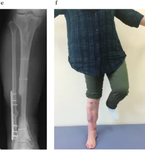
Fig. 2 . Case 1.
(a) Preoperative plain radiograph of the right lower leg showing infected nonunion after in- tramedullary nailing performed at another hospital ; anteroposterior (AP) view. The maximum length of the tibial bone defect was 96 mm.
(b) Preoperative angiography showing vascular occlusion of the fibular and anterior tibial ar- teries, and only the posterior tibial artery is patent at the distal part of the lower leg, which is called a ‘single
-artery leg.’
(c) Postoperative view of affected limb after the FVFG procedure.
(d) Postoperative plain radiograph (AP view) af- ter the FVFG procedure.
(e) Postoperative plain radiograph (AP view) at 24 months after the FVFG procedure shows complete bone union.
(f) The patient was able to stand on only the af- fected leg at 48 months after receiving the FVFG. The fistula had closed, and no sign of infection was observed.
a b
c d
e f