Fukushima Medical University
福島県立医科大学 学術機関リポジトリ
This document is downloaded at: 2021-11-08T00:11:44Z
Title Ornithine decarboxylase activity as a prognostic marker for colorectal cancer
Author(s)
Hoshino, Yutaka; Terashima, Shinya; Teranishi, Yasushi;
Terashima, Masanori; Kogure, Michihiko; Saitoh, Takuroh;
Osuka, Fumihiko; Kashimura, Seigo; Saze, Zenichiroh; Gotoh, Mitsukazu
Citation Fukushima Journal of Medical Science. 53(1): 1-9
Issue Date 2007-06
URL http://ir.fmu.ac.jp/dspace/handle/123456789/202
Rights © 2007 The Fukushima Society of Medical Science
DOI
Text Version publisher
Fukushima J. Med. Sci., Vol. 53, No.1, 2007
[Original Article]
ORNITHINE DECARBOXYLASE ACTIVITY AS A PROGNOSTIC MARKER FOR COLORECTAL CANCER
YUTAKA HOSHINO!), SHINYA TERASHIMN), YASUSHI TERANISHP), MASANORI TERASHIMN), MICHIHIKO KOGUREl), TAKUROH SAITOHl),
FUMIHIKO OSUKN), SEIGO KASHIMURAl), ZENICHIROH SAZEl) and MITSUKAZU GOTOHl)
1) Department of Surgery I, Fukushima Medical University School of Medicine
2) Department of Surgery, Fujita General Hospital ')Department of Surgery, Southern Tohoku General Hospital
(Received January 22, 2007, accepted March 5, 2007)
Abstract: Ornithine decarboxylase (ODC) is a key enzyme in the biosynthesis of polyamines, which are essential for cell proliferation. ODC activity was measured in 47 colorectal cancer patients, 5 patients with adenoma of colorectum and 4 healthy volunteers. Mean ODC activities of cancer tissue, non-cancerous mucosa from cancer-bearing colorectum, adenoma tissue, and normal mucosa from healthy volunteers were 435±392, 154±173, 295±202, 103±60 pmol C02/h/mg protein, respectively. ODC activity of cancer tissue or adenoma tissue was significantly higher than that of the others. Among colorectal cancer patients, ODC activity in cancer tissue was correlated with T factors, lymph node metastasis and stages.
Patients with tumors that had high ODC activity (;;;;;350 pmol C02/h/mg protein) showed a poor 10-year survival rate. These results suggest that ODC activity may be a useful marker for patients' prognosis after surgery.
Key words: ornithine decarboxylase activity, colorectal cancer, biological marker
INTRODUCTION
Ornithine decarboxylase (ODC) is a key enzyme in the biosynthesis of polyamines, which are essential for cell proliferation. Since the ODC activity is elevated during the promotion stage in carcinogenesis of animal models!), the ODC activity can be used as a biomarker of potential malignancy. Recent studies have demonstrated that ODe activity in cancer tissue is high in carcinoma of the stom-
£~ ft,~~&~,~~ ~,~~$~,*¥~~,*.~M,*~~x~,~M~ft,~~~
-R~, ~.~-
Correspondence to: Hikarigaoka 1, Fukushima City, Fukushima Prefecture.
E-mail: [email protected]
1
2 Y. HOSHINO et al.
ach
2 - 4),lung
5),breast
6.7l,head and neckS), skin
9),and colorectum
10- 14).However, high
ODC activity was not always correlated with poor prognosis in patients bearing one of these cancers
I4).Here, we show that the high ODC activity of tumors correlates with lymphatic as well as distant metastasis and also with 10-year survival of patients after surgery.
SUBJECTS AND METHODS
Four normal colorectal specimens were obtained from healthy volunteers includ- ing five men and one woman with informed consent. Seven specimens of adenoma of colorectum (three mild atypia and four severe atypia) were obtained from 5 male patients. Forty-eight specimens of cancer tissue as well as 80 non-cancerous adjacent specimens ware obtained from 47 patients including 29 males and 18 females with a mean age of 63 years (range 32-82). All patients had undergone elective tumor resection with lymph node dissection at Fukushima Medical Univer- sity Hospital, between 1991 and 1994. The median postoperative follow-up time was greater than 10 years.
Among 47 patients with colorectal cancers, there were 6 Tis, 4 T1, 7 T2, 14 T3 and 17 T4. According to TNM staging criteria
I5),patients were subdivided into stage 0(7), stage 1(10), stage 1I(10), stage III(15) and stage IV(6), respectively. Lymph node metastases were present in 22 patients (N1; 15, N2; 7), while 6 patients had distant metastases. Histopathologically, these tumors were subdivided into G1(14), G2(29) and G3(5), respectively. None of the patients received preoperative chemo- therapy or radiotherapy. Postoperative follow-up data were obtained by periodic examinations at the out patient clinic, or from mailed questionnaires.
ODe
activity assay
All specimens were obtained by the endoscopic biopsy, and were immediately
frozen at -80·C. ODC activity assay was performed within two weeks. ODC
activity was assayed by modified Furihata's method
I6).Frozen specimens were
homogenized in 50 mM sodium phosphate buffer 3 ml (pH
=7.2) containing 0.1 mM
pyridoxal phosphate and 0.1 mM EDT A, using Polytron R Model K with PT AlOS
generator. The homogenates were centrifuged at 30,000 G for 15 min at 2"C, and the
supernatants, 0.2 ml, used as enzyme extracts Supernatant remnants were used to
determine the protein content by micro-assay using UVDEC-660R. Enzymes were
assayed in the out side of a double test tube with rubber cap, and released 14C02 was
trapped in Protozol (Scintilamine R -OH) 200 pI, in the inside of test tube. Enzyme
extracts were incubated at 37"C for 60 min after addition of DL- [1-14C] ornithine
hydrochloride 80 pI; 0.5 pCi (58.4 mCi/mmol, NEC-469), 50 mM sodium phosphate
buffer (pH
=7.2) containing 0.2 mM pyridoxal phosphate, 50 pM EDT A and 0.5 mM
dithiotreitol 720 pI (Total 1.0 mI). The enzyme reaction was terminated by addition
of 2 M citric acid 0.4 ml to the outside of the test tube, and incubation was continued
ODC ACTIVITY AS A PROGNOSTIC MARKER 3
for another 30 min at 3TC. Protosol was then transferred to the scintillator (Scintisol R EX - H: 10 ml), and its radioactivity was determined in a scintillation counter (LSC-3500, Aloka). The results were expressed as pmol C02/h/mg protein.
Statistical analysis
The relationship between ODC activity and various clinicopathological vari- ables was examined individually using Mann-Whitney's U test or one way Analysis of Variance with Fishier's exact test when appropriate. The primary statistical outcome in the study of prognosis was overall survival measured from the data of resection. Overall survival was calculated according to the method of Kaplan- Meier. The difference in survival among the clinicopathological variables, or between low and high ODC activity was tested using Willcoxson-Graham tests.
The relative importance of the prognostic factors extracted by the univariate analysis, was assessed in a multivariate analysis by the Cox proportional hazards regression model. The differences were considered significant when the p-value was less than 0.05. The program used for these analyses was the StatView 4.5J package (Abacus Concepts Inc., Berkeley, CA).
RESULTS
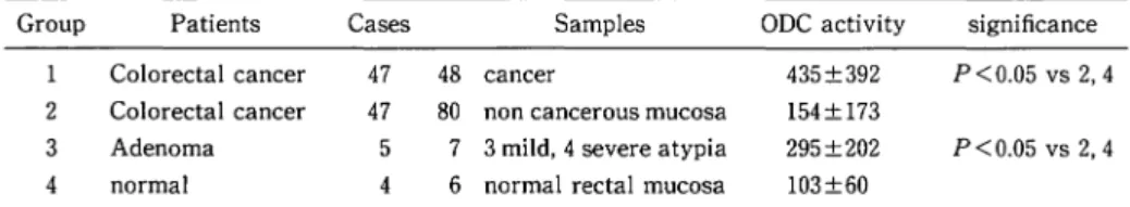
ODC activities in normal mucosa, adenoma and non-cancerous mucosa from cancer-bearing colorectum were 103±60, 295±202, 154±173 pmol C02/h/mg pro- tein, respectively (Table 1). ODC activity in cancer tissue was 435±392 pmol C02/
h/mg protein. Significant difference was noted between cancer tissue and non- cancerous or normal colon tissue. The same was true for adenoma over non- cancerous or normal colon tissue, although the levels tended to be low as compared to those of cancer tissue. Thus, cancer tissue had significantly higher ODC activity than the normal appearing mucosa from cancer bearing patients or from healthy volunteers.
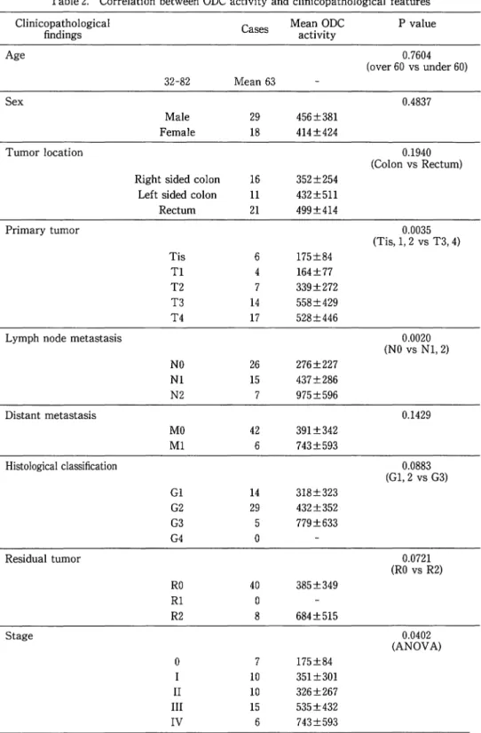
ODC activity in cancer tissue and clinicopathological features were compared and summarized in Table 2. Mean ODC activities increased in association with elevation of T factors (Tis, Tl, T2, T3, T4) giving values of 175±84, 164±77, 339±
272, 558±429, 528±446 pmol C02/h/mg protein, respectively. ODC activities in T3 and T4 were significantly higher than those of Tis, Tl and T2. This was the case for N status (NO, Nl, and N2) giving values of 276±227, 437±286, 975±596 pmol
Table l. ODC activity in normal mucosa, adenoma and cancer tissue Group Patients Cases Samples ODC activity significance
1 Colorectal cancer 47 48 cancer 435±392 P<0.05 vs 2,4 2 Colorectal cancer 47 80 non cancerous mucosa 154±173
3 Adenoma 5 7 3 mild, 4 severe atypia 295±202 P<0.05 vs 2,4 4 normal 6 normal rectal mucosa 103±60
4 Y. HOSHINO et at.
Table 2. Correlation between ODC activity and clinicopathological features Clinicopathological
Cases Mean ODC P value
findings activity
Age 0.7604
(over 60 vs under 60) 32-82 Mean 63
Sex 0.4837
Male 29 456±381
Female 18 414±424
Tumor location 0.1940
(Colon vs Rectum) Right sided colon 16 352±254
Left sided colon 11 432±511
Rectum 21 499±414
Primary tumor 0.0035
(Tis, 1, 2 vs T3, 4)
Tis 6 175±84
Tl 4 164±77
T2 7 339±272
T3 14 558±429
T4 17 528±446
Lymph node metastasis 0.0020
(NO vs Nl, 2)
NO 26 276±227
Nl 15 437±286
N2 7 975±596
Distant metastasis 0.1429
MO 42 391±342
Ml 6 743±593
Histological classification 0.0883
(Gl,2 vs G3)
Gl 14 318±323
G2 29 432±352
G3 5 779±633
G4 0
Residual tumor 0.0721
(RO vs R2)
RO 40 385±349
Rl 0
R2 8 684±515
Stage 0.0402
(ANOVA)
0 7 175±84
I 10 351 ±301
II 10 326±267
III 15 535±432
IV 6 743±593
ODC ACTIVITY AS A PROGNOSTIC MARKER 5
e02/h/mg protein, respectively. ODe activities in NI and N2 were significantly higher than that of NO. Mean ODe activities in cancer tissue of patients with or without distant metastasis were not significantly different. Mean ODe activity tended to increase in association with the degree of adenocarcinoma differentiation (GI, G2 and G3) or in patients with residual tumor, however it was not statistically significant. Mean ODe activity increased in association with elevation of TNM stages
(0,I, II, III, IV) giving values of I75±84, 35I±301, 326±267, 535±432, 743±
593 pmol e02/h/mg protein, respectively, reaching a statistically significant difference with the ANOVA test.
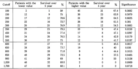
Table 3. Effect of cut off values of ODC activity on 5 year overall survival after colorectal resection
Cutoff Patients with the Survival over
% Patients with the Survival over
% Significance
lower value 5 year higher value 5 year
100 2 1 50 46 31 67.4 0.5682
150 12 9 75 36 23 63.9 0.5079
200 17 12 70.6 31 20 64.5 0.6625
250 22 16 72.7 26 16 61.5 0.381
300 26 20 76.9 22 12 54.5 0.0883
350 29 23 79.3 19 9 47.4 0.0159
450 31 24 77.4 17 8 47.1 0.0297
500 34 26 76.5 14 6 42.9 0.0179
550 36 27 75 12 5 41.1 0.0197
600 37 27 73 11 5 45.5 0.0817
650 38 28 73.7 10 4 40 0.036
800 39 28 71.8 9 4 44.4 0.1015
850 40 29 72.5 8 3 37.5 0.0405
900 41 29 69 6 3 50 0.3158
1,050 46 32 69.6 2 0 0 0.0069
1,700 47 32 68.1 1 0 0 0.0718
survival ate (%)
100
80
ODC activity < 35060
P=0.022940
ODe activity ~ 35020 0
0 20 40
60 80
100 120 MonthsFig. 1. Ten year overall survival rates of patients showing high or low ODC activity
(~350 or <350 pmol C02/h/mg protein) of cancer tissue.
The patients with high ODC activity showed significantly lower survival rates than those with low ODC (47.4% vs 79.3%, p<0.03)
6 Y. HOSHINO et at.
Patient overall survival was compared with ODe activity by changing cutoff points between the high and low ODe activity groups. Table 3 shows the relation- ship of overall survival to the different threshold points of ODe activity in patients with colorectal cancer. Several cutoff points between 350 and 550 pmol e02/h/mg protein proved to be significantly associated with overall survival. Since the most significant association was observed at the cutoff value of 350 pmol e02/h/mg protein
(p=0.0159), this value was applied to draw 10 year over all survival curves between the low and high value groups (Fig. 1). The patients with high ODe activity had significantly lower 10-year survival rates than those with low ODe
Table 4. Clinicopathological findings and long-term survival (univar·
iate analysis)
Clinicopathological findings Cases 5 year survivial rate P value
Sex 0.939
Male 29 65.5
Female 18 66.7
Tumor location 0.433
Colon 27 70.4
Rectum 21 61.9
Primary Tumor 0.001
Tis,
n,
T2 17 100.0T3, T4 31 48.4
Regional Lymph Nodes 0.001
NO 25 88.0
N1, N2 22 45.5
Distant Metastasis 0.0001
MO 42 76.2
M1 6 0.0
Histological Grading 0.009
G1, G2 43 72.1
G3 5 20.0
Residual Tumor 0.0001
RO 40 80.0
R2 8 0.0
ODC activity 0.02
<350 29 79.3
~350 19 47.4
Table 5. Clinicopathological findings and long-term survival (multivariate analysis)
Clinicopathological findings P value Hazard ratio 95% CI
ODC activity' 0.9745 0.977 0.239 - 3.996
Histological classification 0.7979 1.212 0.278 - 5.281 Distant Metastasis 0.0905 8.941 0.708-112.949
Residual Tumor 0.0004 0.003 -1.193-0.075
• : Cutoff 350 pmol C02/h/mg protein
ODC ACTIVITY AS A PROGNOSTIC MARKER 7
activity (47.4% vs 79.3%,
p<0.03).Univariate analysis demonstrates a significant association of tumor stage, lymph node metastasis, distant metastasis, histological grading, tumor residual and ODe activity with five year survival (Table 4).
Multivariate analysis did not demonstrate a significant prognostic factor (Table 5).
DISCUSSION
Recently, polyamines have attracted the attention of many investigators, because of the relation of cell growth to polyamine synthesis. Among the enzymes involved in polyamine synthesis, special attention has been paid to ornithine decar- boxylase (ODe) activity, which has been shown to be first metabolizing, rate-limiting and sensitive to various stimuli
I7 - 20).ODe activity is generally elevated in tumor tissue compared with normal tissue in both experimental and human cancers
3 - 9,2I,22).Also, there are a few reports concerning ODe activity in human colorectal can- cer
IO - 14).The major findings of our study were the following; 1) An approximately four fold higher ODe activity in cancer tissue than the normal mucosa from non-colo- rectal cancer patients, 2) Increased ODe activity in adenoma, 3) ODe activity in cancer tissue was correlated with T factors, lymph node metastasis and stages, and 4) Patients with tumors that had high ODe activity (;?;350 pmol e02/h/mg protein) showed poor 10-year survival rates. Our results indicated that colorectal cancer tissue had an approximately four fold higher ODe activity than the normal mucosa from non-colorectal cancer patients, and even the normal appearing mucosa in those with colorectal cancer had 1.5 fold higher ODe activity than that of patients without colorectal cancer. This data confirms earlier findings by Berdinskikh et al.
3 )that these differences in ODe activity may be related to differences in proliferative activity.
When ODe activity in cancer tissue was compared with clinicopathological
findings, it was significantly higher in patients with deep tumor invasion, lymph node
metastasis, or low grade differentiation.
Ithas been reported that there is a
negative correlation between ODe activity and progression or stage of colorectal
cancer
I4 ).On the other hand, a positive correlation between ODe activity and TNM
classification was reported
13).The difference in tissue samples (ie. biopsy or resect-
ed specimens), may partly explain this discrepancy. As ODe has a short half life on
the order of 10 minutes, we compared ODe activity in endoscopic specimens with
that in surgical samples from the same 5 patients. ODe activity in biopsy speci-
mens was approximately two to ten fold higher than that in operative specimens
(data not shown). Porter et al.
13 )reported similar results with a comparison of
biopsy and surgical specimens, and recommended that ODe activity be measured
using biopsy samples. The time required for surgical tissue resection may be
sufficient to account for substantial loss in enzyme activity. In this study, we
measured ODe activity using biopsy samples
I3 ).More detailed investigations are
8 Y. HOSHINO et ai.
also recommended to ascertain the problems including heterogeneity of the tumor and contamination of the inflammation or fibrous tissue.
In the present study, higher
ODeactivity correlates with poor prognosis, sugges- ting various inhibitors against
ODeactivity or inducers for catabolic enzyme (the spermidine/spermine-N(l)-acetyltransferase; SSAT) of polyamine may have some role on chemoprevention as well as on colon cancer progression
23 - 2B).Thus, careful follow-up and intense postoperative therapy are required for patients with color- ectal cancer showing high
ODeactivity.
REFERENCES
1. Furihata C, Sato Y, Matsushima T, Tatematsu M. Induction of ornithine decarboxylase and DNA synthesis in rat stomach mucosa by methylglyoxal. Carcinogenesis, 6: 91-94, 1985.
2. Terashima S, Teranishi Y, Hoshino Y, Ogata M, Inoue H, Motoki R. Ornithine Decarboxylase Activity in Human Stomach Cancer. Recent Advances in Management of Digestive Cancers, Tokyo, 212-214, 1993.
3. Berdinskikh NK, Ignatenko NA, Zaletok SP, Ganina KP, Chorniy VA. Ornithine decarboxylase activity and polyamine content in adenocarcinomas of human stomach and large intestine. Int J Cancer, 47: 496-498, 1991.
4. Lundell L, Rosengren E. Polyamine levels in human gastric carcinoma. J Gastroenter- 01, 21: 829-832, 1986.
5. Cohen DJ, Verma AK. Ornithine decarboxylase activity as a biochemical marker in individuals predisposed to lung cancer. J Investigative Surgery, 2: 103-106, 1986.
6. Romano M, Santacroce MA, Bonelli P, Cecco L, Cerra M. Differences in polyamine metabolism between carcinomatous and uninvolved human breast tissues. Int J Biolog- ical Markers, 1: 77-80, 1986.
7. Love RR, Astrow SH, Cheeks AM, Havighurst TC. Ornithine decarboxylase (ODC) as a prognostic factor in operable breast cancer. Breast Cancer Res Treat, 79: 329-334, 2003.
8. Weiss RL, Calhoun KH, Ahmed AE, Stanley D. Ornithine decarboxylase activity in tumor and normal tissue of head and neck cancer patients. Laryngoscope, 102: 855-857, 1992.
9. Scalabrino G, Pigatto P, Ferioli ME, Modena D, Puerari M, Caru A. Levels of activity of the polyamine biosynthetic decarboxylases as indicators of degree of malignancy of human cutaneous epitheliomas. J Investigative Dermatology, 74 : 122-124, 1980.
10. Nishioka K, Grossie VB, Chang TH, Ajani JA, Ota DM. Colorectal ornithine decar·
boxylase activity in human mucosa and tumors; Elevation of enzymatic activity in distal mucosa. J Surg Oncol, 47: 117-120, 1991.
11. Lamuraglia GM, Lacaine F, Malt RA. High ornithine decarboxylase activity and polyamine levels in human colorectal neoplasia. Ann Surg, 204: 89-93, 1986.
12. Narisawa T, Takahashi M, Niwa M, Koyama H, Kotanagi H, Kusaka N, Yamazaki Y, Nagasawa 0, Koyama K, Wakizaka A, Fukaura Y. Increased mucosal ornithine decarboxylase activity in large bowel with multiple tumors, adenocarcinoma, and adenoma. Cancer, 63: 1572-1576, 1989.
13. Porter CW, Herrera-Ornelas L, Pera P, Petrelli NF, Mittelman A. Polyamine Biosyn- thetic activity in normal and neoplastic human colorectal tissues. Cancer, 60 : 1275- 1281, 1987.
14. Matsubara N, Hietala OA, Gilmour SK, Yum KY, Litwin S, Watts P, Brennan EJ, O'Brien TG. Association between high levels of ornithine decarboxylase activity and
ODC ACTIVITY AS A PROGNOSTIC MARKER 9
favorable prognosis in human colorectal carcinoma. Clin Cancer Res, 1: 665-671, 1995.
15. UlCC: TNM Classification of Malignant Tumours, Fifth Edition. A John Wiley &
Sons, INC., Publication, 66-69, 1997.
16. Furihata C, Sato Y, Hosaka M, Matsushima T, Furukawa F, Takahashi M. NaCl induced ornithine decarboxylase and DNA synthesis in rat stomach mucosa. Biochem Biophys Res Com, 121: 1027-1032, 1984.
17. Gillette JH, Mitchell JLA. Ornithine decarboxylase; a biochemical marker of repair in damaged tissue. Life science, 48: 1501-1510, 1991.
18. Wang YJ, Johnson LR, Tsai YH, Castro GA. Mucosal ornithine decarboxylase, polyamines, and hyperplasia in infected intestine. Am J Physiol, 260: G45-51, 1991.
19. Luk GD, Baylin SB. Ornithine decarboxylase as a biologic marker in familial colonic polyposis. New Engl J Med, 311: 80-83, 1984.
20. Garewal HS, Sampliner R, Alberts D, Steinbronn K. Increase in ornithine decar·
boxylase activity associated with development of dysplasia in Barrett's esophagus. Dig Dis Sci, 34: 312-314, 1989.
21. Klekner A, Rohn AG, Schillinger G, Schroder R, Klug N, Ernestus Rl. ODC mRNA as a prognostic factor for predicting recurrence in meningiomas. J Neurooncol, 53: 67-75, 2001.
22. Mafune K, Tanaka Y, Mimori K, Mori M, Takubo K, Makuuchi M. Increased expres·
sion of ornithine decarboxylase messenger RNA in human esophageal carcinoma. Clin Cancer Res, 5: 4073-4078, 1999.
23. Devens BH, Weeks RS, Byrns MR. Polyamine depletion therapy in prostate cancer.
Prostate Cancer Prostatic Dis, 3: 275-279, 2000.
24. Seiler N. Thirty years of polyamine-related approaches to cancer therapy. Retrospect and prospect. Part 1. Selective enzyme inhibitors. Curr Drug Targets, 4 : 537-564, 2003.
25. Thomas T, Thomas TJ. Polyamines in cell growth and cell death: Molecular mech- anisms and therapeutic applications. Cell Mil Life Sci, 58: 244-258, 2001.
26. Hu HY, Liu XX, Jiang CY, Lu Y, Liu SL, Bian JF, Wang XM, Geng Z, Zhang Y, Zhang B. Ornithine decarboxylase gene is overexpressed in colorectal carcinoma. World J Gastroenterol, 11: 2244-2248, 2005.
27. Nemoto T, Kubota S, Ishida H, Murata N, Hashimoto D. Ornithine decarboxylase, mitogen-activated protein kinase and matrix metalloproteinase-2 expressions in human colon tumors. World J Gastroenterol, 11: 3065-3069,2005.
28. Schipper RG, Deli G, Deloyer P, Lange WP, Schalken JA, Verhofstad AA. Antitumor activity of the polyamine analog N(l), N(ll)-diethylnorspermine against human prostate carcinoma cells. Prostate, 44: 313-321, 2000.