Acta Med. Nagasaki 41: 66-70
Diagnostic Validity of Combining HTLV-1 Serology and Immunophenotyping in Adult T-Cell Leukemia
Kazuyuki SUGAHARA1), Kazuto TSURUDA1), Yasuaki YAMADA1),2), Youichi HIRAKATA1),2), Sunao ATOGAMI3), Hisashi SOHDA3), Kunihiro TSUKAZAKI3), Masao TOMONAGA3) and Shimeru KAMIHIRA1),2)
1) Central Diagnostic Laboratory 2) Department of Laboratory Medicine
3) Department of Haematology, Atomic Disease Institute, Nagasaki University School of Medicine, 1-7-1 Sakamoto, Nagasaki 852, Japan.
Adult T-cell leukemia (ATL) is heterogeneous and some- times equivocal to other T-cell neoplasms. Detecting anti- HTLV-1 antibodies is significant for a first screening not only for HTLV-1 infection but also for the HTLV-1-related disorders of ATL and TSP/HAM. The purpose of the present study was to investigate the diagnostic validity of HTLV-1 serology in ATL. The serologic results by a gelatin particle agglutination (PA) assay were highly sensitive (100%) and specific (99.5%) for the results of polymerase chain reaction (PCR) assay in 666 healthy blood donors who live in an area endemic for the HTLV-1 virus. Of 7,536 hospitalized patients, 189 patients with ATL were serologically screened. There were 1,140 patients (15.2%) infected by chance with HTLV-1, showing specificity, sensitivity, positive predictive value (PV), and negative PV of 84.3%, 100%, 14.2%, and 100%, respectively. Since the low positive PV (14.2%) was useless, we tried combining the anti-HTLV-1 assay with the immuno- phenotyping necessary for the diagnosis of lymphoid neo- plasms. This combination gave nearly 100% positive and negative PV, and could prove to be useful in diagnosing ATL with the probability of 98%, especially for epidemiologic studies.
Key words : sensitivity, specificity, predictive value, ATL, HTLV-1, anti-HTLV-1
Introduction
The human T-cell leukemia virus type-1 (HTLV-1) was first identified as the causative agent of adult T-cell leukemia (ATL) in 1980 by the Gallo group'). Since then, several sero-epidemiologic studies") have elucidated the close etiological relationship between the HTLV-1 virus and disorders such as tropical spastic paraparesis/HTLV- 1-associated myelopathy (TSP/HAM) and others of unknown etiology. There are many apparently healthy persons without any clinical symptoms who are infected with HTLV-1-6'. Among these carriers, only 1 of 1,000- 1,500 develops ATL each year, and the cumulative occur- rence rate of this disease is estimated at about 3 to 5% in
the carrier's lifetime"). With the development of immuno- phenotypical diagnosis of lymphoid neoplasms, the dis- crimination of ATL from T-cell neoplasms without HTLV- 1 has recently been proposed by the international lym- phoma study group (REAL classification)"). Although they have classified T-cell neoplasms into ATL and non- ATL, it is often difficult to discriminate between ATL and other T-cell neoplasms using routine laboratory meth-
11-13)
Although the commercially available kits for HTLV-1 serology are known to precisely detect persistent HTLV-1 infections'), there are no studies on the diagnostic validity of antibody detection in diagnosing ATL.
Accordingly, we were prompted to quantitate the validity of the serologic HTLV-1 test as not only a marker of persistent infection but also as a diagnostic tool for ATL. In the present study, using a gelatin particle aggluti- nation (PA) test, we reviewed the clinical HTLV-1 serol- ogy of 7,536 patient samples, including 189 ATL patients who were diagnosed by immunophenotyping and molecular analysis. The objectives of this study were to : (1) dis- criminate ATL immunophenotypically and molecularly from other T-cell neoplasms without HTLV-1 in an area endemic for the virus, (2) quantitatively evaluate the diagnostic validity of HTLV-1 serology in ATL, and (3) establish objective minimal tests to be used in the diagno- sis of ATL using common laboratory assays.
Materials and Methods
Subjects.
Serum samples were collected from 666 healthy blood donors of residents in an area endemic for HTLV-1 and from 7,536 patients who were admitted or referred to our hospital from January 1991 to November 1995. The diagno- sis of peripheral T-cell lymphoma/leukemia including ATL was based on a complete hematological battery of May-
Grunwald stained smears, hematoxylin-eosin and immuno- histology, and immunophenotyping with flow cytometric techniques. ATL was diagnosed according to the Japan Lymphoma Study Group (JLSG) guidelines"). In some of the T-cell neoplasm cases, especially those resembling ATL, the diagnosis of ATL was reached after Southern blot analysis of HTLV-1 monoclonal integration. Cells for immunophenotyping B- or T-lineage were obtained from heparinized peripheral and bone marrow aspirated blood and biopsied lymph nodes.
Detection of anti-HTLV-1 antibody and the provirus Anti-HTLV-1 antibodies were first screened by a gelatin PA kit (Serodia-HTLV-1, Fuj iRebio, Tokyo)'). Some
samples, including the indeterminates, were screened again using an ELISA kit (Kyowa Medix, Tokyo) and Western blot kit (FujiRebio) as supplementary confirming tests.
All assays were done according to the manufacturer's instructions and evaluated by the criteria of the kits. To detect HTLV-1 provirus in peripheral blood from the blood donors, a commercially available PCR kit (Amplicor HTLV-1 and -2 Detection kit, Roche, USA) was used.
Molecular analysis of HTLV-1 integration
Determination of the HTLV-1 monoclonal integration in leukemic cells from the blood or lymph node suspensions was performed as previously described"). Briefly, high molecular-weight genomic DNA was extracted from peripheral and bone marrow mononuclear cells and lymph node suspension cells in patients with T-cell neoplasm and anti-HTLV-1. Next, 10 Leg of DNA was were digested with EcoRl restriction enzyme and were size-fractionated on 0.7% agarose gels. They were then transferred by the Southern blot technique onto a nylon membrane. Hy- bridization to randomly primed 'p- or digoxigenin-labeled DNA probes of the pX region was performed.
Phenotypical examination of tumor cells
We cytophenotypically and immunophenotypically examined all of the lymphoid neoplasms. ATL cells were cytologically defined by their morphologic features on the blood film stained with May-Grunwald Giemsa, character- ized by polymorphic mature cells with convoluted or poly-lobulated nuclei, per the FAB criteria") and JLSG guidelines").
Immunophenotyping was performed by standard flow cytometry in a Cytoron (Ortho Diagnostics KK, Tokyo) by using single- and double-labeling with the following monoclonal antibodies (MoAb) : CD2, CD3, CD4, CD5, CD7, CD8, HLA-DR, CD25, CD10, CD19, and CD20. A marker was considered positive when it stained more than 30% of the cells above the controls. The immuno-
phenotypes were classified into two main categories:
typical (CD4+ . CD8-) and unusual (CD4 and/or CD8 antigen abnormalities), based on the criteria of Kamihira et ally. The statistical analysis was performed using the FISHER software package (Nakayama Syoten, Tokyo).
Results
1) Anti-HTLV-1 detection in blood donors and patients.
HTLV-1 serologic tests have been routinely conducted in our hospital since 1986 by the PA assay. In the present study, we re-evaluated the PA assay in comparison with the PCR method, and quantitated the diagnostic validity in practical samples from patients with various diseases.
As shown in Table 1, only 2 of the 666 samples were discrepant, i. e., seropositive and PCR-negative.
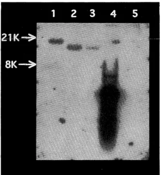
Of the two samples, Western blot analysis revealed a negative finding in one sample (no. 491) and indeterminate in another one (no. 433) which had strong p19 and faint p24 bands (Fig. 1).
The sensitivity and specificity between PA and PCR were calculated as 100% (95% CI, 89.8-100) and 99.5% (95% CI, 98.9-100), respectively. The PA assay actually gave posi- tive results in 1,329 (17.6%) and negative results in 6,307
(82.4%) of the 7,536 routine patient samples tested. We then reviewed the diagnosis of the subject patients from the files of our hospital.
2) T-cell neoplasms and anti-HTLV-1 distribution
Out of the 7,536 patients, 208 were diagnosed as having T-cell neoplasms ; 189 patients with ATL and 19 with T-cell malignant lymphoma (T-ML), which included 5 cases of Sezary's syndrome and mycosis fungoides. All of the 189 ATL, 4 (21.5%) of the 19 T-ML, and 1,136 (15.5%) of the other patients were seropositive for HTLV-1, as shown in Table 2.
One hundred sixty four (86.5%) of ATL had immuno- phenotypically prototypic helper-T immunophenotype with CD2+, CD3+, CD4+, CD7-, CD8-, CD25+, and HLA-DR+.
Unusual cases with immunophenotype of CD4 and CD8 Table 1. Serologic (PA) and PCR (Roche) results in 666 healthy blood donors who live in an area endemic for HTLV- 1 infection.
subjects tests serology PCR
(+) (-) HTLV-1 HTLV-2 (- ) healthy donors
N = 666 45 621 43 0 623
(%) (6.8%) (93.2%) (6.5%) (0.0%) (93.5%) sensitivity = 100% (95%CI, 89.8-100)
specificity = 99.5% (95%CI, 89.8-100)
Fig 1. Western blot analysis of the two samples discrepant between the PA and PCR assays.
Lane 1 : positive control Lane 2 : weak positive control Lane 3 : negative control
Lane 4 : sample of no. 491 (negative) Lane 5 : sample of no. 433 (indeterminate) Lane 6 : a representative positive sample
Table 2. Serological anti-HTLV-1 Distribution by the PA assay in ATL, peripheral T-cell Lymphomas not associated with HTLV-1, and other disorders.
anti-HTLV-1
Diseases Total
a) ATL* 189 0 189
(1) proto-typic 164 0
(2) unusual CD8 4 0
DP 13 0
DN 8 0
b) T-ML without 4 15 19
HTLV-1
c) Others 1136 6192 7328
Total 1329 6207 7536
'
, Diagnosis of equivocal ATL was made molecularly with demonstration of HTLV-1 monoclonal integration.
DP, double-positive type ; DN, double-negative type
Fig 2. Southern blot analysis of HTLV-1 integration status in patients with seropositive T-cell neoplasm.
Lane 1 to 3 : monoclonal integration one band correspond- ing to ATL
Lane 4: control (pX DNA fragment obtained from the cloned vector)
Lane 5: no demonstrable band corresponding to T-cell neoplasm infected by chance with HTLV-1
abnormalities were observed : CD8-positive type (4 ; 2.1%), CD4 and CD8-double positive (DP) type (13 ; 6.9%), and CD4 and CD8-double negative (DN) type (4 ; 2.1%). All but 1 of the 19 T-ML cases had common helper-T phenotypes with CD3+ and CD4+, similar to that of prototypic ATL. In the ATL cases with diagnostic difficulty, including 25 unusual ATL cases with aberrant phenotype and T-ML infected by chance with HTLV-1, whether or not integration with HTLV-1 had occurred was confirmed by molecular techniques. They were subse- quently distributed as shown in Table 2. Figure 2 shows an example of Southern blot analysis on monoclonal in- tegration band in ATL but not in T-ML. The non- monoclonal band corresponds to T-ML infected by chance with HTLV-1 (lane 5).
3) Diagnostic value of combining serology and immuno- phenotyping in ATL
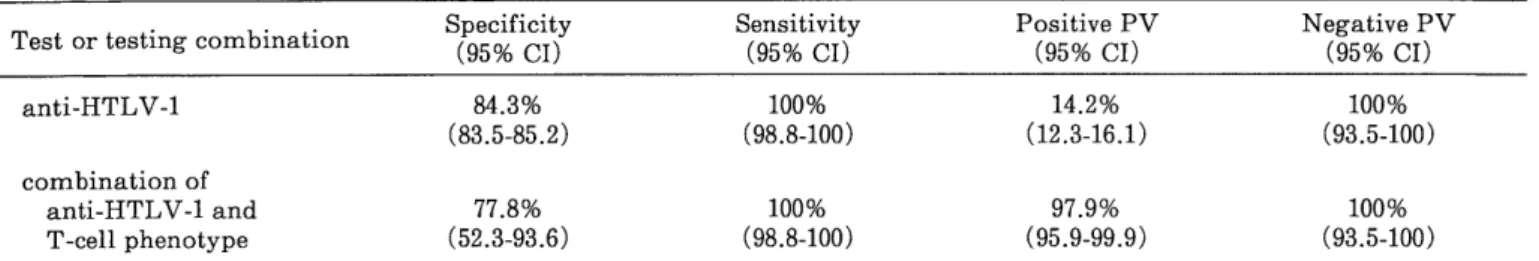
Based on the raw data of Table 2, specificities, sensitivi- ties, and PVs of the diagnostic tests with Bayes' rule') were computed as, shown in Table 3. As a diagnostic tool for ATL, the anti-HTLV-1 antibody test was 100% sensi- tive, but its positive PV was extremely low (14.2%). Since
these findings imply no relevance as a practical diagnostic tool, we then tried combining the serologic test with immunophenotyping, one test of the routine hematologic battery. The combination provided high positive and negative PVs, nearly 100%, with a 95% confidence inter- val.
Table 3. Summary of Specificities, Sensitivities, and Predictive Values of HTLV-1 alone Combination of anti-HTLV-1 and T-cell Phenotype in ATL versus non-ATL.
Test or testing combination Specificity (95% CI) Sensitivity (95% CI) Positive (95% CI) PV Negative PV (95% CI)
anti-HTLV-1 84.3% 100% 14.2% 100%
(83.5-85.2) (98.8-100) (12.3-16.1) (93.5-100)
combination of
anti-HTLV-1 and 77.8% 100% 97.9% 100%
T-cell phenotype (52.3-93.6) (98.8-100) (95.9-99.9) (93.5-100)
PV, predictive value : CI, confidence interval
Discussion
HTLV-1 was first identified as the causative agent of ATL. It has since become increasingly evident that the virus is also associated with other disorders such as TSP/
HAM, uveitis, arthropathy, infectious dermatitis, and polymyositis2-4.ls). Many apparently healthy people are carriers of HTLV-1, and they may develop one of these disorders. Thus, detecting the antibodies against HTLV-1 is of great importance to screen illness due to the HTLV-1 virus, particularly in areas endemic for ATL and TSP/
HAM. Considering the cost-and time-benefit and the objectivity of ATL diagnosis, we quantitated the diagnos- tic validity of the PA serologic test in diagnosing ATL.
Our present results re-confirmed the relevance of the PA assay to detect the viral infection with the highest sensi- tivity (100%) and specificity (99.5%) comparable to that of PCR results, by testing a sufficient number of serum samples from blood donors who live in an area endemic for the virus. We targeted blood donors to obtain a variety of HTLV-1 infection state, eg. carriers with different sero- status such as seronegative or partially antibody positive against the HTLV-1 antigens'). The PA and PCR results were discrepant for only two of our samples, suggesting false positive in the PA. However, the sample designated as "indeterminate" according to the WHO criteria of WB21> could not be excluded as a possible true infection, because about 10% of ATL cases have been reported to have no env-antibodies, by the present authors (data not shown)') and others'). This may imply a defective virus or immunological tolerance to the antigens in some individu- als characterized by an MHC habit. Recent studies dis- closed very rare ATL cases') and a healthy carrie') with seronegative/proviral-positive status.
Our present data demonstrated that all ATL cases were correctly screened serologically. In general, althogh proto- typic ATL can be easily diagnosed by ordinary hematologic tests, some ATL cases are difficult to diagnose because of diverse clinical manifestations and/or aberrant immuno- phenotypes and unusual morphology of the tumor cells.
About 10% of ATL patients admitted to our hospital had the phenotypically difficult diagnosis, as presently and previously described","). However, Southern blot analysis,
the result of which is a diagnostic "gold standard" of ATL2) can not be done in all cases because of time and cost constraints. Considering this, the negative PV of 100%
presented by the serologic test is very useful in the sero- logical screening of all cases for ATL at the first clinical step. In contrast, the positive PV of 14% is too low, implying that there is a risk of falsely labeling a patient as having ATL. Accordingly, since the serology in laboratory tests has limited diagnostic value, we need to adopt another objective marker for improving specificity. The clinical diagnosis of lymphoid neoplasms is primarily based on clinical manifestations, cyto- and histo- morphology, and immunophenotypes. We chose immuno- phenotyping, one of the most objective and reliable tests for T-cell neoplasms. When combined with serology, both positive and negative PVs became nearly 100%, implying more clinical significance of the two tests for making a correct diagnosis of ATL.
In conclusion, we demonstrated that : (1) HTLV-1 serology by the PA test can sensitively detect HTLV-1 infection both in ATL and non-ATL cases, and (2) detec- tion of anti-HTLV-1 has limited diagnostic value for ATL, but the combination of anti-HTLV-1 testing and immuno- phenotyping could prove to be useful, with 98% probabil- ity. These two tests in combination should be used and evaluated further, particularly for epidemiologic study across the world.
Reference
1) Poiesz BJ, Ruscetti FW, Gazdar AF, Bunn PA, Minna JD, Gallo RC:
Detection and isolation of type-C retrovirus particles from fresh and
cultured lymphocytes of a patient with cutaneous T-cell lymphoma.
Proc Natl Acad Sci USA 77: 7415-7419, 1980.
2) Gessain A, Barin F, Vernant JC, Gout 0, Maurs L, Calender A, de The G : Antibodies to human T-lymphotropic virus type-1 in patients with
tropical spastic paraparesis. Lancet ii : 407, 1985.
3) Osame M, Usuku K, Izumo S, Ijichi N, Amitani H, Igata A : HTLV-1 associated myelopathy, a new clinical entity. Lancet i : 1031, 1986.
4) Mochizuki M, Watanabe T, Yamaguchi K, Takatsuki K, Yoshimura K, Shirao M, Nakashima S, Mori S, Araki S, Miyata N : HTLV-1
uveitis : a distinct clinical entity caused by HTLV-1. Jpn J Cancer Res
83: 236-239, 1992.
5) Tajima K, Tominaga S, Suchi T, Kawagoe T, Komoda H, Hinuma Y, Oda T, Fujita K : Epidemiological analysis of the distribution of
antibody to adult T-cell leukemia virus-associated antigen (ATLA):
possible horizontal transmission of adult T-cell leukemia. Jpn J Cancer
Res 73: 893-901, 1982.
6) Kinoshita K, Hino S, Amagasaki T, Yamada Y, Kamihira S, Ichimaru M, Munehisa, T, Hinuma Y : Development of adult T-cell leukemia/
lymphoma (ATL) in two anti -ATL-associated antigen-positive
healthy adults. Jpn J Cancer Res 73: 634-635, 1981.
7) Kamihira S, Yamada Y, Ikeda S, Momita S, Atogami S, Sohda H, Tomonaga M, Tokudome S : Risk of adult T-cell leukemia developing
in individuals with HTLV-1 infection. Leukemia and Lymphoma 6:
437-439,1992.
8) Murphy EL, Hanchard B, Figueroa JP, Gibbs WN, Lofters WS, Campbell M, Goedert JJ, Blattner WA: Modeling the risk of adult
T-cell leukemia/lymphoma in persons infected with human T-
lymphotropic virus type-1. Int J Cancer 43: 250-253, 1989.
9) Tokudome S, Tokunaga 0, Shimamoto Y, Miyamoto Y, Sumida I, Kikuchi M, Takeshita M, Ikeda T, Fuziwara K, Yoshihara M, Yanagawa T, Nishizumi M : Incidence of adult T-cell leukemia/
lymphoma among human T-lymphotropic virus type-1 in Saga, Japan.
Cancer Res 49: 226-228, 1989.
10) Harris NL, Jaffe ES, Stein H, Banks PM, Chan JKC, Cleary ML, Delsol G, Wolf-Peeters CD, Falini B, Gatter KC, Grogan TM,
Isaacson PG, Knowles DM, Mason DY, Muller-Hermelink HK, Pileri
SA, Piris MA, Ralkiaer E, Wranke RA: A revised European-American
classification of lymphoid neoplasms : A proposal from the Inter- national Lymphoma Study Group. Blood 84: 1361-1392, 1994.
11) Kamihira S, Sohda H, Atogami S, Toriya K, Yamada Y, Tsukasaki K, Momita S, Ikeda S, Kusano M, Amagasaki T, Kinoshita K, Tomonaga
M : Phenotypic diversity and prognosis of adult T-cell leukemia.
Leukemia Res 16: 435-441, 1992.
12) Kamihira S, Sohda H, Atogami S, Fukushima T, Toriya K, Miyazaki Y, Yamada Y, Ikeda S, Tomonaga M : Morphological characteristics
of adult T cell leukemia cells with aberrant phenotypic diversity.
Leukemia and Lymphoma 12: 123-130, 1993.
13) Yamaguchi K : Human T-lymphotropic virus type-1 in Japan. Lancet 343: 213-216, 1994.
14) Karopoulas A, Silverster C, Dax EM: A comparison of the perform- ance of nine commercially available anti-HTLV-1 screening assays. J
Virol Methods 45: 83-91, 1993.
15) Shimoyama M, and members of Lymphoma Study Group : Diagnostic
criteria and classification of clinical subtypes of adult T-cell leuke- mia/lymphoma. Br J Haematol 79: 428-439, 1991.
16) Ikeda M, Fuzino R, Matsui T, Yoshida T, Komada H, Imai J : A new agglutination test for serum antibodies to adult T-cell leukemia virus.
Jpn J Cancer Res 75: 845-848, 1984.
17) Ikeda S, Momita S, Kinoshita K, Kamihira S, Moriuchi Y, Tsukasaki K, Itoh M, Kanda T, Moriuchi R, Nakamura T, Tomonaga M :
HTLV-1 carriers with molecularly detectable monoclonal proliferation
of T lymphocytes have high probability of developing adult T-cell leukemia (ATL), but mostly show benign clinical course. Blood 82:
2017-2024, 1993.
18) Bennet JM, Catovsky D, Daniel MT, Flandrin G, Galton DAG, Gralnick HR, Sultan C : Proposals for the classification of chronic
(mature) B and T lymphoid leukemia. J Clin Pathol 42:567-584,1989.
19) Lesser ML, Fishman-Javitt MC, Stein HL: Issues in the evaluation of magnetic response imaging technology : diagnostic accuracy and costly
benefit ratio. In : Fishman-Javitt MC, Stein HL, Lovecchio JL, eds.
Imaging of the pelvis MRI with correlation to CT and ultrasound.
Boston ; Little, Brown, 263-278, 1990.
20) Miyata H, Kamahora T, Iha S, Katamine S, Miyamoto T, Hino S : Dependency of antibody titer on provirus load in human T-
lymphotropic virus type-1 carriers : An interpretation for the minor
population of seronegative carriers. J Infect Dis 171 : 1455-1460, 1995.
21) WHO: Acquired Immunodeficiency Syndrome (AIDS) Proposed WHO criteria for interpreting results from Western blot assays for HIV-1,
HIV-2, and HTLV-1/HTLV-2. WHO, WER 65: 281-283, 1990.
22) Kamihira S, Atogami S : Detection of anti-HTLV-1 antibodies in the modified Western blot supplementing recombinant env-antigens (in
Japanese). Rinsyo and Biseibutu 22: 615-618, 1995.
23) Kiyokawa T, Yamaguchi K, Nishimura Y, Fukuoka N, Watanabe T, Takatsuki K : Western blot criteria. Lancet ii : 64-65, 1991.
24) Mohamed A, EL-Farrash, Salem HA, Kuroda MJ, Morizono K, Kannagi M, Harada S : Isolation of human T-cell leukemia virus
type-1 from a transformed T-cell line derived spontaneously from
lymphocytes of a seronegative Egyptian patient with mycosis
fungoides. Blood 1842-1849, 1995.
25) Yoshida M, Miyoshi I, Hinuma Y : Isolation and characterization of retrovirus from cell lines of human adult T cell leukemia and its
implication in the disease. Proc Natl Acad Sci USA 79: 2031-2035, 1982.