Acta med. Nagasaki. 18: 1-17
Pathological Study on Development of Chronic Glomerulonephritis
Hiroyuki TAKAKI
Department of Pathology, Nagasaki University School of Medicine Nagasaki, Japan
Received for publication, June 5, 1968
For the study of chronicity, malignancy or development of diffuse glomerulonephritis, 60 autopsy cases of chronic glomerulonephritis were selected and their intrarenal vascular changes were compared with respective clinical data and clinical course.
In arteries measuring over 200µ in diameter, the degree of arterioscle- rosis was totally proportional to age, duration of illness, blood pressure, etc., and the degree of fibrous simple thickening, fibrous proliferative thickening and necrosis was proportional to duration of illness but not necessary to other factors.
As to histological findings, glomerular changes were in proportion to vascular changes in intensity. Malignant phase of chronic glomerulonephritis
was noted in 20 cases and in most of these cases the duration of illness from onset of nephritis to death was relatively long. Such malignant cases rapidly increased in number since 1963 and there were noted some cases of death due to cerebral vascular insufficiency.
INTRODUCTION
Concerning diffuse glomerulonephritis, many studies have been carried out beginning with the presentation of BRIGHT'S disease in 18274) and including VOLHARD'S classification in 193128). Moreover, due to the rapid progress of renal function tests" and application of percu- taneous renal biopsyll)21), sectioning, staining12.)13), electromicroscopes 2)22)23) and immunological methods"') 151111 in recent years , observation has been made from new-aspects and new knowledge is being added to the conventional understanding. On the other hand, concerning the relationship between the kidney and hypertension, many studies have been made on juxta glomerular apparatus, renin-angiotensin system, 3)14)26) etc ., and fairly good results are being obtained. Despite those efforts, however, many important factors still remain unknown. The
*高 木 寛 之
matter of chronicity as well as malignancy of chronic glomerulone- phritis is an example of unknown problems. In consideration of the fact that hypertension is believed to be one of the factors of chronicity or malignancy of glomerulonephritis, a study was made by the author on the relationship between the two from standpoint of histological changes especially on arteriolar and arterial changes and in comparison with the clinical course or clinical data.
MATERIALS AND METHODS
Of the 4816 autopsy cases handled by the Department of Pathology, Nagasaki University School of Medicine, from 1948 to 1965, 60 cases (1.2%) with definite diagnosis of chronic glomerulonephritis were selected as the sample of this study. The diagnosis of chronic glomer- ulonephritis was made in distinction from "malignant phase" of essential hypertension in histological consideration of the diffused distribution of glomeralar changes and proliferative changes of glomerular cells and in. reference with the clinical course especially with the symptoms of glomerulonephritis at onset.
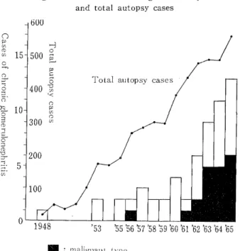
Quetionable cases and thoses with insufficient clinical data were excluded from the sample. Chronological. change: Among the 60 cases of sample, incidence of glomerulonephritis increased annually along with the increase of total autopsy cases.' The trend has been remar- kable since 1962 (See Fig. 1).
Fig. 1 Cases of chronic glomerulonephritis
and total autopsy cases
: malignant type
Fig. 2 Cases of chronic glomerulonephritis
and age
Age distribution: The age at death is 33 years and 4 months in average ranging from 3 years and 1 month to 68 years and 10 months.
Except for the under 10 and over 50, age distribution is approximately uniform even though date was so- mewhat high in the age group of 20's. (Fig. 2).
Sex distribution: There were 42 cases in male and 18 cases in fem- ale. The ratio of male to female was 7 : 3. Clinical course:
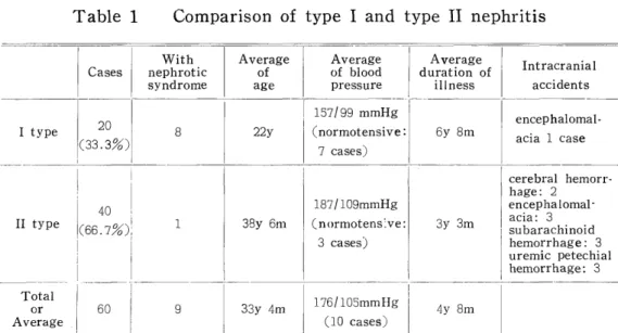
(1) Acute glomerulonephritis was experienced (ELLIS' type I") by 20 cases (33.3%) and not experienced (ELLIS' type II") by 40 cases (66.7%)
(2) Nephrotic syndrome was experienced during the course by 9 cases 8 cases of type I nephritis and 1 case of type II nephritis.
(3) Blood pressure was normal in 10 cases but hypertensive in all other cases.
(4) Duration of illness or the period from onset to death varied from a minimum of 20 days to a maximum of 34 years with the average of 4 years and 8 months.
(5) Cause of death: Though it is somewhat dangerous to deter-
Table 1 Comparison of type I and type II nephritis
With Average Average Average
Cases nephrotic of of blood duration of Intracranial
syndrome age pressure illness accidents
157/99 mmHg encephalomal-
I type 20
0 8 22y (normotensive: 6y 8m acia 1 case
(33 .3/0) 7 cases)
cerebral hemorr-
hage: 2
40 187/ 109mmHg encephalomal-
II type (66.7/0 ), o 1 38y 6m (normotens:ve: 3y 3m subarachinoid acia: 3
3 cases) hemorrhage : 3
j uremic petechial
hemorrhage: 3
Total 176/105mmHg
or 60 9 33y 4m 4y 8m
Average (10 cases)
Table 2 Macroscopically visible cerebral accidents Cerebral accidents
Cerebral hemorrhage ... 2 cases Encephalomalacia ... 4 cases
Subarachinoid hemorrhage ... 3 cases
Uremic petechial hemorrhage ... 3 cases
Total llcases (among 39 cases)
mine the immediate cause of death only from the clinical history and pathological findings, most of the cases died of uremia. Other possible causes of death for 39 cases with which intracranial study was made so-called "hypertensive cerebral hemorrhage" in 2 cases, encephalo- malacia. in 4 cases, subarachinoid hemorrhage in 3 cases, and uremic petechial hemorrhage in 3 cases. Besides these, hypertensive cardiac failure was noted in 2 cases and complication of bacterial pneumonia in 4 cases. The above is summarized in Table 1 and 2.
For preparation of histological spesimens, the kidneys including cortex and medulla fixed by 10% formalin solution were made into sections measuring 3 cm2 or more insize, embedded in paraffin, sliced to be about 4,u in thickness, and stained by hematoxylin-eosin, Wei- gert's elastica stain, van Gieson stain, PAS stain, PAM stain and Mallory-Azan stain.
In determination of vascular changes, intrarenal arterioles were classified into small arterioles measuring less than 200p in diameter and larger arteries measuring more than 200,E in diameter and 100 peices of former and 20 pieces of the latter were selected for each of the right and left kidneys, and the total of vascular changes in each artery or arteriole was determined as 0- +3. As to glomerular changes, 50 glomeruli were selected for each of the right and left kidneys and the total of glomerular changes in each glomerulus was determined as 0
+3.
RESULTS
A. Histological changes on arterioles (<200p).
Among the cases of chronic glomerulonephritis that were autopsied, there was no case without any vascular change, except for the cases that died at an early stage due to complication. Moreover, vascular changes were fairly advanced in the cases with hypertension. Most remarkable changes were noted on arterioles especially in afferent ar- terioles and ampulla portion of glomerular capillary.
(1) Fibrous simple thickening.
This is a repressentative arteriolar change in the cases of benign hypertension. Hyalinous or fibrous thickening was noted evenly throu-
ghout the arteriolar wall (photo. 1). Changes in endothelial cells were hardly observed. Fibrous simple thickening was noted in various deg- rees in all the cases including normotensive cases but excluding the cases of short duration of illness and early death due to compli- cation.
(2) Fibrous proliferative thickening.
As compared with fibrous simple thickening, hyalinous changes were poor, the arteriolar wall was evidently thickned fibrously and the myofiber were proliferated resulting in the narrowing of the arter- iolar cavity which resemble the findings of onionskin lesion observed in systemic lupus erythematosus (photo. 2). Such changes are regarded by some investigators to be fibrous simple thickening. However, the author dared handle these separately and compared these with other data.
(3) Necrosis.
There was noted so-called "fibrinoid necrosis" involving materials which were homogenously swollen on the entire circumference of the arteriolar wall and stained deeply by PAS stain. There was another type of change in the subintimal space involving materials which were stained deeply by eosin and PAS positive. This type of change is be- lieved to be the initial stage of the former. These two types of change were described as necrosis (photo. 3, 4 and 5).
(4) Angitis.
In addition to the above three arteriolar changes, angitis was a peculiar finding though the incidence was rare. In the afferent arterioles
of two cases (one age 68 years and 10 months and the other age 37 years and 9 months) the cells of the wall around fibrinoid necrosis were proliferated outwards in nodular shape. Infiltrated cells were also noted in the region (photo. 6).
B. Histological changes on larger arteries (>200,a).
On larger arteries in the renal parenchyma measuring over 200a in diameter,intimal thickening and elastosis in various degrees were generally noted. In other words, usual arteriosclerosis was the only finding and there was no necrosis nor angitis (photo. 7).
C. Change in ampulla portion of glomerular capillary.
In addition to the vascular changes, degenerative changes of glo- meruli and extracapillary proliferative change previously stated in cases with chronic glornerulonephritis, changes in ampullar potion of glomer- ular capillary should not be overlooked. The changes were thickening of the wall, narrowing of the internal cavity due to cell proliferation (photo. 9), dilatation, infiltration of serum components (photo. 11), hemorrhage, thrombosis (photo. 10) and angitis acompanied with fibr- inoid necrosis distributed diffusely on the afferent arterioles (photo. 6).
As the changes in the ampullar portion increased in intensity, the
changes in the peripheral area of glomerular tufts were also intensified resulting in hemorrhage, thrombosis, necrosis and crescent. Such cha-
nges are the findings often observed in "malignant type" which is des- cribed below.
D. Malignant phase of chronic glomerulonephritis8' (malignant type).
The above-stated arteriolar glomerular changes such as necrosis, angitis and proliferative glomerulitis are the findings of so-called "ma-
Table 3 Malignant phase of chronic glomerulonephritis
Arteriolar Changes ' Glomemlar
4.1
(<200%c) N I changes
a O
R U
O rn U U (n CUd
a aI C; p, aA 0
cd a~ b0 o C. , o ° m -. W (n
4- o rd .~ to o a~ 3, . U a~ o
° ° 0 y ° aU' U o A Cz
CZ r.
° W o U v ~ r 0 Q) M. as
z 'J.- Z 4-1 Q Z 4 U 4"
4~ 4.1
w w~ ~ 41
I
3877* F 41y4m 22y 160/100 +2 +1 +1 +2 +1 +2 encephalomalacia
5374 M 44y8m 4y 200/120 +2 +3 +2 +1 +3 +3
6145 F 68y 10m 28y 1701 90 + 1 + 2 + 1 + + 1 + 1 + 1
6229 M 26y10m 5y 210/130 +2 +3 +2 +3 +1 +2 encephalomalacia
6370 F 22y7m 4y2m 180/110 +3 +3 +2 +2 +1 +1
6421* M 14y2m 2y9m 180/120 +1 +3 +2 +1 +1 +3
6476 M 21y1m ly6m 200/140 +2 +2 +1 +2 +1 +3
6684 F 46y2m lly 200/130 +3 +2 +1 I +2 +2 +1
6717* M 27y 24y 200/120 +3 +3 +3 +3 +1 +1
6867 M 46y8m 6m 260/120 +2 +1 +1 +3 +1 +2 cerebral hemorrhage
6952* M 32y5m 5y 200/120 +2 +1 +1 +1 +2 +2
7011 M 37y9m 3y 250/120 +3 +2 +2 + +2 +3 +2
7122 M 67y2m 2y 210/130 +2 +3 +2 +3 +2 +1
7126 M 37y7m 5y 190/110 +3 +2 +3 +3 +1 +2
7286 M 51y7m l0y 250/150 +3 +1 +1 +3 +2 +2 cerebral hemorrhage
7289 M 48y11m ly 210/130 +2 +2 +1 +1 +1 +3 subarachinoid hemorrhage
7328 M 30y 6y 200/130 +2 +2 +1 0 +1 +1
7345 F 53y11m 3y 2101120 +2 +2 +1 +2 +3 +1
7566 F 45y6m 2y6m 260/ 180 +2 +2 +2 +2 +2 +1 encephalomalacia
7701 F 42y 3y6m 230/120 +2 +2 +1 +2 +1 +2
Average 40y4m 7y2m l 209/125 I+2.2\+2.1I+1.6i ~+2.0I+1.4 +1.81 -
Non-malignant type
3y 160/ 90 +1.6I+0.5 +1.0 +0.9+0.2 +0.8
Average 29y10m
Total
Average 33y4m 4y8m 176/105 +1.8 +1.0+1.2 + 1.2 +0.6i + 1.1
*type I nephritis
lignant nephrosclerosis" itself. Further, cases having remarkable car- diac or cerebral symptom are regarded as "malignant phase of chronic glomeru.lonephritis".
Among the 60 cases of sample, malignant type was observed in 20 cases. As shown in Table 3, male-female ratio was about 2:1. In comparison with the ratio of 7:3 in total cases, incidence of malignant type is high in females. The age at death, duration of illness, blood pressure and histologic findings were all higher in degree in malignant type than in non-malignant type. As to vascular changes, not only necrosis and fibrous proliferative thickening but also fibrous simple thickening and arteriosclerosis were more advanced in most cases of malignant type (Table 3). Moreover, the stronger were the vascular changes, the more advanced were glomerular changes especially extra- capillary changes in most cases. However the degree of these changes was not necessarilly in parallel with the blood pressure.
Chronologically, malignant type showed a trend of rapid increase after 1963 as shown Fig. 1.
E. Comparison between vascular changes and clinical course.
The relationship between vascular changes and each of age, dur- ation of illness and blood pressure was investigated in all cases.
Age: An apparent correlation was noted between arteriosclerosis of larger arteries (>200p) and age, but advanced (+1 or more) fibrous simple thickening, necrosis and fibrous proliferative thickening showed no correlation with age though many cases in younger group had no such vascular change. Fibrous simple thickening 0 was noted only in two young cases who died at an early stage due to complication. There were some cases of fairly advanced age that were absolutely free from necrosis and fibrous proliferative thickening. In other words, the ch- anges in small arterioles measuring less than 200it had no relation with age. Most cases of malignant type showed changes of +2 more (Fig.
3).
Duration of illness: Irrespective of the size of arteries and nature of changes, there was a close correlation between all vascular changes and duration of illness, i.e. the longer was duration, the more advan- ced were the vascular changes. Particularly in small arteriolar changes (fibrous simple thickening, necrosis and fibrous proliferative thicken- ing), the patterns of +2 and +3 were totally reversed as compared with Fig. 3 (Fig. 4).
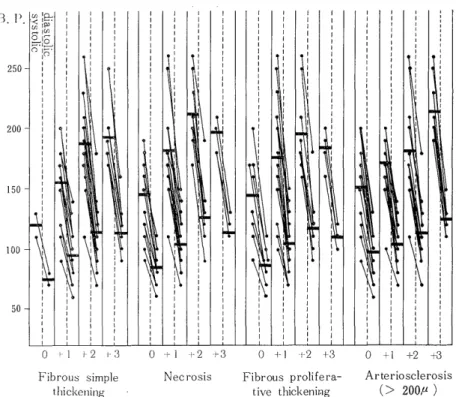
Blood pressure: Each vascular change was almost parallel with blood pressure both systolic and diastolic. However, as shown in Fig.
5, fibrous simple thickening and arteriosclerosis (>200p), and necrosis and fibrous proliferative thickening respectively showed a similar patt- ern. While in the former group the blood pressure was in complete correlation with the degree of vascular change, the latter group showed
Age Fig. 3 Vascular changes and age
Fibrous simple Necrosis Fibrous prolifera- Arteriosclerosis
thickening five thickening ( > 200, )
• malignant type 0 non-malignant average
Fig. 4 Vascular changes and duration of illness
Fibrous simple Necrosis Fibrous prolifera- Arteriosclerosis
thickening five thicl:enine (> 200,E )
Fig. 5 Vascular changes and blood pressure
Fibrous simple Necrosis Fibrous prolifera- Arteriosclerosis
thickening tive thickening (> 200I-1
a decrease of blood pressure as the vascular change increased from +2 to +3. Blood pressure over 200 mmHg was frequent in cases with +2 and +3 of fibrous simple thickening and arteriosclerosis (>200,u) but
in cases with +1 and +2 of necrosis and fibrous proliferative thicken- ing.
From this point of view, vascular changes may be classified into following 3 groups: 1) arteriosclerosis (>200t) which is closely related to age and blood pressure, 2) fibrous simple thickening which is related only to blood pressure, and 3) necrosis and fibrous proliferative thi- ckening which are little related to either factor.
DISCUSSION
As the causes of chronicity and malignancy of diffuse glomer:ulone- phritis, many factors have been indicated by investigators, such as
the degree of attack at onset, intrinsic progress of renal changes, continuation of the primary lesion or reinfection, inadequate care of patient, family factor and also development or extension of vascular changes.
The author paid attention to hypertension and vascular changes and compared them with clinical course and other factors in 60 autopsy cases,
Clinical course: The 60 autopsy cases were classified according to ELLIS ' classification into 20 cases (33.3%) of type I nephritis, and 40 cases (66.7%) of tyge II nephritis. Generally type I nephritis is belie-
ved to be relatively well curable with satisfactory prognosis 25121). In the present study, autopsy cases of type I nephritis occuped 1/3 of the total cases, which is not so rare. However, it was reported by SARRE24' that acute nephritis with nephrotic syndrome results in poor prognosis.
Of the 20 cases of type I nephritis in the present study, 8 cases (40%) indicated nephrotic syndrome during the course, and it is not contra- dictory that type I nephritis indicated relalively high percent of the total cases. In the group of type II nephritis, only 1 case (2.5%) sh- owed nephrotic syndrome.
In comparison with type II nephritis was longer in duration of illness but lower in age at death, onset being seen in young subjects.
These findings were in conformity with many other reportsts'24' . Concerning blood pressure, there was a great difference between type I and type II. The difference was 10 mmHg in diastolic pressure and 30 mmHg in systolic pressure. Since type II nephritis is often accompanied with hypertension from the onset, the difference between two types is understandable.
However, as to the difference between systolic and diastolic pre- ssure, the author has no answer at present. Intracranial changes were of course more frequent in the cases of type II nephritis which were often accompanied with intensive hypertension.
Arterial changes: Arteriosclerosis in the arteries measuring over 200,u showed correlation with age. Arteriosclerosis in the arterioles measuring under 200,1 and fibrous simple thickening had correlation with blood pressure as reported by other investigators") 17) . However, necrosis and fibrous proliferative thickening, had no correlation with age, and cases with +3 degree of there vascular changes indicated evidently lower valve of blood pressure than cases with +2 degree.
All these arterial and arteriolar changes showed a close correlation with duration of illness. This evidence suggests that the vascular ch- anges are not simply due to age and blood pressure but rather affected by the continuation of nephritis29'.
Among various arteriolar changes in chronic glomerulonephritis, fibrous proliferative thickening which is rare in other organs and ob- served mostly in the arterioles measuring under 200,i was given a spe- cial consideration in this study. In the past fibfous proliferative thi- ckening had been regared as arteriosclerosis in the same light with fibrous simple thickening. On the other hand, this was considered by BELL and CLAUSONI) , MURPHY and GILL'9', and HUCKELIQ' as a charac- teristic change of malignant nephrosclerosis and described as "concen- tric arteriolitis". The author can not agree that fibrous proliferative
thickening is regarded simply as arteriosclerosis in the same light with fibrous simple thickening, and moreover, the expression of arteriolitis is not adequate since this type of thickening is entirely different mor- phologically from periarteritis or panarteritis. The various data of fibrous proliferative thickening described above shows the same pattern as necrosis rather than arteriosclerosis (>200k) or fibrous simple thi- ckening. It is deemed from the above that fibrous proliferative thicken- ing is not a mere sclerotic change but a peculiar change with some potentiality in itself and/or in "nephritic ground and environment
Malignant phase of chronic glomerulonephritis :
Recently, cases with clinical symptoms of chronic glomerulonephr- itis accompanied with malignant hypertension and remarkable cardiac and cerebral disorders who would finally die of uremia have drawn atten- tion of those concerned as malignant phase of chronic glomerulonephr- itis8' in comparison with malignant phase of essential hypertension. In the present study, the author selected among 60 cases of sample 20 cases with the above-stated clinical symtoms and histological findings of so-called "malignant nephrosclerosis". The results of study in this aspect may be summarized as follows.
(1) There were 13 male cases and 7 female cases. The rate of female cases was higher than in total cases. (2) Type I nephritis was noted in 4 cases and type II nephritis in 16 cases. (3) In comparison with non-malignant group, age at death was higher by 10 years and 6 months. (4) The duration of illness was over twice as long as that in non-malignant group. (5) Blood pressure and histologic findings were greatly different from those in non-malignant group. (6) Cases of ma- lignant type have rapidly increase since 1963.
Whereas the mean valve of the duration of type II nephritis was extremely shorter than that of type I nephritis the duration of the ma- lignt type with included mainly type II nephritis was twice longer.
This indicates that a long duration of the nephritis may be an impor- tant factor development of malignant type and the rapid increase of the cases of malignant type since 1963 may be due mostly to the longe- vity effect of the therapies, such as rest in early stage, treatment aga- inst the primary lesion and complication, and administration of depre- ssor drugs, diuretica, adrenal cortical hormone and so forth. However, the data of the author failured to show any special evidence to support this presumption.
In conclusion, the author emphasized that so-called "malgnant type appears during the course of chronic glomerulonephritis due to advanced arteriolar changes and that cases of death due to cerebral accident particulary cerebral hemorrhage had been increasing20'.
SUMMARY
With 60 autopsy cases of chronic glomerulonephritis, histological findings of the kidney, particulary intrarenal arterial and arteriolar changes were compared with clinical course.
The arteries were classified by diameter into those over 200 and those under 200,x. Whereas the former showed only usual arterioscle- rosis, the latter demonstrated fibrous simple thickening, fibrous pro- liferative thickening, necrosis and angitis, though the last one was noted only in 2 cases.
The degree of vascular changes was compared with clinical data shch as age, duration of illness and blood pressure. Arteriosclerosis of larger arteries (>200,~) was proportional to age. On the contrary, all vascular changes were totally proportional to duration of illness. Grade of both fibrous simple thickening and arteriosclerosis (>200,u) was parallel to blood pressure. On the contrary grade of both fibrous pro- liferative thickening and 'necrosis was proportional to blood pressure, i.e. Cases having high degree vascular changes showed rather lower blood pressure.
The author consisered fibrous proliferative thickening not simply as a sclerotic change but as a change close to necrosis or angitis.
Malignant phase of chronic glomerulonephritis was noted in 20 cases (33.3%), 16 of which were of type II nephritis. In comparison with non-malignant type, malignant cases showed higher values in blood pressure and higher degree of histological vascular changes, and moreover they were higher in age by 10 years and 6 months and longer in duration of illness by 4 years and 2 months. Malignant phase of chronic glomerulonephritis rapidly increased in incidence since 1963.
In view of the above, it is indicated that the patients of chronic glomerulonephritis may survive longer due to the advancement of treatment and rehabilitation, and accordingly it may be anticipated that hypertension and more intensive vascular changes would develop resulting in more cases of death due to cerebral vascular insufficiency such as cerebral hemorrhage.
REFERENCES
1) BELL, E.T. & CLAWSON, B.J.: Arch. of Path. 5: 937, 1928.
2) BERGSTRAND, A. & BUCHT, H.: Z. Zellforsch. 48: 51, 1958.
3) BRAUN-MENENDEZ, E., FASCIOLO, T.C., LELOIR, L.F., and MUNOZ, J.M.: J.
Physiol., 98: 283, 1940.
4) BRIGHT, R.: Reports of Medical Cases, London, Vol. 1, 1827 & Vol. 2, 1831.
5) COCHRANE, C.G.: J. Exp. Med. 118: 489, 1963.
6) DIXON, F.J., VAZQUE, J.J., WEIGLE, W.O. & COCHRANE, C.G.: Am. J. Path.
65: 18 1958.
7) ELLIS, A.: LANCET, 242: 1, 1942.
8) FISHBERG, A.H., : Hypertension and Nephritis, 5th ed. Lea and Febiger 1,
Philadelphia. 1954.
9) HARGITAY, B. & KUHN, W.: Zschr. f. Electrochem. u. angew. physik . Chem.
55: 539, 1951.
10) HuCKEL, R.: Virch. Arch, 276: 447, 1930.
11) IVERSEN, P. & BRUN, C.: Am. J. Med. 11: 327, 1951.
12) JONES, D.B.: Am. J. Path. 29: 33, 1953.
13) JONES, D.B.: Am. J. Path. 33: 313, 1957.
14) KOHLSTEDT, K.G., PAGE, I.H., and HELMER, O.M, : Am. Heart Jour., 19: 92, 1940.
15) MASUGI, F.: Nephritis, Neiraku Shobo., Tokyo, 1948. (Japanese) 16) MATSUOKA, S.: Nihon -Byori gakka ish i, 54: 333, 1965. (Japanese).
17) MORITZ, A.R., & OLDT, M.R.: Am. J. Path. 13: 679, 1934.
18) MOVAT, H.Z. & FERN NDO, N.V.P.: Am. J. Path. 42: 41, 1963.
19) MURPHY, F.D. & GILL, J.: Arch. Int. Med. 46: 75, 1939.
20) NISHIMORI, I. & TAKAKI, H.: Rinshoto-Kenkyu, 42: 2256, 1965. (Japanese).
21) PEREZ ARA, A.: Bol. Ligacontra el Cancer, 25: 121, 1950.
22) RINEHART, J.F., FARQUH'_R, M.G., JING, C.H. and ABDUL-HAJ, s.k.: Am.
J. Path. 29, 21, 1953.
23) RINEHART, J.F.: Arch. Path. 59: 439, 1955.
24) SARRE, H.: Nierenkrankheiten, G. Thieme, Stuffgart 1959.
25) TAKATSU, T.: The Japanese J. of Nephrol. 4: 13, 1962.
26) TIGERSTEDT, R., & BERGMANN, P.G.,: Skand. Arch. Physiol. 8: 223, 1898.
27) Tsuji, S.: The Japanese J' of Nephrol. 4:1, 1952.
28) VOLHARD, F.: Handbuch der inneren Medizin 2 Auf1. 6 Bd. II Julius springer, 1931.
29) YASHIMA, G.: Saishin-fgaku, 19: 3062, 1064. (Japanese).
Photo. 1 fibrous simple thickening, H-E stain (10X40)
Photo. 2 fibrous proliferative thickening, H-E stain (10x16)
Photo. 3 subintimal necrosis, H-E stain (10 x 40)
Phto. 4 fibrinoid necrosis, PAS stain (l0 x 40)
Phto. 5 fibrinoid necrosis, PAS stain (l0 x 40)
Photo. 6 angitis, H-E stain (l0 x 16)
Photo. 7 arteriosclerosis (>2009), H-E stain (10 x 16)
Photo. 8 fibrinoid necrosis of afferent arteriole and hyaline droplet in glomerular tuft. H-E stain (10x16)
Photo. 9 thickening, cell proliferation and narrowing in ampullar portion of glomerular capillary
Photo. 10 hemorrhage and early stage of thrombosis in ampullar portion of glomerular capillary
Photo. 11. dilatation and infiltration of serum component in ampullar portion of glomerular capillary.
Photo. 12 proliferation of capsular and glomerular cells.