Acta Med. Nagasaki 30 :156-167
Electron Microscopic Study on the Kidney of Adrenal Regeneration Hypertension Rats
-A Special Reference to the Glomerular Foam Cell and Nephrotic Syndrome-
Minoru HONDA
Department of Pathology, Atomic Disease Institute, Nagasaki University School of Medicine
Nagasaki, Japan
Received for publication, May 15, 1985
Adrenal regeneration hypertension was produced in Wistar rats, WKY and SHR by the SKELTON's method. We observed glomerular foam cells in the kidneys of those exp- erimental rats. Glomerular foam cells were seen in the rats that were markedly hyperten- sive and in a hypercholesterolemic state, and in those that showed a high excretion of protein in the urine. We assumed that these experimental rats exhibited the state of nephrotic syndrome.
Moreover, we found glomerular foam cells in all of the Wistar rats and WKY fed a high cholesterol diet, that is, a five percent cholesterol and two percent cholic acid-including diet by the SKELTON'S method, but glomerular foam cells were noticed in only about one-third of rats fed basic food by the SKELTON's method. Glomerular foam cells were located in the mesangial matrix or the subendothelial space of the glomerular capillary. An electron microscopic study of the glomerular foam cells revealed that the cell origin was probably circulating monocyte and mesangial cell, but was strongly suspected to be circu- lating monocytes.
INTRODUCTION
Adrenal regeneration hypertension (ARH) was established by SKELTON, but the mechanism of the disease has not yet been elucidated. At present, Spontaneously Hy- pertensive Rats (SHR) tend to be used instead of ARH, as an animal model of human hypertension, although, from the point of view of endocrinology, ARH is still useful. In
A part of this study was presented at the Winter Annual Meeting of the Japan Atherosc- lerosis Society, Jan. 18, 1985.
本田 実
this study, the hypertension by using ARH was examined, and, through the examination, the experimental rats prompted nephrotic syndrome. Foam cells were discovered in the renal glomerulus. The electron microscopic observation of the foam cells was investigated, and its pathogenesis and cell origin were discussed.
MATERIALS AND METHODS
Three strains of female rat weighing 50g to 70g (Wistar rats, WKY (from Japan Charles River), and SHR] were used for the investigation. The operation was carried out according to the SKELTON' S method (right nephrectomy-adrenalectomy, left adrenal enucleation), and hypertension was produced by the administration of 1 % saline. Each strain of rat was divided into the following four groups : 1) first group : a control group given basic food (F2 food from Funabashi farm) and tap water ; 2) second group : given a high cholesterol diet of 5% cholesterol and 2% cholic acid mixed with basic food and tap water ; 3) third group : received the SKELTON-method operation and given the basic food and 1% saline ; 4) fourth group : received the SKELTON-method operation and given the high cholesterol diet and 1 % saline. The experimental period was three weeks for SHR (because the operated SHR could not live more than four weeks) and seven to eight weeks for Wistar and WKY. The blood pressure was measured every week by the tailcuff method using a RAT automatic blood pressure recorder USM-105-R type. The urine protein was measured by the TCA method using 24 hour-stored urine 3 to 5 day before sacrifice. After 12 hours-fasting, the blood was obtained from the abdominal aorta of all rats under light anesthesia by ethylether, and the separated serum was used in the biochemical analysis. The measurements were carried out by the enzyme method for total cholesterol (T-CH OL) and triglyceride (TG), the heparin-Ca precipitation method for LDL and VLDL, the phosphotungstic acid-Mg precipitation method for HDL-cholesterol (HDL C), the Biuret method for total protein (TP), and the UV enzyme method for BUN.
After weighing, the extracted organs were fixed in 10% formaline and stained with Hematoxyline Eosin (HE), Periodic acid Schiff (PAS), Periodic acid Methenamine Silver (PAM), Azan-Mallory (AM), Phosphotungstic acid hematoxyline (PTAH), Elastica van Gieson (EVG), etc, and then observed by light microscopy. For the observation of the foam cells, Oil red 0 stain and various immunohistochemical stains such as factor VIII associated antigen, S-100 protein and al-antitrypsin were added. A part of the kidney was immersion fixed in 1.5% glutaraldehyde, postfixed in 1% osmic acid, given a double stain of uranyl acetate and lead citrate, and observed with a JEM-100B type electron microscope.
RESULTS
1) Blood pressure : The blood pressure was under 140mmHg in all three strains of rat in the first and second group, and 171 to 206mmHg in the third and fourth group of
Wistar and WKY. The blood pressure in the fourth group was lower than in the third group of Wistar and WKY, and a significant difference (p<0.05) was observed among the Wistar rats. In the third and fourth group of SHR, despite the short period of 3 weeks, hypertension of over 209mmHg was observed, and unlike Wistar and WKY, the blood pressure in the fourth group was higher than in the third group and a significant difference of p<0.05 was observed (Fig. 1).
Fig. 1. Systolic blood pressure at the terminal stage Mean±SD; *p<0.001 vs Group I
*p<0.05 vs Group III
2) Urine protein : Less than 10mg a day was measured in all three strains of rat in the first and second group and 14mg a day was the largest amount. The average in the third group was 136mg (80-19.8mg) a day for Wistar and 131mg (22-293mg) a day for WKY. The average in the fourth group was 216mg (60-360mg) a day for Wistar and 123mg (68-200mg) a day for WKY.
3) Organ weight : The mg organ weight/100g body weight ratio is shown in Table 1 . The weight of the brain, heart, and kidney had increased in the third and fourth groups of all three strains of rat. The weight of the liver had increased in the second and fourth groups of all three strains of rat. The weight of the adrenal gland (unilateral re- generating adrenal gland) of Wistar and WKY in the third and fourth group had also in
Table 1. Organ weights mg/100gm body weight
Rat Group n Brain Heart Thymus Liver Kidney Adrenal gland
Wistar I 9 757±20 350± 18 147±20 3626±199 274± 8 11±1
II 8 857±20 379± 7 151±12 5670±195*** 347± 5 9±0
III 21 1022±71** 566± 36 *** 156±13 4918±281** 979±106.*** 18±1***
IV 13 982±43*** 588± 23*** 235±27* 6543±329*** 784± 26*** 20±1***
WKY I 6 933±20 454± 5 99±13 2916± 47 283± 4 11±1
II 8 1293±50*** 449± 12 112±10 5941±215*** 377± 10 14±1
III 6 939±23 758±108** 106±11 3513±234* 736± 78*** 22±3***
IV 8 1153±43*** 607± 45** 113±12 6182±121*** 664± 25*** 23±1***
SHR I 6 1107±41 530± 40 226±23 4639±319 362± 21 15±2
II 8 1658±65*** 461± 17 123± 9** 5637±157** 422± 13 21±1**
III 6 1392±76** 685± 31** 208±33 5012±165 791± 67*** 17±2
IV 7 1333±72** 734± 19*** 217±26 5785±380* 722± 33*** 19±2**
afean±SE; *p<0.05, **p<0.01, ***p<0.001 vs Group I
creased. The thymus of Wistar and WKY in the second, third, and fourth groups showed a tendency toward increase in weight, while the thymus of SHR showed a tendency toward
decrease in weight.
4) Serum lipid level and others : T-CHOL was 882 and 999 mg/dl in Wistar and WKY of the fourth group respectively, and there was a markedly high level in the three
strains of rat in the second and fourth group as compared to the first group. In all three
strains of rat, HDL-C of the third group showed a tendency to increase in level, but that
Table 2. Serum lipids level (mg/dl), TP (g/dl) and BUN (mg/d')
Rat Group n T-CHOL TG LDL VLDL HDL-C TP BUN
Wistar I 6 72± 4 48± 8 66± 9 2± 1 53± 4 6.2±0.1 24±2
II 8 414± 49*** 182±22* 139±19 725± 76*** 37± 2 6.9±0.2* 17±1***
III 9 115± 28 139±32* 113±27 20± 10 114±14 6.7±0.3 27±2
IV 12 882±123*** 135±28* 305±42*** 1286±175*** 30± 3*** 5.4±0.2* 17±1***
WKY I 6 107± 5 33± 1 75± 6 4± 2 78± 5 6.2±0.1 16±0
I I 8 678± 54*** 110±10*** 223±62** 1509±141*** 46± 4 *** 6.9±0.2* 16±2
III 6 133± 14 59± 7*** 227±5C** 10± 3* 80± 6 6.2±0.1 20±2
IV 8 999± 91*** 95±13*** 186±36** 1835±158*** 43± 3*** 5.7±0.3* 16±3
SHR I 6 71± 8 118±18 89± 5 3± 1 41± 3 6.4±0.2 16±1
II 7 628± 21*** 159±14 199±44* 1455± 37*** 38± 1 6.8±0.5 29±1***
III 6 116± 11** 165±29 167±38* 69± 63 59±10* 6.1±0.5 29±3***
IV 7 221± 62* 181±47 326±70** 259±207 53± 8 6.2±0.2 36±4***
Mean±SE; *p<0.05, **p<0.01, ***p<0.001 vs Group I
of the fourth group showed a significant decrease. TG, LDL and VLDL showed a similar tendency. TP showed a significant decrease in the fourth group of Wistar and WKY, and the rats which showed an especially low level had a tendency toward increase in ex- cretion of urinary protein. No definite tendency was seen in the level of BUN (Table 2 ).
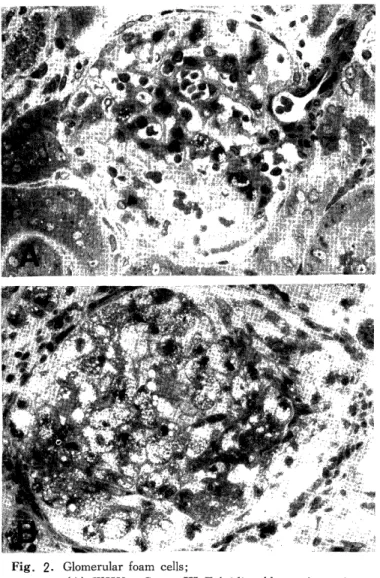
5) Light microscopic observation of the kidneys : The most characteristic finding was fibrinoid necrosis of the small arteries and afferent arteriole, which is seen in human malignant hypertension, and it extended over all or part of the glomerular tuft and fell into a degenerative necrosis. Many of the glomeruli were swollen and had foam cells. In the third group of rats foam cells appeared in small numbers in glomeruli which had strong changes (Fig. 2 A), although in the fourth group they appeared in glomeruli which showed less change, and had a tendency to appear in larger numbers (Fig. 2 B).
Foam cells were usually positive for Oil red 0 stain, negative for PAS reaction, and also
Fig. 2. Glomerular foam cells;
(A) WKY, Group III,Toluidine blue stain. X380 (B) Wistar, Group IV, Toluidine blue stain. X420
negative for immunohistochemical stains (used by DAKO PAP kit) like VIII associated antigen, S-100 protein, al-antitrypsin, etc. The tubule epithelium had a strong hyaline droplet degeneration, was filled with cast in the tubules, and became thyroid-like in appearance. On rare occasions, cast with cholestelline crystal was seen.
6) Frequency rate of foam cells : The rats which revealed even one foam cell in the glomerular tuft by light by light microscopic examination were designated as positive. In the first and second group, all three strains of rat were negative. All of the fourth group of Wistar and WKY, one third of the third group of Wistar and WKY, and also one third of the fourth of SHR were positive (Table 3) .
Table 3. Relation between GFC and serum T-CHOL
Rat Group n GFC(+) rat GF'C(-j rat T-CHOL (mg/dl) (+j rat
Wistar 1 9 0 159<
I I 7 0 355:b
111 20 7 202-c 207>
IV 13 13 384 >
WKY I 6 0 120 <
II 8 0 368>
111 6 2 165< 124>
IV 7 7 442 >
SHR I 7 0 104 <
II 7 0 574>
III 12 0 148<
IV 7 2 154 < 381>
GFC(+) : glomerular foam cell positive
GFC(-) glomerular foam cell negative
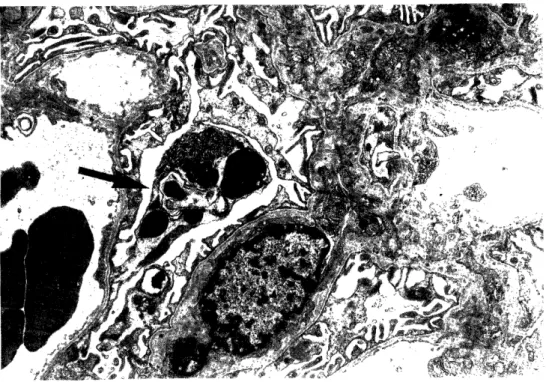
7) Electron microscopic observation of the glomeruli : In the glomerulus there was no thickening of the glomerular basement membrane and no abnormal deposits were seen in or outside the basement membrane or mesangial area, although there were glomerular basement membrane changes continuing to the mesangial area. Tortuosity of the basement membrane and enlargement of the subendothelial space were seen in rats with marked hypertension. Numbers of electron dense bodies and myeline figures were seen in the ep- ithelium. Fusion of foot processes was also seen in some parts although this was not very conspicuous (Fig. 3 ). Mesangial matrix had increased and foam cells were mainly seen in the mesangial area. It is conceivable that some mesangial cells rounded basemeet me- mbrane became foam cells, or that some circulating monocytes having abundant lysosomes and mitochondria, pressed upon the mesangial cells and became foam cells (Fig. 4). In the latter case, clear but very narrow spaces could sometimes be seen between the cytoplasm of foam cells and the mesangial matrix (Fig. 5 ). In severely damaged glomeruli, although the proximity with mesangial cells was unclear, it is obvious that foam cells originated from
Fig. 3. Change of epithelial cell;
Myeline figure and dense body (arrow). X6000
Fig. 4. Foam cell in mesangial area;
Foam cell (F) and mesangial cell (M). X9000
Fig. 5. Foam cell originated from circulating monocyte;
Slit-like spaces between foam cell and mesangium
matrix (arrows). X9000
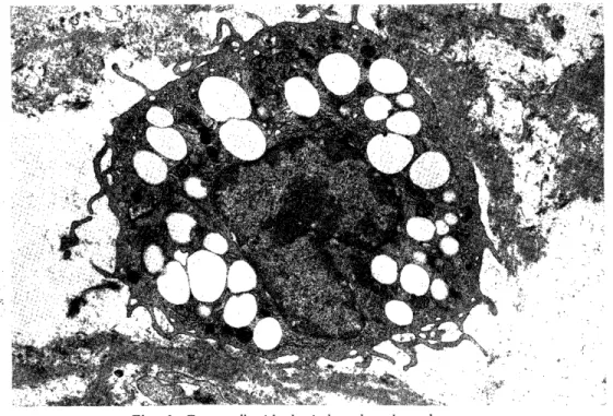
Fig. 6. Foam cell with the indented nucleus, lysosomes, mitochondria and cytoplasmic processes. X9000
circulating monocytes because of the characteristic indentation of the nucleus, numbers of cytoplasmic processes, the existance of lysosomes and mitochondria, and so on (Fig. 6 ).
However, there were few foam cells which could be proved to be originated from circu- lating monocytes. In most cases, it was impossible to distinguish whether they were circu- lating monocytes or mesangial cells.
DISCUSSION
In 1955 SKELTON13) established ARH, which is easily reproducible, but the me- chanism of the disease has not yet been elucidated. At present SHR are used as an an- imal model of human hypertension instead of ARH, although ARH is often used in en- docrinological studies. Although etiology is not investigated in this study, SKELTON15) stated that an enzyme abnormality was caused in the regenerating adrenal gland because large quantities of deoxycorticosterone accumulated due to a deficiency of 11 (3-hydroxylase, and HALL et al.'), GOMEZ-SANCHEZ et al.4) detected 19-nor-deoxycorticosterone in the urine of adrenal regenerating experimental rats and reported that this substance, which cannot be found in healthy rats, had a function of increasing the blood pressure which was several times as strong as deoxycorticosterone. In any case, ARH is undoub- tedly caused by a severe hormone abnormality and it is classified in the category of endocrinological hypertension.
As seen in the detailed reviews by SKELTONI3)14) and NISHIMORI10)11), ARH is a highly reproducible method. The blood pressure, organ weights, and the light micro- scopic findings are almost standardized and this investigation obtained the same results.
We discuss here the fact that adrenal regeneration hypertension rat (ARH rat) revealed nephrotic syndrome and that foam cells appeared in the renal glomerulus.
(1) Pathogenesis of foam cells : It has been reported that foam cells in the human kidney appear in ALPORT' S syndromes'), Membranoproliferative glomerulonephritis9>12>
Diabetes Mellitus"), Toxemia of pregnancy2), and transplanted kidney''. These are all representative of diseases that prompt nephrotic syndrome, and it has been stated that hypercholesterolemia and proteinuria are found in these diseases. Although there are very few reports about glomerular foam cells in experimental animals, TAKEBAYASHIl6) found them in ARH rats and WATANABE et al.") found them in rabbits with Masugi's nephritis fed a high cholesterol-diet. Diagnostic criteria have been established for ne- phrotic syndrome in humans but not for the rat or the rabbit. In WATANABE'S study hypercholesterolemia and proteinuria were confirmed, and the rabbits were assumed to be in a nephrotic state.
In the present study, the rats in which foam fells appeared in the glomeruli showed remarkable hypertension and also had hypoproteinemia, proteinuria, and hypercholesterol- emia, which are similar to the symptoms of nephrotic syndrome seen in humans. Com- pared to the first and second group of rats which had less than 10mg/day proteinuria, the third and fourth group rats had over 100mg/day proteinuria, which is a large amount
of proteinuria for the rat. Moreover, in the fourth group of Wistar and WKY, glomerular foam cells appeared in all rats. Serum TP was significantly decreased, but T-CHOL showed a remarkably high level.
When the cause of foam cells appearing in diseases with nephrotic syndrome is considered, the existence of hypercholesterolemia along with glomerular lesion, seems to contribute in some part. FUJINAMI et al.1) found foam cells in the transplanted kidney and has presented the significance of hypercholesterolemia caused by steroid hormone used in inhibition of rejective response. MCKENZIE et al.") prescribed an anticholesterol- emic agent to a patient who had hypercholesterolemia from nephrotic syndrome and chronic nephritis and has reported that foam cells disappeared by decreasing cholesterol though histologic appearance of glomerulus deteriorated and nephritic appearance remained.
In the present study, the same result was obtained. In the fourth group of Wistar and WKY, foam cells appeared, even when glomerular damage was mild, because the serum cholesterol level became remarkably high. On the other hand, foam cells were not found in the third group, that the serum cholesterol was not very high, unless the glomerulus, was severely damaged. As seen in the second group, foam cells did not ap- pear by only hypercholesterolemia.
As KIHARA et al .6 has stated, foam cells are rarely seen in cases of disease of the hepatobiliary system accompanied by hypercholesterolemia where no remarkable changes are seen in the glomerulus.
Therefore, it is thought that glomerular damage and the existence of hyperchole- sterolemia are important factors in the appearance of glomerular foam cells.
(2) Cell origin of foam cells : We discuss here the origin of foam cells from this experimental viewpoint. Most of the foam cells observed in this study were located in the mesangial area : some of them existed surrounding by the mesangial matrix and others existed pressing the endothelium at the subendothelial space. Therefore, mesangial cells and foam cells can be said to be closely related. A few foam cells could originated from the circulating monocyte because of the characteristic morphological features and the nar- row space between the cytoplasm of foam cells and the mesangial matrix. In this experi- ment foam cells originating from the capillary endothelium or epithelium were not found.
It is thought that the foam cells originated from both mesangial cells and circulating monocytes, but especially from circulating monocytes.
As has been said before, there have only been a few reports about glomerular foam cells in experimental animals. TAKEBAYASHI'6 has indicated the existence of vacuolated cells in the mesangial area and stated that they were mesangial cells. WATANABE et al.17) has stated that the foam cells originated from both mesangial cells and circulating mono- cytes. GEER et al. 3) has observed the ARH rat by electron microscopy, though he has not reported the existence of glomerular foam cells.
CONCLUSION
1) ARH rats prompt nephrotic syndrome, and foam cells appear in the renal glo- merulus.
2) The appearance of glomerular foam cells depends upon two conditions : glomer- ular damage and hype rcholesterolemia. The latter condition, that is, hypercholesterolemia, is more important.
3) It is thought that glomerular foam cells originate from both circulating mono- cytes and mesangial cells, and especially from circulating monocytes.
ACKNOWLEDGEMENT
The author wishes to express his sincere gratitude to Prof. Issei NISHIMORI, De- partment of Pathology, Atomic Disease Institute for his kind guidance in this study and review of this paper. Thanks are also due to Associate Prof. Ichiro SEKINE, Depart- ment of Pathology, Atomic Disease Institute, Associate Prof. Masao KISHIKAWA, De- partment of Pathology, Scientific Data Center of Atomic Bomb Disaster, and Associate Prof. Nobuo TSUDA, Pathology Division, Department of Laboratory Medicine, Nagasaki University School of Medicine. SHR were provided by Prof. Masayori OZAKI, Second Department of Pharmacology, Nagasaki University School of Medicine. The author is also grateful to all the staff members, Department of Pathology, Atomic Disease Institute, for their kind collaboration.
REFERENCES
1) FUJINAMI, T., MOROZUMI, K . , YOSHIDA, A., SOUMIYA, S., SAKUMA, N . , HAYAHSI, K . , YOKOI, J., IWASE, T., and TAKADA, M.: Arterial changes in the
transplanted kidney. J. Jap. Atheroscler. Soc. 10: 67-73, 1982 (Japanese).
2) FURUKAWA, T., SHIGEMATSU, H., AIZAWA, T., OGUCHI, H., and FURUTA, S.: Residual glomerular lesions in postpartal women with toxemia of pregnancy.
Acta. Pathol. Jpn. 33: 1159-1169, 1983.
3) GEER, J. D., MCGILL, H. C., NISHIMORI, I. and SKELTON, F. R.: A develop- mental study of adrenal regeneration hypertension. Lab. Invest. 10: 51-75,1961.
4) GOMEZ-SANCHEZ, C. E., HOLLAND, O. B., MURRY, B. A., LLOYD, H.A. and MILEMICH, L.: 19-nor-deoxycorticosterone: A potent mineralocorticoid isolated from
the urine of rats with regenerating adrenals. Endocrinology 105: 708-711, 1979.
5) HALL, C. E., GOMEZ-SANCHEZ, C. E., HOLLAND, O. B. and NASSETH, D.:
Influence of 19-nor deoxycorticosterone on blood pressure saline consumption, and
serum electrolytes, corticosterone, and renin activity. Endocrinology 105: 600-604,
1979.
6) KIHARA, I. and AKANUMA, N.: ALPORT's syndrome and the foam cell.
-Pathological significance of renal disease with the foam cell- Tr . Soc. Pathol. Jpn.
64 (Suppl.): 36-52, 1975 (Japanese).
7) KRICKERSTEIN, H. I., GLOOR, F. J. and BALOGH, K. Jr.: Renal pathology in hereditary nephritis with nerve deafness. Arch. Pathol. 82: 506-517, 1966.
8) MCKENZIE, I. F. C. and KINCAID-SMITH, P.: Foam cells in the renal glomerulus.
J. Pathol. 97: 151-154, 1969.
9) NEUSTEIN, H. B., O'BRIEN, J. S., ROSSER, R. J. and FILLERUP, D. L.:
Chronic nephritis and renal foam cells. -Cholesterol ester storage- Arch. Pathol. 93:
503-509, 1972.
10) NISHIMORI, I., MURATA, S., MIURA, A. and KARASHIMA, S.: Studies on experimental hypertension (Report V). Brain lesions in adrenal regeneration hyper-
tension. Nagasaki Igk. Z. 36: 400-405, 1961 (Japanese).
ii) NISHIMORI, I.: The role of adrenal cortex on adrenal regeneration hypertension
and onset of hypertension. Saishin Igaku 22: 1162-1169, 1967 (Japanese).
12) ROSEN, S., PIRANI, C. L. and MUEHRCKE, R. C.: Renal interstitial foam cells.
A light and electron microscopic study. Am. J. Clin. Pathol. 45: 32-41, 1966.
13) SKELTON, F. R.: Development of hypertension and cardiovascular renal lesions during adrenal regeneration in the rat. Proc. Soc. Exper. Bloi. Med. 90: 342-346, 1955.
14) SKELTON, F. R.: Experimental.. .hypertensive vascular disease accompanying adrenal regeneration in the rat. Am. J. Pathol. 32: 1037-1053, 1956.
15) SKELTON, F. R.: Recent studies on adrenal regeneration hypertension. Tr. Soc.
Pathol. Jpn. 54: 21-22, 1965.
16) TAKEBAYASHI, S.: Ultrastructural studies on glomerular lesions in experimental
hypertension. Acta. Pathol. Jpn. 19: 179-200, 1969.
17) WATANABE, T., HATTORI, F. and TANAKA, K.: An experimental study on the origin of foam cells in glomerulonephritis. Acta. Pathol. Jpn. 32: 371-383, 1982.