いわゆるBuruli ulcerの一例
山下裕人,千馬正敬,板倉英世
長崎大学熱帯医学研究所病理学部門
A Histopathological Study of the so-called "Buruli ulcer"
Hiroto YAMASHITA, Masachika SENBA, and Hideyo ITAKURA (Department of Pathology, Institute for Tropical Medicine, Nagasaki University)
Abstract: A histopathological study of the so-called "Buruli ulcer" was reported. The patient was a 10-year-old African boy in Kenya suffering from ulcer formation. The ulcer was 10cm in diameter and located over the left hip. It was undermined and covered by overhanged epithelia at the ulcer edge. There was edema or lytic change in the dermis which formed gaps between the epidermis and dermis in adjacent regions. Mi- croscopically, inflammatory cells infiltration, granulation tissue, fibrosis, and several epitheloid cell tubercles were observed at the base of the ulcer. In the ulcer edge, active reactions of Langhans' giant cells, epitheloid cell tubercles, lymphocytes, and plasma cells were seen. In the surrounding non-ulcerated areas, zonal acellular necrosis, which was pathognomonic findings of "Buruli ulcer", was found in the lower dermis and sub- cutaneous fat tissues. It seemed that the original site of the zonal necrosis was subcuta-
neous fat tissues. Neither bacteria nor fungi were found in our case.
Tropical Medicine, 22(3), 161-165, October, 1980
Buru①i ulcerほ1948年MacCallum らによって 報告された皮膚の漬癌性病変であり, Mycobacte‐
rium (M・) ulceransが起炎菌であることが知られ ている・本症ほ主としてUganda (Clanceyet al・, 1961), Congo (Janssens et al., 1959), Nigeria (Gray et al., 1957)で報告されているが, Mexico (Aguilar et al., 1953)や Malaya (Pettit et a①・, 1966)での報告例もある.本邦でほ皮膚を侵
す非定型抗酸菌としてほM・ marinum (中嶋ら, 1972), M・ Kansasi・i (滝野ら, 1973), M. fortui・‑
turn (飯島ら, 1978)などがあるが M. ulcerans の報告はない. Buruli ulcer ほ北米においても稀 であり, Nigeria にて本症に羅患した人がアメリ
カ合衆国にて治療をうけたが診断不明のまま膝下切 断術をうけたとの報告がある(Farber and Tsang, 1967)・同様の事態が本邦においても起り得ること ほ充分考えられる.我々はケニア国ナクルにおいて Buruli ulcerの一例を経験したので,その病理組 織像を中心に報告し,皮膚慢性肉芽性病変の鑑別の 一助としたい・
症例:患者ほ10才アフリカ人男子・病歴は不明であ るが左腎部の潰癌にて来院したものである.痛み, 局所熱感等ほなく全身状態は良好であった・
潰療の大きさは径10c皿であった・周囲との境界は 鮮明であり,いわゆるpunched‑out ①esionを呈し ていた(Figン1)ン潰蕩底ほ平担であり肉芽組織の
長崎大学熱帯医学研究所業績第1 , 007号 Received for publication, August 13, 1980
もりあがりは少なく灰白色の色調であった.潰癌ほ 下掘れ状であり表皮ほ潰蕩辺縁部で反転して内下方 にめくれこんでいる・特徴的な所見は潰蕩周辺の非 潰癌部の皮下組織に空隙が存在することであり,演 癌底より非潰蕩部の真皮に向けて麦粉甜子を差し込 むと銀子ほズブズブと容易に非潰蕩部の表皮下に入 り込んでいく(Fig・2).この空隙部は辺縁部より 約10cmの長さにわたって存在している・
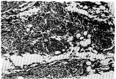
組織所見:潰蕩底は四層よりなっている.最上層ほ いわゆる肉芽組織である.局所的にほ出血巣や壊死
巣も認められる.壊死巣の周辺にはラ氏型巨細胞や 好中球も認められるが,その数ほ少ない・第二層は 約4ooμmの厚さの新生した線経組織からなる線経 化層である・この線経化層は所々で上層の肉芽組織 によって離断されている(Figン 3)・第三層は本来 の結合組織層である・最下層は脂肪織層であり,こ の部にほ径約1ooμm の類上皮細胞結節を数個認め る(Fig・ 4)・これより下の層は採取されていないン 潰痴辺縁部では表皮の再生も認められる・非潰蕩部 の表皮よりのびてきた再生上皮ほ潰痴辺綾部で反転
Fig・1・Macroscopic appearance ofBuruliulcer.Aboutl0cm
in diameter・
Fig. 2. Edema or lytic change of the dermis in non-ulcerated
areas was demonstrated easily by a insertion of a clamp
into the lesion.
し下掘部の内表面をおおっている・この再生上皮ほ 異型性のない偏平上皮であるがメラニン色素や附属
Fig. 3. Inflammatory tissue and fibrosis in the ulcer base. The healing process was started in the ulcer base,
x200, H. E. stain.
Fig. 4. Several epitheloid cell tubercles in subcutaneous tissue in ulcer base. Tuberculosis and sarcoidosis must be differentiated.
xlOO, H.E. stain.
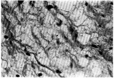
器ほ認められない(Figン 5)ン この辺綾部でほ真皮 においてラ氏型巨細胞,類上皮細胞結節, l十//バj求 や形質細胞の浸潤,浮陣をみとd)る・非潰揚部では 表皮は釘脚の延長を示L,真皮上層でほ浮任をみと める.本疾患の特徴的な所見は非潰癌部の真皮下層 から脂肪織層にかけて認められる・そこでほ細胞反 応のない壊死層があり,その厚さほ約1mmである・
それを取り囲むような配置で異物型巨細胞,ヲ氏巨 細胞,類上皮細胞結節が認められる・好中球の浸潤 ほごくわずかである(Fig・ 6)・潰痴部から更に少 し離れた部位では皮下脂肪織間の浮腫状の拡大が目 立つ(Fig. 7)・個々の脂肪細胞のなかにほ細胞が その輪郭の)部をなくし変性に陥っているものもあ る(Fig・ 8)・血管には著変ほなく真菌,細菌等ほ 見出せなかった・
考 案
Buruli ulcerの組織発生ほ Connor and Lunn (1966)によれば, 1・皮下脂肪織に無細胞性壊死 がおこり,それが周辺の皮下組織や,また真皮や表 皮に広がる・ 2.表皮の壊死がおこり,つぎに表皮 が脱落し潰痴が形成される・この時,表皮よりも皮 下組織の方が病変が進行しているので潰痴は下掘れ 状を呈する. 3・潰癌形成後3週間後位から反応性 の細胞浸潤が始まり次第に治癒へと向う,とされて
Fig. 5. Overhanging of the epitheliumatthe ulcer edge. xlOO, H.E. stain.
Fig. 6. Almost acellular necrosis in the dermis of the non-ulcerated areas.
The necrosis showed zonal spreading in the lower dermis and subcutaneous fat tissue, x 200, H. E. stain.
Fig. 7. Edematous widening of fatty tissue in non-ulcerated areas. Then,
zonal necrosis seemed to begin. x40, H.E. stain.
Fig. 8. Degeneative change in the individual fat tissue in non-ulcerated areas. Ghost out line of fat cells was seen. x200.
いる・脂肪織よりさらに深部の組織の病変も報告さ れている(Dodge, 1964)つ 抗酸菌は壊死層に認め られるが,組織学的にほ見出されないことも多い・
しかし,そのような症例でも組織惨出液から〃・
ulcerans が培養できることが多い(Lunn et a①,, 1965, Connor and Lunn, 1966)・それゆえCon‑
nor and Lunn (1966)は抗酸菌の検出ができない 症例でも組織学的な特徴だけで本症と診断できると している・我々の症例も抗酸菌を検出できなかった が,組織学的には特徴的な皮下脂肪織の無細胞性壊 死を見出したので本症と診断した・
M. ulceransほM・ intercellulareに属している が皮膚抗酸菌症の起炎菌として本邦で最も多いもの ほM・ mannumである(水野, 1975) ・しかし M. marinumによる病変の組織像ほ肉芽性反応が 主体であり無細胞性壊死がないことから鑑別は容易 である(Connor and Lunn, 1966)・その他本症と 鑑別すべきものとして結核,スポロトリコ‑シス,
ザルコイド‑ジス等が考えられるが,菌の証明,下
掘れ状潰疹,無細胞性の壊死層の存在によって容易 に鑑別できると思われる・
M. ulceransの感染経路ほまだ不明である.人か ら人への直接感染は考えられず(Uganda Buru①i group, 1971),またM. ulceran∫がin vitr。で 32‑33〇Cで培養されること(Clancey, 1964)負 類に本症に類似した病変がみられること(Parisot and Wood, 1960)などより河川や,そこに棲息す
る魚等を通じての感染が考えられているが,まだ確 証ほない(Barker, 1973) ・ Buruli ulcer と最も 関係があるのほ気温であると思われる・ Lunn ら (1965)はBuruli u①cerの存在する地方ほ日中ほ 日陰でも温度ほ33〇C まで上昇すると述べている・
Shkolnikら(1980)ほ気温が38。C もあるような 砂漠でもrobeを着た人の皮膚の表面温度ほ 33。C であると報告している・この33。C という温度は M. ulceranのin vitro における至適培養温度と 一致し,気温とBuruli ulcer との関係を示してい
るように思われる・
文 献
1) Aguilar, P. L., Iturribarria, F. M., & Middlebrooks, G. (1953): Un caso de infeccion humana por Mycobacterium ulcerans en el hemisferio occidental nota previa. Int. J. Leprosy, 21, 469-
476.