Analysis of the Physiological Function of Scd6
through its Interaction and Methylation by
Hmt1
著者
PHAM THI KIM LIEN
year
2017
その他のタイトル
Hmt1との相互作用とメチル化によるScd6の生理学的
機能の解析
学位授与大学
筑波大学 (University of Tsukuba)
学位授与年度
2017
報告番号
12102甲第8386号
URL
http://doi.org/10.15068/00150031
筑 波 大 学
Analysis of the Physiological Function of Scd6 through its
Interaction and Methylation by Hmt1
(Hmt1 Scd6 )
2017
TABLE OF CONTENTS
Page
TABLE OF CONTENTS ... i
ABBREVIATION ... iv
LIST OF TABLES ... vii
LIST OF FIGURES ... viii
DISSERTATION ORGANIZATION CHAPTER 1 GENERAL INTRODUCTION ... 1
1.1! Literature Review ... 2
1.1.1 Messenger RNA quality control ... 2
(a) An overview of gene control ... 2
(b) RNA degradation ... 3
(c) Decapping and translation repression ... 5
1.1.2 Processing bodies ... 6
1.1.3 Non-histone protein arginine methylation ... 9
1.1.4 Protein Scd6 ... 12
1.2 Overall Objectives ... 14
CHAPTER 2 MATERIALS AND METHODS ... 16
2.3 Yeast two-hybrid assays ... 19
2.4 Western blot analysis ... 20
2.5 Immunoprecipitation of Scd6Flag ... 21
2.6 Protein purification and GST pull down assays ... 22
2.7 Microscopy ... 22
2.8 Tandem mass spectrometry (arginine methylation mapping) ... 23
2.9 Polysome analysis ... 25
2.10 Statistical analysis ... 26
CHAPTER 3 RESULTS ... 27
3.1 Yeast two-hybrid screening for Scd6 interacting factors ... 28
3.2 Scd6 directly interacts with Hmt1 ... 29
3.3 Scd6 contains asymmetrically methylated arginines in RGG motifs ... 32
3.4 Hmt1 regulates subcellular localization of Scd6 ... 36
3.5 Hmt1-dependent arginine methylation of Scd6 involved in itself subcellular localization ... 38
3.6 Scd6 is important for P-body formation ... 40
3.7 Arginine methylation is not involved in Scd6 function for P-body formation 43 3.8 Scd6 has synthetic effects with Dhh1 on cell growth and P-body formation .. 45
3.9 Scd6 is not a global translation repressor ... 48
3.10 Arginine methylation regulates Scd6 function in relation to Dhh1 on cell growth ... 49
CHAPTER 4 DISCUSSION ... 54
4.1 Hmt1-based arginine methylation could be a reversible modification under environmental stimuli ... 56
4.2 Hmt1 mediates the effect of Scd6 on specific target mRNAs ... 58
4.3 Scd6 might have functional connection to the biological processes that regulated by Hmt1 ... 59
4.4 Arginine methyltransferases has functional and dynamic link with RGG-motif containing mRNP components ... 60
CHAPTER 5 CONCLUSION ... 63
REFERENCES ... 66
ABBREVIATION
3-AT 3-Amino-1,2,4-Triazole
CCR Carbon catabolite repression
cDNA complementary DNA
CEN CENtromere
DCP mRNA DeCaPing
DEAD Asp-Glu-Ala-Asp
DED Defines Essential Domain
Dex Dextrose
DHH DEAD box helicase homolog
DMA DiMethylArginine
DNA DeoxyriboNucleic Acid
EBS Est1-like Bcy1 Suppressor
EDTA EthyleneDiamineTetraacetic Acid
eIF eukaryotic Initiation Factor
GAD Gal4 Activation Domain
GBD Gal4 Binding Domain
GEO Gene Expression Omnibus
GFP Green Flourescent Protein
GST Glutathione S-Transferase
hnRNP heterogenous RiboNucleoProtein
hRAP human RNA-Associated Protein
HRP Heterogenous nuclear RibonucleoProtein
LC Liquid Chromatography
Lsm Like sm
m/z mass-to-charge ratio
MMA MonoMethylArginine
mRFP monomeric Red Fluoresent Protein
mRNA messenger RiboNucleoic Acid
mRNP messenger RiboNucleoProtein
MS Mass Spectrometry
NOT Negative regulator Of Transcription
NPL Nuclear Protein Localization
OD Optical Density
P P Value
P-body Processing body
PAGE PolyAcrylamide Gel Electrophoresis
PAN Poly(A)-binding protein-dependent poly(A) riboNuclease
PAT Protein Associated with Topoisomerase II
PBS Phosphate Buffered Saline
PRMT Protein Arginine Methyltransferase
RBP RNA Binding Protein
RF Arginine Phenylalanine
RGG Arginine Glycine Glycine
RK Arginine Lysine
RPS Ribosomal Protein of Small subunit
S. cerevisiae Saccharomyces cerevisiae
SBP Single-stranded nucleic acid Binding Protein
SC Synthetic Complete
SCD Suppressor of Clathrin Deficiency
SD Synthetic Defined
SD Standard Deviation
SG Stress Granule
TAP Tandem Affinity Purification
TBS Tris-Buffered Saline
TBS-M Tris-Buffered Saline-skinMilk
XRN eXoRiboNulease
LIST OF TABLES
Page Table 1. Strains used in this study ... 18 Table 2. Plasmids used in this study ... 19 Table 3. Result of Yeast two-hybrid screening assay ... 28
LIST OF FIGURES
Page
Figure 1. Gene expression can be controlled at different levels ... 3
Figure 2. General mRNA decay pathways ... 4
Figure 3. mRNP granules; P-bodies and stress granules ... 8
Figure 4. PRMTs methylate RGG/RG-containing proteins ... 12
Figure 5. Protein Scd6 ... 15
Figure 6. Scd6 interacts with Hmt1 ... 31
Figure 7. Arginine residues in RGG motifs of Scd6 are dimethylated in Hmt1-dependent manner ... 34
Figure 8. MS/MS spectra of identified peptides containing asymmetric dimethylarginines ... 35
Figure 9. Hmt1 affected Scd6 subcellular localization ... 37
Figure 10. Subcellular localization of Scd6 mutations in the RGG motifs ... 39
Figure 11. scd6 deletion impairs the accumulation of Dcp2-GFP foci ... 41
Figure 12. Overexpression of Scd6 induces the accumulation of Dcp2-GFP foci ... 42
Figure 13. Scd6 function for P body formation is independent on arginine methylation 44 Figure 14. scd6 dhh1 mutant strain showed synthetic growth defect ... 46
Figure 15. Scd6 and Dhh1 have overlapping functions in P-body formation ... 47
Figure 16. Scd6 was not essential for general translation repression under glucose starvation conditions ... 48
Figure 18. Positive charge at arginine residues is required for Scd6 function on cell
growth at elevated temperature ... 51
Figure 19. dhh1 edc3 scd6 mutant strain showed synthetic growth defect ... 53 Figure 20. Proposal model of Scd6 physiological activities in this study ... 56
DISSERTATION ORGANIZATION
This thesis is organized into five chapters. Chapter 1 is a general introduction providing literature review on the regulation of gene expression, processing-bodies (P-bodies), protein arginine methylation and Scd6 protein followed by overall objectives of this study. In chapter 2, the materials and methods used in this study are presented. Chapter 3 is the results of this study. In chapter 4, the present data and future directions are discussed. Chapter 5 provides general conclusions.
CHAPTER 1
1.1 Literature review
1.1.1 Messenger RNA quality control
(a) An overview of gene control
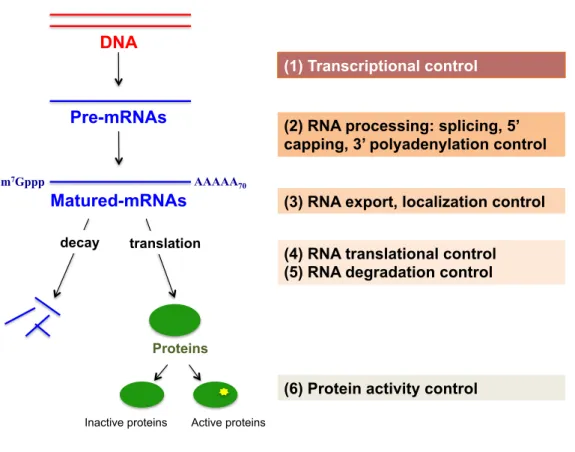
Thousands of DNA sequences contain the information to synthesize proteins, which dictate cell function. Different cell types express different set of genes. On the other hand, cells can change the fraction of expressed genes in response to environmental stimuli. There are many steps in the flow of information from DNA to RNA to protein. Thus a cell can regulate a gene expression at different levels: (1) when and how often a gene is transcribed into RNA (transcriptional control), (2) the splicing and processing of nuclear RNA are controlled (RNA processing control), (3) determining which processed-mRNAs are transported from nucleus to cytoplasm and where mRNAs locate in the cytoplasm (RNA transport and localization control), (4) controlling which mRNAs are translated to proteins (translational control), (5) steady state mRNA levels in cytoplasm are determined by mRNA decay control (mRNA degradation control), (6) the functional capabilities of the proteins are regulated (protein activity control) (Figure 1) (1).
Figure 1. Gene expression can be controlled at different levels.
Messenger ribonucleoprotein (mRNP) complexes, comprising transcripts and RNA-binding proteins (RBPs), regulate gene expression. The lifecycle of mRNP complex also begins with mRNA transcription and stretches to mRNA degradation. The ensuing gene regulatory mechanisms have been clarified by the analyses of compositions and kinetics of mRNP complexes at each of these steps (2).
(b) RNA degradation
The control of cytoplasmic mRNA, which dictated by the degradation rate
DNA Pre-mRNAs m7Gppp AAAAA 70 Matured-mRNAs (1) Transcriptional control
(2) RNA processing: splicing, 5’ capping, 3’ polyadenylation control (3) RNA export, localization control (4) RNA translational control (5) RNA degradation control translation
decay
Proteins
Active proteins Inactive proteins
of mRNAs, is an essential step in the mRNP lifecycle to set the steady state levels of mRNA expression and to respond rapidly to environment changes. In
Saccharomyces cerevisiae (S. cerevisiae), cytoplasmic RNA turnover is regulated
by two main pathways: (i) decapping by Dcp1/Dcp2 decapping complex followed by 5´-3´ degradation by exonuclease Xrn1 and (ii) 3´-5´ degradation by exosome complex. Both of these pathways follows the deadenylation by Pan2/Pan3 complex and/or Ccr4/Not complex (Figure 2) (3–6).
Figure 2. General mRNA decay pathways
m7Gppp AAAAA 70 Matured-mRNAs deadenylation m7Gppp Aoligo m7Gpp p Aoligo m7Gppp Aoligo decapping 3´-5´ degradation 5´-3´ degradation Ccr4 Pan2 Dcp2 Xrn1 Exosome
(c) Decapping and translation repression
Previous studies have suggested that mRNA decay, which modulated by decapping complex, is the predominant pathway of mRNA degradation. Deletion of decapping enzyme led to slow growth or lethality in some strain backgrounds (7, 8). In yeast, mRNA decapping is carried out through a complex of the Dcp1/Dcp2 proteins-decapping enzymes, and several decapping activators. The core set of decapping activators includes Edc1, Edc2, Edc3, Pat1, Dhh1, the Lsm1-7 complex, and Scd6, all of which are conserved proteins in eukaryotes (9–12). Decapping activators promote the formation of decapping complex and stimulate removal cap structure by decapping enzymes (9, 13). An important issue of future research is study how these decapping activators are activated to be functional through how is their interaction network.
All of these factors may not associate and function at the same time. Individual factors activate decapping process in different manners as follows. The cap structure is also involved in promoting translation by recruiting eIF4E/eIF4G translation initiation complex to the 5´ end of mRNA. Therefore, the first step of mRNA decapping process is the loss of cap-binding eIF4E/eIF4G complex from mRNA. Several studies have shown that competition exists
between translation initiation and decapping. Replacing the translation initiation factors by decapping enzymes requires mRBPs referred to as decapping activators. Several decapping activators were indentified to repress translation initiation, thereby enhance decapping process such as Dhh1, Scd6, Stm1, Pat1 (14–17). In particular, dhh1 pat1 double mutant showed the defect in mRNA decapping and was unable to repress translation in response to glucose deprivation - overexpression of Dhh1 or Pat1 led to translational repression (15). Pat1, Dhh1 and Scd6 were shown to block translation before the formation of 48S pre-initiation complex (13), whereas Stm1 inhibited translation after the formation of 80S complex (14). Unresolved issue for these RBPs is how these components are remodeled to form decapping complex after releasing translation complex.
1.1.2 Processing bodies
Actively translating mRNPs associate with polysomes to initiate translation. Various stress conditions, such as glucose starvation and severe heat shock, induce phase transitions of these mRNPs toward the non-translating state and lead to the assembly of most mRNPs into cytoplasmic foci such as processing-bodies (P-bodies) and stress granules (SGs) (18). Although in both
yeast and mammalian cells, P-bodies and SGs frequently overlap, they have distinct assembly dynamics and functions (Figure 3). Proteins required for active translation are found in SGs, whereas P-bodies include protein factors for mRNA decay machinery such as Dcp1/2, Dhh1, Edc3, and Scd6, suggesting functional diversity of these granules as sites of mRNA storage and mRNA degradation, respectively (Figure 3) (18). However, mRNA decay may occur without the formation of large P-bodies (19), and P-body core components have been co-localized with various molecules that are involved in biological processes such as DNA replication and PKA signaling (20, 21). Hence, knowledge of the activities of P-body components may clarify the physiological functions and kinetics of P-bodies.
Figure 3. mRNP granules; P-bodies and stress granules
P-body formation is induced by a variety of stress conditions and almost decapping activators such as Pat1, Dhh1, Edc3 and Lsm4 were recently identified as factors that promote the physical interactions between mRNPs required for PB assembly (22-25). However, it remains poorly understood which the signaling pathways mediating this assembly process. The cAMP-dependent protein kinase (PKA) negatively regulates P-body assembly process upon glucose starvation that is due, at least in part, to the direct phosphorylation of Pat1 (26). In contrast, the inactivation of the Target of Rapamycin Complex 1 (TORC1) and
Lsm1%7 m7Gppp AAAAA Dcp2 Dcp1 Dhh1 Scd6 Edc3 Pat1 Xrn1 m7Gppp AAAAA 70 Lsm1%7 m7Gppp AAAAA Dcp2 Dcp1 Dhh1 Scd6 Edc3 Pat1 Xrn1 Lsm1%7 m7Gppp AAAAA Dcp2 Dcp1 Dhh1 Scd6 Edc3 Pat1 Xrn1 Lsm1%7 m7Gppp AAAAA Dcp2 Dcp1 Dhh1 Scd6 Edc3 Pat1 Xrn1 Lsm1%7 m7Gppp AAAAA Dcp2 Dcp1 Dhh1 Scd6 Edc3 Pat1 Xrn1 Lsm1%7 m7Gppp AAAAA Dcp2 Dcp1 Dhh1 Scd6 Edc3 Pat1 Xrn1 Lsm1%7 m7Gppp AAAAA Dcp2 Dcp1 Dhh1 Scd6 Edc3 Pat1 Xrn1 Lsm1%7 m7Gppp AAAAA Dcp2 Dcp1 Dhh1 Scd6 Edc3 Pat1 Xrn1 Lsm1%7 m7Gppp AAAAA Dcp2 Dcp1 Dhh1 Scd6 Edc3 Pat1 Xrn1 Lsm1%7 m7Gppp AAAAA Dcp2 Dcp1 Dhh1 Scd6 Edc3 Pat1 Xrn1 Lsm1%7 m7Gppp AAAAA Dcp2 Dcp1 Dhh1 Scd6 Edc3 Pat1 Xrn1 m7Gppp AAAAA70 m7Gppp AAAAA70 m7Gppp AAAAA70 m7Gppp AAAAA70 m7Gppp AAAAA70 m7Gppp AAAAA70 m7Gppp AAAAA70 m7Gppp AAAAA70 m7Gppp AAAAA70 m7Gppp AAAAA70 m7Gppp AAAAA70 m7Gppp AAAAA70 m7Gppp AAAAA70 m7Gppp AAAAA70 m7Gppp AAAAA70 m7Gppp AAAAA70 m7Gppp AAAAA70 m7Gppp AAAAA70 m7Gppp AAAAA70 m7Gppp AAAAA70 m7Gppp AAAAA70 m7Gppp AAAAA70 m7Gppp AAAAA70 m7Gppp AAAAA70 m7Gppp AAAAA70 m7Gppp AAAAA70 Lsm1%7 m7Gppp AAAAA Dcp2 Dcp1 Dhh1 Scd6 Edc3 Pat1 Xrn1 Lsm1%7 m7Gppp AAAAA Dcp2 Dcp1 Dhh1 Scd6 Edc3 Pat1 Xrn1 Lsm1%7 m7Gppp AAAAA Dcp2 Dcp1 Dhh1 Scd6 Edc3 Pat1 Xrn1 Lsm1%7 m7Gppp AAAAA Dcp2 Dcp1 Dhh1 Scd6 Edc3 Pat1 Xrn1 Lsm1%7 m7Gppp AAAAA Dcp2 Dcp1Dhh1 Scd6 Edc3 Pat1 Xrn1 Lsm1%7 m7Gppp AAAAA Dcp2 Dcp1Dhh1 Scd6 Edc3 Pat1 Xrn1 Lsm1%7 m7Gppp AAAAA Dcp2 Dcp1Dhh1 Scd6 Edc3 Pat1 Xrn1 Lsm1%7 m7Gppp AAAAA Dcp2 Dcp1 Dhh1 Scd6 Edc3 Pat1 Xrn1 Lsm1%7 m7Gppp AAAAA Dcp2 Dcp1Dhh1 Scd6 Edc3 Pat1 Xrn1 Lsm1%7 m7Gppp AAAAA Dcp2 Dcp1Dhh1 Scd6 Edc3 Pat1 Xrn1 Decapping Translation initiation P-body Stress granule
AMP-activated protein kinase (AMPK) pathways do not affect the formation of P-body (27). Further studies are required to understand completely the regulation of P-body formation under various stress conditions.
1.1.3 Non-histone protein arginine methylation
Analysis of post-translational modifications (PTMs) of mRNP components such as arginine methylation elucidates the structures and functions of these gene expression regulators. It has been shown that the motif termed RGG box, that is rich in arginines and glycines, of several mRNP components influence numerous physiological processes such as transcription, pre-mRNA splicing, DNA damage signaling, mRNA translation and degradation (28). Previous study showed that protein containing the RGG boxes are common substrates of protein arginine methyltransferases (PRMTs) (29, 30). Arginine residues of RGG boxes can be monomethylated or dimethylated. Type I PRMTs (PRMT1-3, CARM1, PRMT6, PRMT8) catalyze the formation of monomethylarginines (MMAs) or asymmetric-dimethylarginines (aDMAs), whereas type II PRMTs (PRMT5, PRMT9) catalyze the formation of monomethylarginines (MMAs) or symmetric-dimethylarginines (sDMAs) (Figure 4A) (28, 30). Heterogeneous nuclear ribonucleoproteins (hnRNPs)
containing N-terminal RNA-binding motifs in conjunction with RGG repeats are major substrates of PRMT1/Hmt1 in mammalian and yeast cells (31). Recently, arginine methylation has been shown to mediate RNA–protein, DNA–protein, and protein–protein interactions (32, 33). For instance, arginine methylation by PRMT1 is critical for the localization of the hRAP55A, Scd6 homologue in mammalian cells (Figure 4B) (34).
In S. cerevisiae, Hmt1 was identified as the major type I PRMT (35). Although hmt1 single deletion did not affect cell viability, it showed cell lethality in genetic backgrounds lacking of cap-binding proteins or harboring npl3-1 allele (36, 37). Global analysis was carried out to understand the involvement of Hmt1 in biological processes, and not only the substrate proteins but also the impact of arginine methylation on their functions were identified. Hmt1 methylates RGG/RG motifs of several RBPs such as Npl3 and Hrp1, thereby regulates their localization and function (Figure 4B) (38). Thus, Hmt1-mediated RGG/RG methylation can negatively or positively regulates protein activities (30, 39).
An important question is whether the dynamic regulatory mechanisms based on the arginine methylation for non-histone proteins exist. Hmt1 methyltransferase activity toward hnRNPs is regulated by environmental
conditions. For example, rapamycin treatment or starvation leads to dephosphorylation of active phosphorylated Hmt1 (40). Moreover, it is also plausible that arginine demethylation reactions can reverse the methylation modifications (41). Recently, it was reported that purified human JmjC lysine demethylases (KDMs) could catalyze demethylation of both histon and non-histon synthetic methylated fragments (42).
Figure 4. PRMTs methylate RGG/RG-containing proteins. (A) Arginine (Arg)
methylation by PRMTs (Type I and Type II) generates monomethylarginine (Arg-MMA) and asymmetric dimethylarginine (Arg-aDMA), or symmetric dimethylarginine (Arg-sDMA). (B) RGG/RG- containing proteins, which are methylated by PRMTs/Hmts.
1.1.4 Protein Scd6
In S. cerevisiae, Scd6 was originally identified as a multicopy suppressor
C H2N NH2+ HN (CH2)3 Arg C HN NH2+ HN (CH2)3 Arg-MMA Me C N NH2+ HN (CH2)3 Arg-aDMA Me Me C HN NH+ HN (CH2)3 Arg-sDMA Me Me Type I Type II Type I Type II
A
B
Npl3 hnRNP)A1 RAP55A/Scd6 RRM RRM SR/RGG RGG LSM14 Ser/Thr RGG FDF RGG RGG RRM RRMof clathrin deficiency (43). Eukaryotic Scd6 family proteins include hRAP55 in human, mTral in mouse, xRAP55 in Xenopus laevis, Trailer Hitch (Tral) in
Drosophila melanogaster (D. melanogaster), CAR-1 in Caenorhabditis elegans,
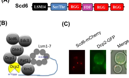
Sum2p in Schizosaccharomyces pombe (44). Scd6 and its homologues are highly conserved, that contain N-terminal Lsm14 domains, serine/threonine (Ser/Thr)-rich regions, central FDF (phenylalanine-aspartate-phenylalanine) motifs and RGG (arginine–glycine–glycine) boxes (Figure 5A) (44). There are several predictions for the functions of Lsm14, Ser/Thr and FDF motifs in RNA metabolism but their actual functions remain to be determined. RGG repeats participate in the regulation of RNA-protein interactions, protein-protein interactions and protein localization (28).
The biological function of Scd6 is not well established although it seems to repress translation initiation. Scd6 directly binds to eIF4G and inhibits the formation of 48S preinitiation complex in vitro (16). In S. pombe, Scd6 functions as a decapping activator, as indicated by the competition with Edc3 to induce decapping enzymes such as Dcp1/2 in vitro (12) (Figure 5B). Tral protein, the D.
melanogaster homologue of Scd6 was shown to interact directly with the
However, in S. cerevisiae, details of the interaction of Scd6 with other decapping activators as well as the regulatory mechanisms for the functions and the locations remain to be determined to understand how this protein contribute in mRNA metabolism.
1.2 Overall Objectives
Scd6 is well known as a decapping activator and translation initiation repressor. However, it remains unknown whether Scd6 regulates general mRNA decapping or affects decapping of only a subset of mRNAs. The role of Scd6 in translation repression has not been confirmed in vivo. Although Scd6 localizes to P-bodies (Figure 5C), the precise dynamic and biochemical function of P-body localization remains to be studied. Moreover, it is restrained to analyze Scd6 activities because scd6 deletion does not show any growth phenotype at normal growth condition. Aiming to understand unresolved issues of Scd6 physiological activities, one of the goals of this study was to identify promising interacting factors of Scd6 that would allow me to assign its functions. In addition, I aim to determine genetic and functional interactions of Scd6 with other decapping activators.
Figure 5. Protein Scd6. (A) The domain architecture of Scd6. (B) Scd6 is a decapping
activator. (C) Scd6 co-localizes with P-body marker, Dcp2.
Lsm1%7 m7Gppp AAAAA Dcp2 Dcp1 Dhh1 Scd6 Edc3 Pat1 Xrn1
Scd6
Scd6-mC herry Dcp2-G FP Merge(A)
(B)
(C)
CHAPTER 2
2.1 Strains, plasmids, and general methods
Escherichia coli DH5α was used for DNA manipulations. The present
yeast strains and plasmids are described in Tables 1 and 2. Cells were grown in yeast extract-peptone dextrose (YPD), synthetic complete medium (SC), and synthetic minimal medium (SD), and in SC media lacking either amino acids or other nutrients (SC-Ura, SC lacking uracil). General procedures were performed as described previously in “Methods in yeast genetics” (46).
Table 1. Yeast strains used in this study
Name Genotype
10B MATα ade2 trp1 can1 leu2 his3 ura3 GAL psi+ HOp-ADE2-HO 3' UTR
10BD MATa/MATα ade2/ade2 trp1/trp1 can1/can1 leu2/leu2 his3/his3 ura3/ura3
10BD-ds MATa/MATα ade2/ade2 trp1/trp1 can1/can1 leu2/leu2 his3/his3 ura3/ura3
DHH1/dhh1∆::CgLEU2 SCD6/scd6∆::CgHIS3
10BD-dsh MATa/MATα ade2/ade2 trp1/trp1 can1/can1 leu2/leu2 his3/his3 ura3/ura3
DHH1/dhh1∆::CgLEU2 SCD6/scd6∆::CgHIS3 HMT1/hmt1∆::CgTRP1
10BD-dse MATa/MATα ade2/ade2 trp1/trp1 can1/can1 leu2/leu2 his3/his3 ura3/ura3
DHH1/dhh1∆::CgLEU2 SCD6/scd6∆::CgHIS3 EDC3/edc3∆::CgTRP1
10BD-s MATa/MATα ade2/ade2 trp1/trp1 can1/can1 leu2/leu2 his3/his3 ura3/ura3
SCD6/scd6∆::CgLEU2
s-DCP2 MATa/MATα ade2/ade2 trp1/trp1 can1/can1 leu2/leu2 his3/his3 ura3/ura3
SCD6/scd6∆::CgLEU2 DCP2/DCP2-GFP::HIS3
d-DCP2 MATa/MATα ade2/ade2 trp1/trp1 can1/can1 leu2/leu2 his3/his3 ura3/ura3
DHH1/dhh1∆::CgLEU2 DCP2/DCP2-GFP::HIS3
ds-DCP2 MATa/MATα ade2/ade2 trp1/trp1 can1/can1 leu2/leu2 his3/his3 ura3/ura3
DHH1/dhh1∆::CgLEU2 SCD6/scd6∆::CgHIS3 DCP2/DCP2-GFP::HIS3
h-DCP2 MATa/MATα ade2/ade2 trp1/trp1 can1/can1 leu2/leu2 his3/his3 ura3/ura3
HMT1/hmt1∆::CgHIS3 DCP2/DCP2-GFP::HIS3
dh-DCP2 MATa/MATα ade2/ade2 trp1/trp1 can1/can1 leu2/leu2 his3/his3 ura3/ura3
DHH1/dhh1∆::CgLEU2 HMT1/hmt1∆::CgHIS3 DCP2/DCP2-GFP::HIS3
10BD-h MATa/MATα ade2/ade2 trp1/trp1 can1/can1 leu2/leu2 his3/his3 ura3/ura3
HMT1/hmt1∆::CgHIS3
h-S MATa/MATα ade2/ade2 trp1/trp1 can1/can1 leu2/leu2 his3/his3 ura3/ura3 HMT1/hmt1∆::CgHIS3 SCD6/SCD6-mCherry::natNT2
10BD-HMT1 MATa/MATα ade2/ade2 trp1/trp1 can1/can1 leu2/leu2 his3/his3 ura3/ura3
HMT1/HMT1-13myc::kanMX6
s-1 MATa ade2 trp1 can1 leu2 his3 ura3 scd6∆::CgLEU2
10B-S MATα ade2 trp1 can1 leu2 his3 ura3 GAL psi+ HOp-ADE2-HO 3' UTR
SCD6-mCherry::natNT2
h-1 MATa ade2 trp1 can1 leu2 his3 ura3 hmt1∆::CgHIS3
ds-1 MATa ade2 trp1 can1 leu2 his3 ura3 dhh1∆::CgLEU2 scd6∆::CgHIS3
dsh-1 MATa ade2 trp1 can1 leu2 his3 ura3 dhh1∆::CgLEU2 scd6∆::CgHIS4 hmt1∆::CgHIS3
10B-DCP2 MATα ade2 trp1 can1 leu2 his3 ura3 GAL psi+ HOp-ADE2-HO 3' UTR
DCP2-GFP::HIS3
s-DCP2-1 MATa ade2 trp1 can1 leu2 his3 ura3 scd6∆::CgLEU2 DCP2-GFP::HIS3
d-DCP2-1 MATa ade2 trp1 can1 leu2 his3 ura3 dhh1∆::CgLEU2 DCP2-GFP::HIS3
ds-DCP2-1 MATa ade2 trp1 can1 leu2 his3 ura3 dhh1∆::CgLEU2 scd6∆::CgLEU2
DCP2-GFP::HIS3
h-DCP2-1 MATa ade2 trp1 can1 leu2 his3 ura3 hmt1∆::CgHIS3 DCP2-GFP::HIS3
dh-DCP2-1 MATa ade2 trp1 can1 leu2 his3 ura3 dhh1∆::CgLEU2 hmt1∆::CgHIS3
DCP2-GFP::HIS3
HMT1-1 MATα ade2 trp1 can1 leu2 his3 ura3 HMT1-13myc::kanMX6
PJ69-4A MATa trp1-901 leu2-3, 112 ura3-52 his3-200 gal4D gal80D LYS2::GAL1-HIS3
Table 2. Plasmids used in this study
Name Description
YCplac33 URA3, CEN-ARS
YCplac33-SCD6 URA3, CEN-ARS, SCD6
YCplac33-SCD6RK URA3, CEN-ARS, SCD6RK
YCplac33-SCD6RF URA3, CEN-ARS, SCD6RF
YCplac33-SCD6FLAG URA3, CEN-ARS, SCD6FLAG
YEplac195 URA3, 2µ
YEplac195-SCD6 URA3, 2µ, SCD6
YEplac195-SCD6FLAG URA3, 2µ, SCD6FLAG
pGBD-c1 TRP1,2µ, ADH1pr-GAL4-BD
pGBD-c1-SCD6 TRP1,2µ, ADH1pr-GAL4-BD-SCD6
pGAD-c1 LEU2,2µ, ADH1pr-GAL4-AD
pGAD-c1-HMT1 TRP1,2µ, ADH1pr-GAL4-AD-HMT1
pCgLEU2 PCR template, C. glabrata LEU2 in pUC19
pCgHIS3 PCR template, C. glabrata HIS3 in pUC19
pCgTRP1 PCR template, C. glabrata TRP1 in pUC19
pFA6a-13myc-kanMX6 PCR template, 13myc-ADH1t::kanMX6
pRS314-EDC3-GFP TRP1, CEN-ARS, EDC3-GFP
pRS316-SCD6-mRFP TRP1, CEN-ARS, SCD6RF-mRFP
pRS316-SCD6RK-mRFP TRP1, CEN-ARS, SCD6RK-mRFP
2.2 Gene deletion and protein tagging
Gene disruption and insertion were performed using PCR-based gene replacement, as described previously (47, 48).
2.3 Yeast two-hybrid assays
PJ69-4A cells harboring pGBD-SCD6 were transformed with the yeast cDNA library pACT (49). Transformants were then plated on SC-Leu-Trp plates and incubated at 30˚C for 4 days. Plates were replica plated onto SC-Leu-Trp-His plates, SC-Leu-Trp-His plates containing 1-mM 3-aminotriazole (3-AT), and SC-Leu-Trp-Ade plates, and were incubated at 30˚C for 3 days. Twenty-three
were isolated from transformants, and were reassessed for interactions with Scd6. Insert DNAs were sequenced.
To confirm the interactions of Scd6 and Hmt1 that were identified in two-hybrid screening analyses, pGAD-c1-Hmt1 was constructed and used as a prey vector. PJ69-4A cells were then co-transformed with pGBD-c1 or pGBD-c1-Scd6 with either pGAD-c1 or pGAD-c1-Hmt1. Transformants were spotted onto SC-Leu-Trp (WL), SC-Leu-Trp-His (WLH), and SC-Leu-Trp-His (WLH) plates containing 1 mM 3-AT and were then incubated for 3 days at 30˚C. 2.4 Western blot analysis
Samples were loaded onto SDS-PAGE or NU-PAGE gels and were then electroblotted onto ImmobilonTM polyvinylidene difluoride membranes (Merck Millipore, USA). Blots were blocked for 1 h at room temperature with TBS-M buffer containing 20 mM Tris-HCl (pH 7.5), 150 mM NaCl, and 5% non-fat dry milk, and were then incubated with 1:1,000-diluted primary antibodies in TBS-M buffer overnight at 4˚C. After three final washes with TBS buffer containing 20 mM Tris-HCl (pH 7.5) and 150 mM NaCl, blots were incubated with secondary antibodies, and were developed using enhanced chemiluminescence detection kits (Merck Millipore, USA).
2.5 Immunoprecipitation of Scd6Flag
Cells were grown in SC-Ura medium at 30˚C to mid-log-phase and were harvested by centrifugation. The cells were then resuspended in XT buffer containing 50 mM HEPES-KOH, (pH 7.3), 20 mM potassium acetate, 2 mM EDTA, 0.1% Triton X-100, 5% glycerol protease inhibitors, phenylmethylsulfonyl fluoride (PMSF), aprotinin, and leupeptin. Glass beads were then added and cells were broken by rigorous vortexing at 4˚C (4 times at 3,500 rpm for 30 s). Lysates were then centrifuged for 10 min at 15,000 g and supernatants were collected. Extracts were incubated with or without 200 µg/mL RNase A (Wako Pure Chemical Industries, Ltd., Japan) at room temperature for 30 min prior to immunoprecipitation.
To immunoprecipitate Scd6Flag, extracts were incubated with anti-Flag antibody coupled to protein G-Sepharose beads (Sigma Aldrich, USA) for 30 min at 4˚C. Scd6Flag-bound beads were then washed three times in XT buffer, and the bound material was eluted with elution buffer containing 0.1 mg/mL Flag peptide in XT buffer for 10 min at 4˚C. Samples were subjected to SDS-PAGE followed by colloidal Coomasie blue staining (Thermo Fisher Scientific). RNA was isolated from immunopreciptated samples using RNeasy Mini kit (Qiagen)
and subjected to UREA-PAGE followed by SYBR-Gold staining (Life Technologies).
2.6 Protein purification and GST pull down assays
GST- and His6- fusion proteins were purified from E. coli strain BL21
(DE3) using glutathione-sepharose beads (GE) and Ni-NTA agarose beads (Novagen), respectively. GST- or GST-Scd6-bound beads were then resuspended in HB buffer containing PBS, 0.5% Tween20, 5 mM MgCl2, 5 mM
2-mercaptoethanol, 1 mM PMSF, and a protease inhibitor cocktail (Complete EDTA-free, Sigma Aldrich, USA). His6-Hmt1 was then eluted from Ni-NTA
agarose beads using a high concentration of imidazole. Proteins were dialyzed and concentrated using Amicon Ultra-2-30K (Merck Millipore Ltd.).
His6-Hmt1 was incubated with either GST- or GST-Scd6 immobilized
beads for 3 h at 4˚C. After washing 5 times with PBS, beads were boiled in SDS sample buffer and samples were then subjected to SDS-PAGE, followed by staining with Coomassie Brilliant Blue and immunoblotting with anti-His antibody (Sigma Aldrich, USA).
2.7 Microscopy
appropriate SC medium. Cells were then collected, washed twice in fresh SC medium with or without 2% glucose, and were then resuspended in fresh SC medium with or without glucose followed by incubation at 30˚C for 20 min. Cells were harvested, washed again and immediately examined for granule formation using a Keyence BZ-X700 microscope (Keyence Corporation, Japan) at room temperature. Experiments were performed a minimum of three times. Fluorescence images were processed and analyzed for numbers of cells with foci formation and signal intensities. More than 200 cells were counted and percentages of cells with foci formation were calculated. Fluorescence intensities along 5-µm lines were measured using the linescan function in MetaMorph software (Molecular Devices, USA) and averages of more than 10 foci from each observation were fitted to a Gaussian distribution.
2.8 Tandem mass spectrometry (arginine methylation mapping)
Scd6Flag was immunopurified as described above and was then subjected to SDS-PAGE and stained using Colloidal Blue Staining Kits (Thermo Fisher Scientific, CA, USA). Gel bands containing Scd6Flag were excised and subjected to in-gel digestion with Chymotrypsin (Sequencing Grade, Promega). The resulting peptides were analyzed using a nanoflow LC-MS/MS system with
LTQ-Orbitrap hybrid mass spectrometer (model XL, Thermo Fisher Scientific, CA, USA) as described elsewhere (50) with some modifications. The peptide mixture was separated using reverse phase chromatography with a 0%–40% gradient of acetonitrile containing 0.1% formic acid over 80 min at a flow rate of 100 nl/min using a Mightysil-RP-18 (3 µm particle, Kanto Chemical, Osaka, Japan) fritless column (45 mm × 0.150 mm i.d.). Eluted peptides were sprayed directly into a LTQ-Orbitrap hybrid mass spectrometer and raw data were acquired using Xcalibur version 2.0.7 (Thermo Fisher Scientific, USA) and were then converted to MGF files using Proteome Discoverer version 1.3 software (Thermo Fisher Scientific). Database searches were performed using MASCOT version 2.2.07 software and the Uniprot S. cerevisiae (strain ATCC 204508/S288c) database with the following parameters: fixed modification, carbamidemethyl (Cys); variable modifications, oxidation (Met); maximum missed cleavages, 1; peptide mass tolerance, 20 ppm; MS/MS tolerance, 0.8 Da. Candidate peptides were selected with probability-based Mowse scores that exceed the threshold, indicating significant homology (p < 0.05; score over 20), and were referred to as “hits.” Precursor peptides containing dimethylarginine residues were detected according to mass increases of 28 Da from those of
unmodified precursor peptides. MS/MS spectra of dimethylated peptides were manually assigned, and side-chain fragmentation of methylated arginine peptides was detected as a neutral loss in MS/MS spectra to determine symmetric (31.04 Da) or asymmetric (45.05 Da) dimethylarginine levels (34, 51).
2.9 Polysome analysis
Cells were grown until they reached an OD600 of approximately 0.8 and
were harvested and resuspended in medium with or without glucose for 20 min. Cultures were washed once and resuspended in lysis buffer containing 80
g/mL cycloheximide, 200 g/mL heparin, 10 mM Tris-HCl (pH 7.5), 0.1 M
NaCl, 30 mM MgCl2, aprotinin and leupeptin. Glass beads were then added, and
cells were broken by rigorous vortexing at 4˚C (4 times at 2000 rpm for 30 s). Lysates were then centrifuged for 10 min at 15,000 g and supernatants were collected. Sucrose gradients of 10%–50% were prepared in solutions containing
1 mM DTT, 50 mM NH4Cl, 50 mM Tris-Acetate (pH 7.0), and 12 mM MgCl2
using a Gradient Station (Biocomp Laboratories Inc.). Lysates were added to sucrose gradients and were centrifuged for 3 h at 27,000 rpm in a Beckman Coulter centrifuge (Optima L-100K) at 4˚C, followed by fractionation using an Incomparable Piston Gradient Fractionator and a Bio-miniUV monitor (Biocomp
Laboratories Inc.). OD254 values were monitored to represent fractionation
results.
2.10 Statistical analysis
Data are presented as means ± standard deviations (SD) of at least three independent experiments. Statistical analyses were performed using student’s t-test or analysis of variance (ANOVA) followed by Tukey’s test and differences were considered significant when p < 0.05.
CHAPTER 3
3.1 Yeast two-hybrid screening for Scd6 interacting factors
Scd6 is a translational repressor that reportedly interacts directly with eIF4G to block translation initiation (16). Scd6 is also known as a decapping activator that contributes to decapping complexes of Scd6, Edc3, Pat1, Dhh1 and decapping enzymes (13). To identify other sets of proteins that associate with Scd6 in cells, I performed yeast two-hybrid screening of yeast gDNA library using Scd6 as bait. DCP1 gene, which encodes a known Scd6-interacting partner, was recovered in this two-hybrid screening, confirming efficiency of my screening method. In addition, DNA fragments encoding other proteins such as Ebs1, Hmt1, Rps28a and Rps28b were recovered in this assay. Scd6-interacting candidates are listed in Table 3.
Table 3. Result of Yeast two-hybrid screening assay
Gene Description
HSE1 Subunit of the endosomal Vps27p-Hse1 complex
GYP1 cis-Golgi GTPase-activating (GAP) protein for yeast Rabs RPS28A Protein component of the small (40S) ribosomal subunit
RPS28B Protein component of the small (40S) ribosomal subunit
ISF1 Serine rich, hydrophilic protein
DCP1 Subunit of Dcp1p-Dcp2p decapping enzyme complex
HMT1 Arginine methyltransferase
EBS1 Protein involved in translation inhibition and nonsense-mediated decay
UTP11 Subunit of U3-containing Small Subunit (SSU) processome complex
HSP82 Hsp90 chaperone
screening. The interaction with Hmt1, an arginine methyltransferase, led me to analyze further as the interaction of Hmt1 and Scd6 might be an enzyme-substrate association.
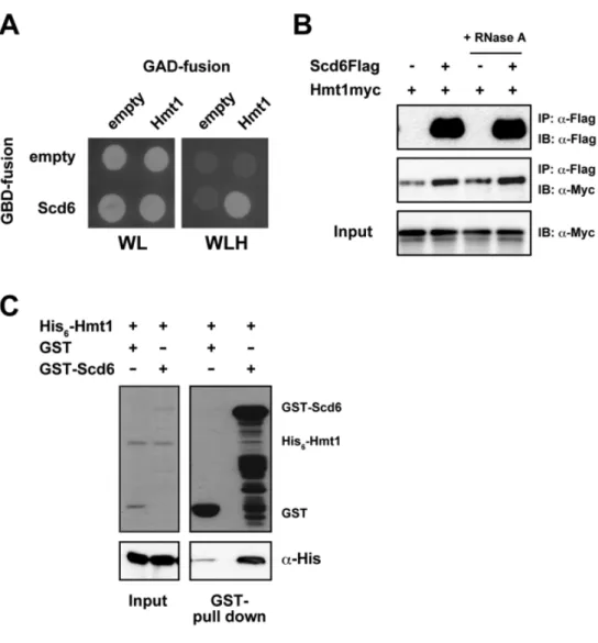
3.2 Scd6 directly interacts with Hmt1
HMT1 was cloned from wild-type gDNA into pGAD-c1 vector and
analyzed for interaction with GBD-Scd6. Cells expressing both GBD-Scd6 and GAD-Hmt1 exhibited the His+ phenotype, suggesting the interaction between Scd6 and Hmt1, confirming my 2-hybrid screening (Figure 6A).
Hmt1 is the major type I protein arginine methyltransferase (PRMT) in budding yeast and catalyzes the production of both mono- and asymmetric methylarginines on histone and non-histone proteins, especially RNA-binding proteins, and thereby regulates the functions and localizations (30, 39). Several RNA-binding proteins require mRNAs to associate with other partners in the complex. Thus, to determine whether mRNA is required for the association of Scd6 and Hmt1 in vivo, I performed co-immunoprecipitation with or without RNase A treatment (Figure 6B). Immunoprecipitation of Scd6Flag from cell extracts with an anti-Flag antibody led to co-precipitation of Hmt1myc, as detected using an anti-Myc antibody in Western blots. I also found that Hmt1
immunoprecipitated with Scd6 regardless of RNase A treatment, suggesting that these proteins co-purify in an RNA-independent manner. Subsequently, I performed in vitro binding assays using recombinant proteins to determine whether this interaction is direct or not. In these experiments, His6-Hmt1 was
significantly bound to GST-Scd6 (Figure 6C), indicating that the Hmt1–Scd6 interaction is direct, and may be akin to enzyme and substrate interactions.
Figure 6. Scd6 interacts with Hmt1. (A) Two-hybrid analysis; the yeast strain PJ69-4A
was co-transformed with pGBD-c1 or pGBD-c1-SCD6 with either pGAD-c1 or pGAD-c1-HMT1, colonies were spotted onto SC-Leu-Trp (WL) and SC-Leu-Trp-His (WLH) plates and incubated for 3 days at 30˚C. (B) Co-immunoprecipitation analysis; Scd6Flag was immunoprecipitated from Hmt1myc strains harboring YCplac33-SCD6FLAG or YCplac33 with an anti-Flag antibody, and probed for Scd6Flag and Hmt1myc using anti-Flag and anti-Myc antibodies. Similar experiments were performed using total extracts treated with RNase A prior
to immunoprecipitation. (C) In vitro binding assays; His6-Hmt1 was incubated with Glutathione
sepharose beads that had been preloaded with equal amounts of GST or GST-Scd6.
3.3 Scd6 contains asymmetrically methylated arginines in RGG motifs
Interactions were observed between Hmt1 and Scd6 in yeast two-hybrid, co-IP, and in vitro binding assays, which suggested an enzyme–substrate interaction. PRMTs bind and methylate RGG repeats in numerous RNA-binding proteins, and Scd6 contains RGG repeats in its carboxy-terminus (16, 31). Accordingly, following immunoprecipitation of Scd6Flag from wild-type and
hmt1 cell lysates using a monoclonal anti-Flag antibody, I found that the
Scd6Flag band from hmt1 cells migrated slightly smaller than that of wild-type cells (Figure 7A), suggesting that bindings of Hmt1 may methylates Scd6.
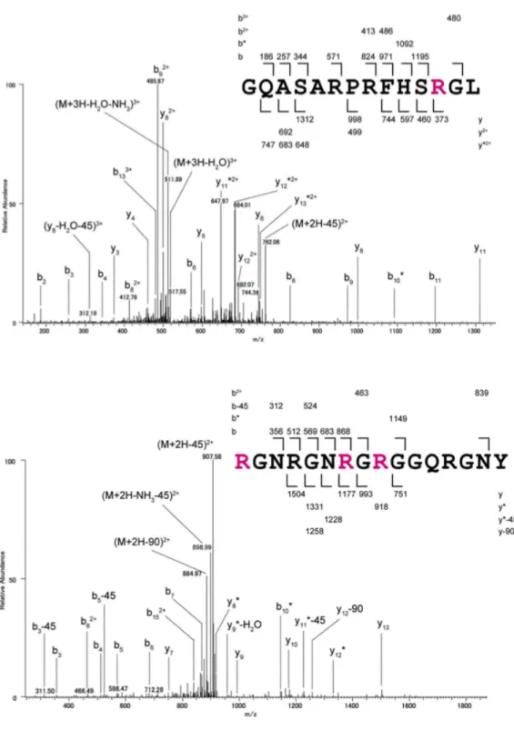
To confirm this possibility and determine whether modifications are present in Scd6, Scd6Flag from wild-type and hmt1 cells were immunopurified as described above and digested with chymotrypsin in gels. Collected peptides were then analyzed by tandem mass spectrometry. Those containing dimethyl arginine were compared. Chymotryptic peptides covering more than 80% of entire protein sequence of Scd6 were detected (Figure 7B). Among these, four peptides contained seven dimethyl arginines in RGG motifs of Scd6 from wild-type cells, but not from hmt1 cells (Figures 7B and C). Peptides containing aDMA or sDMA can be distinguished by neutral losses of dimethylamine (m/z
45.05) and monomethylamine (m/z 31.04), respectively (34, 51). Thus, I assigned spectra to the peptide 277–290, which has single aDMA and to the peptide 298–313, which has three aDMAs (Figure 8).
Figure 7. Arginine residues in RGG motifs of Scd6 are dimethylated in Hmt1-dependent manner. (A) Scd6Flag proteins from wild-type and hmt1 cells were
immunoprecipitated using an anti-Flag antibody and separated using NU-PAGE gels, and then visualized by immunoblotting with anti-Flag antibody. (B) Amino acid sequences of Scd6 are shown and chymotriptic peptides that identified using Tandem mass spectrometry analyses are indicated by underlines. Red letters indicate asymmetric dimethylarginines and residues of RGG motifs are shown in bold. (C) Chromatograms of four chymotryptic peptides of Scd6 containing asymmetric dimethylarginines were compared between wild-type (upper panel) and hmt1 cells (lower panel). Signal intensities of peptides were normalized to total ion current chromatograms and ratios of signal intensities of peptides between wild-type and hmt1 cells are presented as relative abundances of each peptide. NGSEVKDLSIL, an unmodified Scd6 peptide.
Figure 8. MS/MS spectra of identified peptides containing asymmetric dimethylarginines. the peptide 277–290 (GQASARPRFHSRG) and the peptide 298–313
3.4 Hmt1 regulates subcellular localization of Scd6
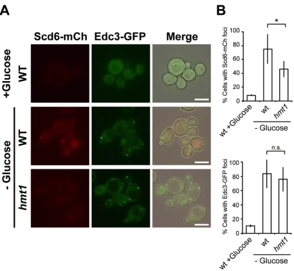
Previous studies have shown that Npl3 and Hrp1 are involved in the export of bulk mRNA from the nucleus and that Hmt1 is important for efficient export of these hnRNP complexes (38). Scd6 is a decapping activator and is accumulated in P-bodies under starvation conditions (16). Thus, I determined whether Scd6 localization is perturbed in hmt1 cells under conditions of glucose deprivation. Scd6 was chromosomally tagged with the red fluorescent protein mCherry in its C-terminus and colocalization with the P-body marker Edc3-GFP was observed in wild-type and hmt1 cells. Scd6-mCherry is functional because it rescued synthetic growth defect of scd6 dhh1 double mutant (data not shown). In wild-type cells, Scd6-mCherry foci were clearly observed upon glucose deprivation and colocalized well with Edc3-GFP. In contrast, disruption of
HMT1 significantly decreased foci formation of Scd6-mCherry without affecting
Edc3-GFP foci formation (Figures 9). These data suggest that Hmt1 is required for targeting of Scd6 to P-bodies under stress conditions.
Figure 9. Hmt1 affected Scd6 subcellular localization. (A) Localization of
Scd6-mCherry and Edc3-GFP; Wild-type (WT) and hmt1 mutant cells carrying endogenous mCherry-tagged Scd6 were transformed with the pRS314-EDC3-GFP plasmid. Cells were then
grown to mid-log phase and were resuspended in medium lacking glucose; Scale bar, 5 m.
(B) Percentages of more than 200 cells with Scd6-mCherry or Edc3-GFP foci from three independent experiments are presented as means ± standard deviations (SD); *, P < 0.05.
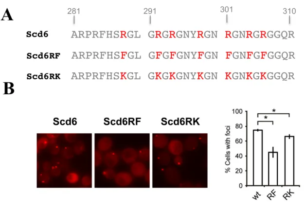
3.5 Hmt1-dependent arginine methylation is involved in Scd6 subcellular localization
Arginine methylation by PRMTs is implicated in nucleocytoplasmic shuttling of some hnRNPs (29), warranting further detailed analyses of the effects of arginine methylation on Scd6 dynamics in granules. Observed methylated arginine residues in Scd6 were substituted with lysine (methylation deficient, Scd6RK) or phenylalanine (methylation mimic, Scd6RF) (52), and were then visualized by tagging mRFP under glucose-deficient conditions. Under these conditions, the percentage of the cells with Scd6RK-mRFP foci was decreased in comparison with those with Scd6WT-mRFP foci, and this was consistent with the defects observed in hmt1 mutant cells (Figure 10). These results show that Hmt1-dependent arginine methylation within the RGG motifs of Scd6 is crucial for Scd6 accumulation in P-bodies under glucose-deprivation conditions. However, more severe defects of foci formation were observed with Scd6RF-mRFP indicating that this substitution may affect protein structure or associations of Scd6 with other components.
Figure 10. Subcellular localization of Scd6 proteins with mutations in the RGG motifs. (A) Substitutions of Scd6; Scd6RF, methylation-mimic substitution; Scd6RK,
methylation-deficient substitution. (B) Scd6-mRFP localization; scd6 cells containing pRS316-SCD6-mRFP, pRS316-SCD6RF-mRFP, or pRS316-SCD6RK-mRFP were grown and observed as in A. (C) Percentages of more than 200 cells with Scd6-mRFP foci from three independent experiments are shown as means ± SD; *P < 0.05.
A
B
Scd6 Scd6RF Scd6RKARPRFHS
R
GL
G
R
G
R
GNY
R
GN
R
GN
R
G
R
GGQR
ARPRFHS
F
GL
G
F
G
F
GNY
F
GN
F
GN
F
G
F
GGQR
281 291 301 310 Scd6 Scd6RFARPRFHS
K
GL
G
K
G
K
GNY
K
GN
K
GN
K
G
K
GGQR
Scd6RK3.6 Scd6 is important for P-body formation
Scd6 was identified as a core component of P-bodies, along with Dhh1, Dcp2, and Edc3 (13), and associations of these components are reportedly essential for P-body accumulation. Previous studies have shown that cells lacking Dhh1 have defective P-body formation (13). Although percentages of cells with Dcp2-foci in scd6 mutant cells under glucose starvation conditions did not differ from those of wild-type cells, signal intensities of these granules in
scd6 cells were lower than those in wild-type cells (Figure 11). Detailed analyses
of wild type cells using linescan showed that foci with strong signal intensities were successfully formed under glucose starvation conditions, but were significantly decreased in scd6 cells (P < 0.01, 0.68-fold). Additionally, Scd6 overexpression induced multiple distinct signals for P-bodies without inducing stress (P < 0.01, 9.32-fold) compared to the cells with non-insert bearing vector (Figure 12). In contrast with a previous study (13), overexpression of SCD6 using the 2 -plasmid backbone did not lead to cell growth defect (data not shown). These results indicate that Scd6 plays important roles in the accumulation of P-bodies.
Figure 11. scd6 deletion impairs the accumulation of Dcp2-GFP foci. Foci formation
of Dcp2-GFP under glucose depletion condition. Wild-type (WT) and scd6 mutant cells carrying endogenous Dcp2 tagged with GFP were grown to mid-log phase and were
resuspended in medium lacking glucose (-Dex); Scale bar, 5 µm. Percentages of cells with
Dcp2-GFP foci among more than 200 cells from three independent experiments are shown as means ± SD. Fluorescence intensities of Dcp2-GFP were measured using the linescan function in MetaMorph, and fluorescence profiles are shown in the right panel.
Figure 12. Overexpression of Scd6 induces the accumulation of Dcp2-GFP foci. Foci
formation of Dcp2-GFP in Scd6–overexpression condition. Dcp2-GFP cells harboring YEplac195 (Empty) or YEplac195-SCD6 (2µ-SCD6) plasmids were grown to mid-log phase in glucose-containing medium (+Dex). Scale bar, 5 µm. Percentages of cells with Dcp2-GFP foci among more than 200 cells from three independent experiments are shown as means ± SD. Fluorescence intensities of Dcp2-GFP were measured using the linescan function in MetaMorph, and fluorescence profiles are shown in the right panel.
3.7 Arginine methylation is not involved in Scd6 function for P-body formation
Under normal growth conditions, overexpression of methylation-mimic and -deficient Scd6 induced the formation of P-bodies to similar degrees as overexpressed wild-type Scd6 (Figures 13A and 13B). No detectable differences in foci formation of Edc3-GFP were observed in hmt1 mutant cells compared with that in wild-type cells under glucose starvation conditions (Figure 9A). Thus, my observations suggest that although arginine methylation of Scd6 by Hmt1 is essential for targeting of Scd6 to P-bodies, it is not required for cooperative contributions of Scd6 and Dhh1 to the formation of P-bodies under physiological and stressed conditions, such as glucose starvation. However, further studies are necessary to determine the involvement of methylation in the regulation of Scd6 function following acute stress conditions such as heat shock.
Figure 13. Scd6 function for P body formation is independent on arginine methylation. (A) scd6 Dcp2-GFP cells harboring YCplac33 (CEN) or YEplac195 (2µ)
plasmids containing wild-type (SCD6), methylation-deficient (SCD6RK), or methylation-mimic (SCD6RF) substitutions were grown to mid-log phase in glucose-containing medium
(+Glucose); Scale bar, 5 µm. (B) Percentages of Dcp2-GFP foci among more than 200 cells
from three independent experiments are shown as means ± SD; *P < 0.05.
A
3.8 Scd6 has synthetic effects with Dhh1 on cell growth and P-body formation
Analyses of Scd6 physiological functions in vivo were restrained because the scd6 mutation did not affect growth under normal conditions. Thus, I performed genetic analyses to investigate synthetic functions of Scd6 with components of decapping activators. Genetic interactions were identified between scd6 and dhh1 alleles. dhh1 scd6 double mutants showed synthetic growth defects compared with those observed in dhh1 single mutants at 25˚C (Figure 14A), and these growth defects became more severe at 37˚C (Figure 14B), suggesting functional redundancies of Scd6 and Dhh1 for cell growth.
As I and others have shown that both Scd6 and Dhh1 have function, in as much as some extent, for P-body formation (15), scd6 dhh1 double mutant showed almost complete defect for the formation of P-bodies under glucose starvation conditions (Figures 15). These data confirm the shared roles of Scd6 and Dhh1 that were observed in in vivo experiments, including roles in P-body formation under stress conditions.
Figure 14. scd6 dhh1 mutant strain showed synthetic growth defect. (A) Growth of
scd6 dhh1 mutant strain; Heterozygous strains carrying mutations in dhh1 and scd6 alleles were
sporulated, and tetrads were dissected onto YPD medium. Growth after 4 days at 25˚C is shown. (B) Growth assays; Wild-type (WT), scd6, dhh1, and scd6 dhh1 mutant cells were spotted onto YPD medium and incubated at 25˚C or 37˚C.
Figure 15. Scd6 and Dhh1 have overlapping functions in P-body formation. (A)
Dcp2-GFP foci formation; Wild-type (WT), scd6, dhh1, and scd6 dhh1 cells in which endogenous Dcp2 was tagged with GFP were grown in glucose-containing medium to the
mid-log phase and were then resuspended in medium lacking glucose; Scale bar, 5 µm. (B)
Percentages of more than 200 cells with Dcp2-GFP foci from three independent experiments are shown as means ± SD; **P < 0.01.
A
3.9 Scd6 is not a global translation repressor
Previously, global translation repression and P-body formation were correlated under conditions of glucose deprivation (15). Accordingly, if Scd6 acts as a general translation repressor, defects in translation repression in scd6 dhh1 double mutants are likely correlated with defects in P-body formation. As a model of global translational repression, glucose deprived cells exhibit declines in translation with polysome profiles (15, 17). In the present study, wild-type and
scd6 dhh1 cells were cultured in glucose-containing medium until the mid-log
phase and were then subjected to glucose deprivation prior to polysome analyses. Under glucose starvation conditions, scd6 dhh1 cells showed similar reductions in polysome fractions to those observed in wild-type cells (Figure 16), suggesting that overlapping cell growth and P-body formation functions of Scd6 and Dhh1 may not be related to global translation repression.
Polysome profiling was performed based on the same total RNA concentrations for all samples. dhh1 scd6 mutant has defect of growth, so the amount of deceased cells were present in polysome profiling was larger than wild-type. Therefore, polysome level decreased in scd6 dhh1 mutant comparing to wild-type cell, both in the media with and without glucose (Figure 16).
Figure 16. Scd6 was not essential for general translation repression under glucose starvation conditions. Wild-type (WT) and scd6 dhh1 cells were grown in rich medium (+Dex)
and were subjected to glucose deprivation (-Dex) and typical polysome profiles (OD254 traces)
are presented. Small and large ribosomal subunits (40S and 60S, respectively), monosomes (80S), and polysomes are labeled.
3.10 Arginine methylation regulates Scd6 function in relation to Dhh1 on cell growth
Next, I investigated the relationship between Dhh1 and Hmt1. Tetrad Sedimentation R el at ive A254nm
WT
scd6 dhh1
10% 50%dhh1 hmt1 double mutant grows much slower than dhh1 single mutant, and was
similar to scd6 dhh1 mutant. hmt1, scd6 and scd6 hmt1 mutants showed normal cell growth phenotype (Figure 17). Thus, Hmt1 and Scd6 may similarly regulate Dhh1-mediated cell growth.
To determined whether Hmt1-dependent arginine methylation of Scd6 is involved in Scd6 and Dhh1 mediated regulation of cell growth, I showed that expression of wild-type Scd6 recovered cell growth in scd6 dhh1 mutant cells at 25˚C (Figure 18). Moreover, suppressive effects of Scd6 methylation-mimic and -deficient substitutions were indistinguishable from wild-type cells at physiological temperature. However, at 37˚C, both wild-type and methylation-deficient Scd6 suppressed cell growth, whereas methylation-mimic Scd6 did not. This result implicates that at elevated temperature loss of positive charge at arginine residues by substituting phenylalanine residues for arginine residues negatively regulates Scd6 function on cell growth; thereby arginine methylation probably leads to the same suppression.
Figure 17. dhh1 hmt1 mutant strain showed synthetic growth defect. Growth of scd6
hmt1 and dhh1 hmt1 mutant strains. Strains that were heterozygous for scd6 hmt1 and dhh1 hmt1 were sporulated, and tetrads were dissected onto YPD medium. Growth was determined
after 4 days at 25˚C.
Figure 18. Positive charge at arginine residues is required for Scd6 function on cell growth at elevated temperature. Growth assays; scd6 dhh1 mutant cells harboring empty
vector (empty) or plasmids containing wild-type, methylation-deficient (SCD6RK), or methylation-mimic (SCD6RF) substitutions were spotted onto YPD medium and incubated at 25˚C or 37˚C.
3.11 Scd6, Dhh1 and Edc3 have synthetic effect on cell growth
Genetic interactions between scd6, dhh1 and edc3 alleles were examined. Both edc3 scd6 and dhh1 edc3 double mutants showed synthetic growth defects. The dhh edc3 scd6 triple mutant had more severe growth defect than those double mutants (Figure 19). This result indicated that these three decapping activators have combined impact on cell growth.
Whereas dhh1 hmt1 double mutant showed synthetic growth defect, the same phenotype was not observed in edc3 hmt1 double mutant (data not shown). This result again suggested that at physiological growth condition, Hmt1-based arginine methylation is not required for Scd6 role for cell growth (Figure 18).
Figure 19. dhh1 edc3 scd6 mutant strain showed synthetic growth defect. Growth of
dhh1 edc3 scd6 mutant strains. Strains that were heterozygous for dhh1 edc3 scd6 were
sporulated, and tetrads were dissected onto YPD medium. Growth was determined after 4 days of incubation at 25˚C.
edc3 scd6
dhh1 edc3
WT
dhh1 scd6
scd6
edc3
dhh1 edc3 scd6
dhh1
CHAPTER 4
In the present study, I demonstrated the roles of Scd6 with Dhh1 and Edc3 on cell growth. Further analysis of Dhh1 and Scd6 interaction revealed their shared functions for the formation of P-bodies and cell growth, particularly under stress conditions. In addition, Hmt1 associated with and methylated Scd6 leading to the efficient targeting of Scd6 to P-bodies. Arginine methylation of Scd6 might negatively regulated Scd6 function on cell growth at elevated temperature (Figure 20). However, molecular details of these arginine methylation-based regulations remain to be addressed in future work. It is possible that arginine methylation is required for Scd6 to regulate its specific mRNA targets. There are increasing evidences that post-translational modifications affect RBPs functions. Likewise, arginine methylation in this study could offer a common mechanism for mRNP components to facilitate their dynamics in the cells.
Figure 20. Proposed model of Scd6 physiological activities in this study
4.1 Hmt1-based arginine methylation could be a reversible modification under environmental stimuli.
Post-transcriptional regulation system and the dynamic of mRNP complexes have the fastest responses to a change of environment compared to other gene expression regulation steps. However, initial studies for the arginine methylation modification suggested that methyl groups were stable on arginine
Scd6 Dhh1 Cell growth Hmt1 Me Scd6 Localization to P-bodies Scd6 Dhh1 Cell growth Me Scd6 at high temperature
A
B
Edc3 P-body formationresidues (30). It remains to be determined that the extent to which arginine methylation is dynamic and whether protein arginine demethylation reactions occur to reverse the effects of the modifications. Recently, it has been shown that purified human JmjC lysine demethylases (JmjC KDMs) can also act as JmjC arginine demethylases (JmjC RDMs) both on histone and non-histone peptides, indicating arginine methylation may be a reversible modification (42). There were significant changes in arginine methylation abundance associated with growth conditions. Particularly, the concentrations of mono-methylarginines and asymmetric-dimethylarginines decreased during heat shock and stationary phase compared to log-phase growth, whereas symmetric-dimethylarginines increased (53). In the present study, under physiological growth condition (25˚C), substitutions of arginine that were identified to be methylated in Scd6 neither affected cell growth nor P-body formation (Figures 13 and 18). However, at elevated temperature (37˚C), suppression of synthetic growth defects in dhh1
scd6 mutant cells was observed with wild-type and the methylation-deficient
Scd6, but not with the methylation-mimic Scd6 (Figure 18). These observations suggest that stress-dependent regulation of Scd6 activities may be due to the dynamic change of Hmt1-mediated methylation. Future studies are needed to
determine whether the methylation of Scd6 is reversible and how the modification is actively regulated upon environmental stimuli. Accordingly, it will be important to identify demethylases that catalyze non-histone arginine demethylation and to assess its biological significance.
4.2 Hmt1 possibly mediate the effect of Scd6 on specific target mRNAs Previous studies suggest that arginine methylation of RNA-binding proteins by PRMTs can modulate binding affinities for RNAs– or protein–protein interactions of targets (30, 39). Compared to wild-type cells, we did not see significant changes in total Scd6Flag-associated proteins or total RNAs in hmt1 cells (data not shown). Thus, arginine methylation of Scd6 may act on specific mRNA targets. However, there is currently limited information on subsets of mRNA targets that coupled to Scd6 accumulation to P-body or function toward cell growth. Therefore, one needs to indentify specific mRNA targets to validate these hypotheses.
Depletion of HMT1 specifically resulted in defect of Scd6 localization whereas remarkable foci formation defects were not observed for other components of P-bodies such as Dcp2 and Edc3 (Figure 9). Scd6 overexpression promoted P-body formation independently of Hmt1-mediated arginine
methylation (Figure 13). Therefore, a goal in future work will be to unveil the significance of arginine methylation by Hmt1 in targeting Scd6 to P-bodies. A set of Scd6-precipitated mRNAs was submitted to the Gene Expression Omnibus (GEO) by Dan Klass in 2010. These data show that mRNAs that are transcribed from sub-telomeric regions, coding mRNAs for proteins of telomere capping maintenance and replication stress response such as MEC1 and TEL1, were substantially accumulated in Scd6-precipitated samples. In addition, it has been shown that protein arginine methylation has roles in DNA repair under replication stress (32). These data suggest that Scd6 may specifically bind to and activates the mRNA decapping under replication stress condition. Arginine methylation on Scd6 could be required to target the complex of these mRNAs to the P-bodies, thereby keeping mRNAs stored rather than to degraded.
4.3 Scd6 could have the functional connection to the biological processes that are regulated by Hmt1.
Recent accumulating evidence indicates that analysis of Hmt1 methyltransferase activity is fundamental to understand protein functions and various molecular processes. In particular, following interactions with Hmt1, Ccr4-Not complexe is involved in mRNA maturation and Npl3-dependent
nuclear export, and likely participate in cell-cycle progression by stabilizing cyclin mRNA in response to environmental stimuli (40, 54). Previous data suggest that Scd6 is functionally involved in Hmt1-targets. For instance, Scd6 and Npl3 played similar roles in the targeting of eIF4G for translation repression (16), and scd6 ccr4 mutant cells had more severe synthetic growth defects than
ccr4 mutant cells (data not shown). Further studies could clarify the involvement
of Scd6 in nuclear export of mRNA, and whether Hmt1 mediates the effects of Scd6 on cell cycle progression, as it does for Npl3 and Ccr4-Not.
4.4 Arginine methyltransferases has functional and dynamic link with RGG-motif containing mRNP components
P-body localization of Scd6 has been shown under conditions of glucose deprivation (16), and previous studies suggest that P-bodies contain multiple proteins of mRNA decay machinery (55). The present data show that Hmt1 binds and methylates Scd6. Hmt1 was previously shown to catalyze arginine methylation of mRNP components with RGG motifs such as Npl3, Sbp1, and Ded1, which are reportedly localized in RNA granules (30). In addition, the P-body component Ebs1 (56) was recovered in my two-hybrid analysis, also recovered in proteomic analysis of Hmt1-TAP associating proteins (57). These
observations suggest the presence of a functional link between Hmt1 and components of P-bodies (58). Hmt1 and Scd6 interaction may facilitate the above consequences.
Several arginine residues within the RGG motif of Scd6 were methylated in Hmt1 dependent manner. Moreover, our mass spectrometry analyses revealed the presence of DMAs in Scd6 peptides from wild-type cells, and these modifications were still observed in samples from hmt1 mutant cells (data not shown). Although Scd6 was not recovered in proteomic analyses using a methylarginine antibody, the present data indicates that Scd6 is hyper methylated by additional methyltransferase. Hence, future assessments of degrees of methylation and identification of responsible enzyme(s) may further reveal how arginine methylation could regulate the function of mRNP components.