69
Yonago Acta medica 2008;51:69–72
Abbreviations: CT, computed tomography; FDG, 18F-fluorodeoxyglucose; PET,positron emission tomography
Inflammatory Pseudotumor of the Lung Identified by
18
F-Fluorodeoxyglucose Positron Emission Tomography:
A Patient Report
Yuzo Takagi, Hiroshige Nakamura*, Ken Miwa*, Yoshin Adachi*, Shinji Fujioka*, Tomohiro Haruki*, Yuji Taniguchi* and Yasushi Horie†
Center for Clinical Residency Program, *Division of General Thoracic Surgery and †Pathology Division, Tottori University Hospital, Yonago 683-8504 Japan
The patient was a 71-year-old man with an abnormal shadow on chest X-ray. Computed tomography (CT) of the chest showed a tumor of 38 mm in size in the upper lobe of the left lung S3. A CT-guided lung biopsy was performed, but no malignancy was observed. 18F-fluorodeoxyglucose (FDG) positron emission tomography (PET) revealed a high
FDG uptake in the tumor: the maximum and mean values of the standard FDG uptake in the early phase were 7.3 and 5.3, respectively, and those in the latter phase, 8.3 and 5.9, respectively. The hilar and mediastinal lymph nodes also showed positive for high FDG uptakes. We strongly suspected lung cancer, and performed a left upper lobecto-my by video-assisted thoracic surgery. The pathological diagnosis was an inflammatory pseudotumor of the lung: it is a rare disease but often requires differentiation from lung cancer. Literature has been few on FDG-PET about inflammatory pseudotumor of the lung. Differentiation of the disease from lung cancer was especially difficult in the pres-ent patipres-ent, because both lymph nodes and the tumor showed high FDG uptakes.
Key words: 18F-fluorodeoxyglucose positron emission tomography; lung inflammatory
pseudo-tumor; video-assisted thoracic surgery
Inflammatory pseudotumor of the lung has been more frequently detected due to the recent progress in diagnostic imaging, but it is difficult to differen-tiate from lung cancer. In many patients, the diag-nosis is confirmed with surgery. We have encoun-tered a male patient highly suspected of lung can-cer due to his high uptake of 18
F-fluorodeoxyglu-cose (FDG) in FDG-positron emission tomography (PET). We diagnosed his disease as inflammatory pseudotumor of the lung via video-assisted thoracic surgery. Few literature focuses on FDG-PET and inflammatory pseudotumor of the lung. The pres-ent patipres-ent showed a high FDG uptake also in the lymph node, which made us confused in differenti-ating from lung cancer. We herein present the case
of our patient, with bibliographical considerations as well.
Patient Report
This 71-year-old man had undergone medical ex-aminations every year, but no abnormalities were detected. In July 2007, he had general fatigue and hemosputum, and visited nearby hospital where an abnormal shadow in the left upper lung was found on examination. He was referred to our Univer-sity Hospital. Laboratory findings at the time of admission were a white blood cell count of 6,100/ μL without any left shift of the nucleus, and the
70
Y. Takagi et al.
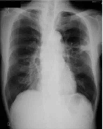
C-reactive protein level of 0.52 mg/dL. General or biochemical examinations of the blood showed no other abnormalities, and all of the tumor markers were negative. Chest X-ray showed a nodular den-sity with approximately 40 mm in size in the left upper lung (Fig. 1). Chest computed tomography (CT) visualized a 38 × 32 mm tumor in size hav-ing broad contact with the parietal pleura in the left upper lobe S3 (Fig. 2). The periphery of the tumor
was irregular, and the boundary was relatively well defined. Lymph nodes were enlarged at both the hilar and the mediastinal regions. In FDG-PET (Fig. 3), we observed a high FDG uptake just matching the tumor. The maximum and mean values of the standard FDG uptake in the early phase were 7.3 and 5.3, respectively, and those in the latter phase, 8.3 and 5.9, respectively. In ad-dition, high FDG uptake was also observed in both the hilar and the mediastinal regions, so lung
Fig. 1. Chest X-ray showed an irregular-circumscribed
abnormal shadow in the left middle field of the lung.
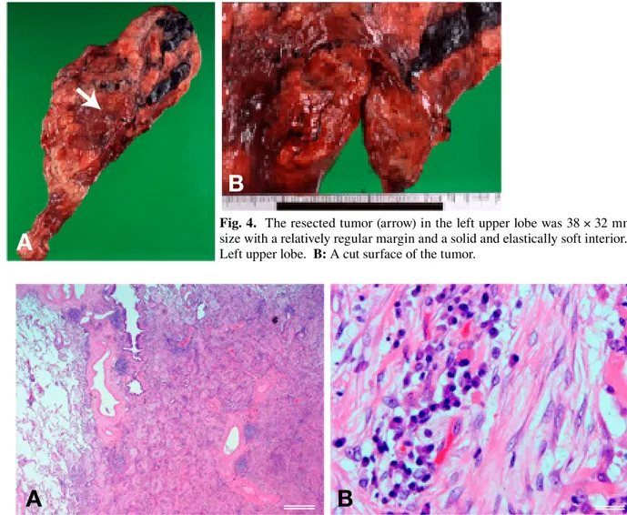
cancer and metastases to the lymph nodes were strongly suspected. The CT-guided lung biopsy resulted in nonspecific inflammatory findings, and no malignant cells were observed. However, on considering FDG-PET findings, lung cancer could not be ruled out. Therefore, thoracoscopic surgery was performed after obtaining informed consent from the patient. The tumor had firmly adhered to the parietal pleura, and a needle biopsy exhibited no malignant cells on pathological tissues frozen intraoperatively. We suspected an inflammatory pseudotumor, and performed a simple left upper lobectomy by video-assisted thoracic surgery. The resected tumor (Fig. 4) was 38 × 32 mm in size, sectionally xanthochromatic with mild anthraco-sis. Its shape was relatively regular in the periph-ery, well defined, solid and elastically soft. In the pathological tissue (Fig. 5), we found a lesion that was mainly comprised of organized pneumonic fi-brosis, with inflammatory cell infiltration involving lymphocytes and plasma cells. When the image of the area was enlarged on the monitor, we observed inflammatory cells, fibrosis and spindle-shaped mesenchymal cells, which led us to the diagnosis of inflammatory pseudotumor, fibrous histiocytic type of the lung. The patient’s postoperative course was good, and no recurrence has been observed.
Fig. 3. FDG-PET revealed a high FDG uptake just
matching the tumor, the hilar and mediastinal lymph nodes (arrows). FDG, 18F-fluorodeoxyglucose; PET,
posi-tron emission tomography.
Fig. 2. Chest CT revealed an irregular tumor, 38 32
mm in size, having broad contact with the parietal pleura in the left upper lobe S3. CT, computed tomography.
71
Pulmonary inflammatory pseudotumor
Fig. 5. Pathological findings showed inflammatory cell infiltration involving lymphocytes and plasma cells, fibrosis
and spindle-shaped mesenchymal cells with hematoxylin and eosin staining of low power field (A) and high power field (B). Bar = 500 µm in A and 50 µm in B.
Fig. 4. The resected tumor (arrow) in the left upper lobe was 38 32 mm in
size with a relatively regular margin and a solid and elastically soft interior. A: Left upper lobe. B: A cut surface of the tumor.
Discussion
Inflammatory pseudotumor of the lung is a solitary, non-neoplastic, space-occupying lesion in which collagenic fiber, inflammatory cells and mesenchymal cells are blended together in various amounts (Bernardi et al., 1983). In 2000, Cerfolio et al. reported that among 56,400 patients treated with chest surgery over 50 years at the Mayo Clinic, inflammatory pseudotumor of the lung was observed in only 23 patients (0.04%), of which 18 patients (78%) were involved with symptoms such as coughing, hemosputum, shortness of breath and chest pain (Cerfolio et al., 2000). In imaging
modalities, the pseudotumor is seen as an isolated and well-defined shadow with a round to oval shape, and clinical differentiation from lung cancer is often difficult (Ishida et al., 1989; Kobashi et al., 2006). In the present patient as well, we suspected lung cancer from the clinical symptoms and image findings, and performed the CT-guided lung biopsy, but no malignant cells were observed. However, FDG-PET of the tumor further showed a high FDG uptake, so we highly suspected lung cancer again and considered performing video-assisted thoracic surgery. Recently, FDG-PET has been widely used as an adjunct for the diagnosis of lung cancer (Ung et al., 2007). A higher level of the standard FDG uptake is correlated to poorer prognosis
A
A
B
72
Y. Takagi et al.
(Berghmans et al., 2008). However, FDG-PET often shows positive even for inflammatory lung disease. Of 66 patients with lung nodule shadows that were positive in FDG-PET, 7 patients were false positive for inflammatory lung disease: one of the 7 had an inflammatory pseudotumor of the lung (Higashi et al., 2001). Because inflammatory pseudotumor of the lung is a rare disease, application of FDG-PET has been seldom reported (Slosman et al., 1994). But, FDG-PET shows high FDG uptake for inflammatory pseudotumor of the lung, and differentiation from liver cancer or lung cancer is difficult (Kawashima et al., 2006). In addition, it is characteristic of FDG-PET that the hilar and the mediastinal lymph nodes also show positive, as observed in the present patient. FDG-PET is also useful in diagnosing metastasis of lung cancer to lymph nodes (Melek et al., 2008), which makes differentiation of lung pseudotumor from lung cancer more difficult. Even though inflammatory pseudotumor of the lung is benign, 3 of 5 patients with incomplete resection had a recurrence (Cerfolio et al., 2000), or 3 of 18 treated patients had a recurrence (Melloni et al., 2005). Therefore, we had no choice but to perform a left upper lobectomy in the present patient, because sufficient resection margins are required. The postoperative course was good, and we believe that this was an appropriate choice. Cases of recurrence after complete resection have been reported (Dehabreh et al., 1999) with some multiple tumor cases (Lee et al., 2005; Kobashi et al., 2006). We emphasize that the present patient will require a careful follow-up. Along with the progress in diagnostic imaging, inflammatory pseudotumor of the lung has been increasingly observed. The characteristics of FDG-PET findings should also be fully understood.
References
1 Berghmans T, Dusart M, Paesmans M, Hossein-Foucher C, Buvat I, Castagne C, et al. Primary tumor standardized uptake value (SUV max) measured on fluorodeoxyglucose positron emission tomography (FDG-PET) is a prognostic value for survival in
non-small cell lung cancer (NSCLC): a systematic review and meta-analysis (MA) by the European Lung Can-cer Working Party for the IASLC Lung CanCan-cer Stag-ing Project. J Torac Oncol 2008;3:6–12.
2 Bernardi RS, Lee SS, Chen HP, Stines GJ. Inflam-matory pseudotumors of the lung. Surg Gynecol Obstet 1983;156:89–96.
3 Cerfolio RJ, Allen MS, Nascimento AG, Deschamps C, Trastek VF, Miller DL, et al. Inflammatory pseudo-tumors of the lung. Ann Thorac Surg 1999;67:933–936. 4 Dahabreh J, Zisis C, Arnogiannaki N, Katis K. In-flammatory pseudotumor: a contcroversial entity. Eur J Cardiothorac Surg 1999;16:670–673.
5 Higashi KK, Ueda Y, Sakuma T, Seki H, Oguchi M, Taniguchi M, et al. Comparison of FDG PET and TI SPECT in evaluation of pulmonary nodules. J Nucl Med. 2001;42:1497–1498.
6 Ishida T, Oka T, Nishino T, Tateishi M, Mitsudomi T, Sugimachi K. Inflammatory pseudotumor of the lung in adults: radiographic and clinicopathological analysis. Ann Thorac Surg 1989;48:90–95.
7 Kawamura E, Habu D, Tsushima H, Torii K, Kawabe J, Ohsawa M, et al. A case of hepatic inflammatory pseudotumor identified by FDG-PET. Ann Nucl Med 2006;20:321–323.
8 Kobashi Y, Fukuda M, Nakata M, Irei T, Oka M. Inflammatory pseudotumor of the lung: clinico-pathilogical analysis in seven adult patients. Int J Clin Oncol 2006;11:461–466.
9 Lee HL, Kim L, Lee KH, Kim KH, Kim CW. A case of pulmonary inflammatory pseudotumor: recurrence appearing as several consolidative le-sion after complete resection. Kor J Intern Med 2005;20:168–172.
10 Melek H, Gunluoglu MZ, Demr A, Akin H, Olcmen A, Dincer SI, et al. Role of positron emission tomogra-phy in mediastinal lymphatic staging of non-small cell lung cancer. Eur J Cardiothorac Surg 2008;33:294– 299.
11 Melloni G, Carretta A, Ciriaco P, Arrigoni G, Fieschi S, Rizzo N, et al. Inflammatory pseudotumor of the lung in adults. Ann Thorac Surg 2005;79:426–432. 12 Slomsman DO, Spilliopoulos A, Keller A, Lemoine
R, Besse F, Couson F, et al. Quantitative metabolic PET imaging of a plasma cell granuloma. J Thorac Imaging 1994;9:116–119.
13 Ung YC, Maziak DE, Vanderveen JA, Smith CA, Gulenchyn K, Lacchetti C, et al. 18
F-Fluorodeoxyg-lucose positron emission tomography in the diagno-sis and staging of lung cancer: a systematic review. J Natl Cancer Inst 2007;99:1753–1767.
Received June 20, 2008; accepted July 22, 2008
Corresponding author: Hiroshige Nakamura, MD