Tyrosine Phosphorylation Regulates the Expression of Major
Histo-compatibility Complex Antigens on a Human Lung Cancer Cell Line by
Interferon−gamma
Yoshimasa Suzuki, Yoshiyuki Tanaka, Hiroshige Nakamura and Shigetsugu Ohgi
Second Department of Surgery, Faculty of Medicine, Tottori University, Yonago 683-0826, Japan Expression of major histocompatibility complex (MHC) antigens on cancer cells is essential for cell-mediated immune function. However, these molecules are reduced on cancer cells enabling them to escape from host immune surveillance. It is well known that interferon-gamma (IFN-γ) upregulates the expression of MHC molecules and restores the immunogenicity of cancer cells. Nevertheless, the mechanism by which IFN-γ modulates MHC expression on cancer cells is not clear. Therefore, in this report, we examined the role of tyrosine protein kinases in IFN-γ-induced MHC expression in a human lung adenocarcinoma cell line, HLC-1. We found that a tyrosine protein kinase inhibitor, herbimycin A, inhibited both IFN-γ-inducible MHC class I and class II expression, as assessed by flow cytometry. Additionally, assessment of tyrosine phosphorylation of cellular substrates by confocal laser microscopy using an anti-phosphotyrosine monoclonal antibody (mAb) revealed that IFN-γ induced protein tyrosine phosphorylation within 5 min of treatment. Herbimycin A inhibited this
IFN-γ-induced tyrosine phosphorylation. Thus, tyrosine phosphorylation plays an important
role in IFN-γ-induced MHC class I and class II expression on HLC-1 cells.
Key words: human lung cancer; interferon-gamma; major histocompatibility complex; tyrosine phosphorylation
Abbreviations: FCS, fetal calf serum; FITC, fluorescein isothiocyanate; IFN-γ, interferon-gamma; JAK, Janus kinase; mAb, monoclonal antibody; MHC, major histocompatibility complex; PBS, phosphate-buffered saline; PE, phycoerythrin; STAT, signal transducer and activator of transcription
Major histocompatibility complex (MHC) antigens on cancer cells are involved in a variety of immune functions affecting tumor immunity (Klein and Klein, 1985). MHC class I and II molecules present antigens to CD8+ and CD4+ T cells in the form of peptide fragments within their molecule-binding grooves, respec-tively (Matsumura et al., 1992; Madden et al., 1993; Stern et al., 1994). MHC class I mole-cules have been shown to act as restriction elements in the lysis of target cells by cytotoxic T lymphocytes (Lurquin et al., 1989), while MHC class II molecules present antigens to helper T cells and regulate autologous T helper cell activation (Eckels et al., 1983). Cancer cells, however, have reduced expression of MHC molecules, enabling them to escape from the
host’s immunosurveillance system (Goodenow et al., 1985; Festenstein and Garrido, 1986). Therefore, it is important to elucidate the mech-anisms modulating the expression of MHC molecules on cancer cells.
IFN-γ can restore the expression of MHC molecules on many types of malignant cells. Nevertheless the mechanisms of IFN-γ-induced MHC expression on cancer cells have not been elucidated, especially in the context of MHC class I expression. Recent biochemical studies indicate that tyrosine phosphorylation of Janus kinases (JAK) is an important step in the signal transduction system activated by IFN-γ (Shuai et al., 1993a; Darnell et al., 1994). Further, Hobart and colleagues (1997) suggested that interferon regulatory factor 1 might regulate the
expression of MHC class I and class II genes of mice. Therefore, in this report we examined the role of tyrosine phosphorylation in the signal transduction of IFN-γ-induced MHC expres-sion in a human lung adenocarcinoma cell line, HLC-1, using the tyrosine kinase inhibitor herbimycin A.
Materials and Methods Cells
HLC-1, a human lung adenocarcinoma cell line, was cultured in a complete medium consisting of RPMI 1640 (Kyokuto Pharmaceutical Indus-trial Co., Ltd., Tokyo, Japan) supplemented with 10% heat-inactivated fetal calf serum (FCS) and penicillin (100 mg/mL).
Reagents
Herbimycin A, an inhibitor of protein tyrosine kinase, was purchased from Wako Pure Chemi-cal (Osaka, Japan). Recombinant human IFN-γ was kindly supplied by Shionogi Pharmaceutical Co., Ltd. (Osaka).
Antibodies
The following monoclonal antibodies (mAb) were used: fluorescein isothiocyanate (FITC)-labeled W6/32 (Serotec, Oxford, United Kingdom) against MHC class I; phycoerythrin (PE)-labeled I3-RD1 (Coulter Immunology, Hialeah, FL) against MHC class II; FITC-labeled 6D12 (MBL, Nagoya, Japan) against phosphotyro-sine; MsIgG1-FITC (Coulter Immunology), FITC-labeled anti-mouse immunoglobulin G1, and MsIgG1-RD1 (Coulter Immunology), PE-labeled anti-mouse immunoglobulin G1, as controls for the FITC- and PE-labeled mAbs, respectively.
Induction of MHC class I and II antigens
HLC-1 cells were cultured in the tissue culture dish (Sumitomo Bakelite, Tokyo, Japan), 1 ×
105 cells/dish at 37°C for 24 h in a humidified
atmosphere of 5% CO2 in air. The number of
cells and their viability were evaluated by the trypan blue exclusion method. Cells were pre-treated for 2 h with or without various concen-trations of herbimycin A before the addition of IFN-γ. After 1 h of incubation with or without IFN-γ (1000 U/mL), cells were washed 3 times with FCS-free medium and cultured in the complete medium for an additional 48 h.
Flow cytometric analysis
Following incubation, HLC-1 cells were har-vested with 0.02% EDTA (Cosmo Bio, Tokyo) and then washed 3 times with phosphate-buffered saline (PBS). The cells were incubat-ed with an appropriate dilution of mAb W6/32 and I3-RD1 or FITC and MsIgG1-RD1 for 30 min at 4°C. After being washed 3 times with PBS, the cells were analyzed by flow cytometry with the FACSort system (Becton Dickinson, Mountain View, CA). Data analy-ses by fluorescence intensity were based on
calculating 1 × 10⁴ cells per sample by the
LYSIS II software (Becton Dickinson). These series of experiments were repeated 3 times with equivalent results. The results were present-ed as mean ± SE.
Laser microscopic analysis
HLC-1 cells were cultured in a glass-bottomed microwell dish (MatTek Corp., Ashland, MA),
1 × 10⁴ cells/dish at 37°C for 24 h in a
humid-ified atmosphere of 5% CO2 in air. Cells were
pretreated for 2 h with or without herbimycin A (0.1 mg/mL) before the addition of IFN-γ (1000 U/mL). After being stimulated with IFN-γ, cells were fixed with 70% ethanol. After being washed 3 times with PBS, the cells were incu-bated with an appropriate dilution of mAb 6D12 for 30 min at 4°C. After incubation, cells were washed 3 times with PBS and analyzed by confocal laser microscopy, ACAS Ultima (Meridian Instruments, Okemos, MI). These series of experiments were also repeated 3 times with equivalent results, and the data pre-sented were representative of one such experi-ment.
50 40 30 20 10 0 10 8 6 4 2 0 Results Flow cytometric analysis
Cells were induced to express MHC antigens in a dose-dependent manner following incubation for 48 h in the presence of IFN-γ, 50, 100, 500, 1000 and 2000 U/mL. The maximum expres-sion was observed at IFN-γ concentrations of 1000 and 2000 U/mL (data not shown). The induction of the MHC expression was initially observed after 12 h of exposure to IFN-γ; the expression of MHC molecules gradually intensified during 48 h of incubation (data not shown). IFN-γ at 2000 U/mL affected cell growth and viability, so the cells were stimulat-ed with IFN-γ at 1000 U/mL, followstimulat-ed by cul-ture for 48 h.
HLC-1 cells had fundamentally reduced ex-pression of MHC class I and II molecules. However, when HLC-1 cells were stimulated with IFN-γ, the expression of these molecules increased immensely. IFN-γ-inducible expres-sion of MHC class I and class II molecules was inhibited by herbimycin A. Analysis of these data by mean fluorescence intensity showed
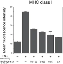
that IFN-γ increased the expression of both MHC class I and II molecules by 3.6 and 1.9 times respectively. Herbimycin A inhibited IFN-γ-inducible expression of MHC class I molecules in a dose-dependent manner and prevented IFN-γ-inducible expression of MHC class II molecules (Figs. 1 and 2).
Laser microscopic analysis
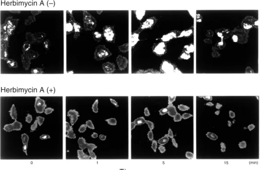
After stimulation with IFN-γ (1000 U/mL), tyrosine phosphorylation of cellular substrates in HLC-1 cells was assessed by confocal laser microscopy. IFN-γ induced protein tyrosine phosphorylation within 5 min (Fig. 3). How-ever, pretreatment of HLC-1 cells with herbi-mycin A (0.1 μg/mL) appeared to prevent tyro-sine phosphorylation. Analyzing these data by mean fluorescence intensity based on
calculat-ing 1 × 10² cells per sample (Fig. 4), protein
tyrosine phosphorylation by IFN-γ was observ-ed within 1 min and peakobserv-ed by 5 min. After 15 min, protein tyrosine phosphorylation returned to baseline levels as assessed by mean fluores-cence intensity. Herbimycin A almost com-pletely inhibited the IFN-γ-inducible tyrosine protein phosphorylation.
Fig. 1. Interferon-gamma (IFN-γ) induced the ex-pression of major histocompatibility complex (MHC) class I molecules on HLC-1 cells. The MHC expres-sion was analyzed by FACsort system after 48 h ex-position to IFN-γ. Herbimycin A (0.0125–0.1 mg/mL) prevented such expression in a dose-dependent manner.
Fig. 2. Interferon-gamma (IFN-γ) induced the ex-pression of major histocompatibility complex (MHC) class II molecules on HLC-1 cells. The MHC expression was analyzed by FACsort system after 48 h exposition to IFN-γ. Herbimycin A (0.0125–0.1 mg/mL) prevented such expression.
MHC class II
Mean fluorescence intensity
IFN-γ (1000 U/mL) Herbimycin A (μg/mL) – + + + + + – – 0.0125 0.025 0.05 0.1
MHC class I
Mean fluorescence intensity
IFN-γ (1000 U/mL) Herbimycin A (μg/mL) – + + + + + – – 0.0125 0.025 0.05 0.1
Herbimycin A (+) Herbimycin A (–) 6 5 4 3 2 1 0
Fig. 3. Effects of herbimycin A on the interferon-gamma (IFN-γ)-induced phosphorylation of tyro-sine. As shown in the upper panel, IFN-γ induced phosphorylation of tyrosine on HLC-1 cells. In the lower panel, pretreatment with herbimycin A ap-peared to inhibit the phosphorylation of tyrosine.
These findings indicate that IFN-γ induced tyrosine phosphorylation of cellular substrates which was inhibited by herbimycin A.
Discussion
Three intracellular signal transduction path-ways have been considered in the induction of MHC antigens by IFN-γ in several types of cells (Koide et al., 1988; Klein et al., 1990; Nezu et al., 1990; Towata et al., 1991; Lahat et al., 1993); that
is, the protein kinase A, Ca2+-calmodulin and
protein kinase C pathways. However, these pathways have not been shown to be decisive on the IFN-γ-induced expression of MHC class I and II antigens. There is only one report which states that tyrosine kinase pathways may be involved in the IFN-γ-induced expression of MHC class II (Ryu et al., 1993). Nevertheless, the detailed signal transduction mechanisms responsible for the IFN-γ-inducible expression of MHC antigens on cancer cells have not been elucidated, especially for MHC class I.
Recent biochemical studies have shown that IFN-γ activated the JAK1 and JAK2 tyrosine kinases which phosphorylate a down stream signal transducer and activator of transcription
Fig. 4. Effects of herbimycin A on interferon-gamma (IFN-γ)-induced phosphorylation of tyro-sine. Analysis of tyrosine phosphorylation by mean fluorescence intensity shows that IFN-γ increased the tyrosine phosphorylation five fold within 5 min. Herbimycin A inhibited the production of IFN-γ-inducible phosphotyrosine.
Mean fluorescence intensity
Time 0 10 20 (min) Herbimycin A (+) Time 0 1 5 15 (min) Herbimycin A (–)
(STAT) in the IFN-γ response signal pathway. Phosphorylated STAT1α dimerizes, translo-cates to the nucleus, and binds specific DNA elements, i.e., IFN-γ activation sites, thereby activating transcription factors, e.g., interferon regulatory factor 1 (Shuai et al., 1993a, 1993b; Silvennoinen et al., 1993; Darnell et al., 1994; Taniguchi et al., 1995).
We used an inhibitor of tyrosine protein kinase, herbimycin A (Uehara et al., 1989; Obinata et al., 1991), to determine whether tyrosine phosphorylation may be involved in the signal transduction for IFN-γ-induced ex-pression of MHC class I and II molecules on HLC-1 cells. However, while herbimycin A is useful in determining whether tyrosine phos-phorylation is involved in the mechanisms for activation by receptor-mediated signal transduc-tion, it is not specific for JAK tyrosine kinases.
We observed that IFN-γ induced the expres-sion of MHC class I and II on HLC-1 cells and the phosphorylation of protein tyrosine in their cellular substrates within 5 min. Herbimycin A inhibited both these expressions and the en-hancement of tyrosine phosphorylation. The findings that IFN-γ activated the protein tyrosine kinases that phosphorylate the cellular substrates and induce MHC class I and II molecules suggest that tyrosine phosphoryla-tion is involved in the inducphosphoryla-tion of the expres-sion of MHC molecules on HLC-1 cells by IFN-γ. Therefore, a JAK-STAT signal trans-duction pathway must be involved in the IFN-γ-induced expression of MHC class I and II molecules. However, we cannot exclude the possibility of involvement by other pathways because herbimycin A did not completely in-hibit IFN-γ-inducible expression of MHC mole-cules. While the expression of MHC molecules is known to be regulated by other cytokines such as tumor necrosis factor alpha and inter-leukin 1 alpha (Seong et al., 1991; Sedlak et al., 1992; Wolchok and Vilcek, 1992), little is known about the signal transduction mecha-nisms which induce MHC antigens by these cytokines. Further, there are some reports which state that c-myc oncogene regulates the expression of MHC molecules on cancer cells (Versteeg et al., 1988; Gaforio et al., 1991). We
think it should also be necessary in the future to clarify the signal transduction pathways by these cytokines and oncogenes for understand-ing the more detailed mechanisms which induce MHC molecules.
The expression of MHC class I and II anti-gens on cancer cells are involved in a variety of immune functions affecting tumor immunity. Cancer cells have reduced expression of MHC molecules enabling them to escape from the host’s immunosurveillance system (Goodenow et al., 1985; Festenstein et al., 1986). There-fore, we think it is important to elucidate both the mechanisms modulating the expression of MHC molecules and the methods restoring the expression of these molecules on cancer cells for improvement of clinical problems about tumor immunity. In the present study, we eluci-dated the fact that tyrosine phosphorylation played an important role as the mechanism which restores the expression of MHC mole-cules on cancer cells.
In conclusion, tyrosine phosphorylation is considered to be an essential factor in the IFN-γ-induced expression of MHC class I and II molecules on HLC-1 cells.
Acknowledgments: The authors wish to express their
sincere gratitude to Emeritus Prof. Tohru Mori, Sec-ond Dept. of Surgery, Faculty of Medicine, Tottori Univ., for his kind advice and valuable suggestions during the course of this study. We would also like to thank Prof. Hisao Ito, First Dept. of Pathology, Faculty of Medicine, Tottori Univ. and Prof. Kenzo Sato, Dept. of Molecular Biology, Faculty of Medi-cine, Tottori Univ., for reading this manuscript and helpful suggestions.
References
1 Darnell JE Jr, Kerr IM, Stark GR. Jak-Stat path-ways and transcriptional activation in response to IFNs and other extracellular signaling proteins. Science 1994;264:1415–1421.
2 Eckels DD, Lake P, Lamb JR, Johnson AH, Shaw S, Woody JN, et al. SB-restricted presentation of influenza and herpes simplex virus antigens to human T-lymphocyte clones. Nature 1983;301: 716–718.
3 Festenstein H, Garrido F. MHC antigens and malignancy. Nature 1986;322:502–503. 4 Gaforio JJ, Pérez M, Algarra I, Mialdea MJ,
(Received December 5, Accepted December 22, 1997)
Ljunggren HG, Garrido F. Differential mRNA levels of c-myc, c-fos and MHC class I in several clones of a murine fibrosarcoma. Int J Cancer 1991;49:906–910.
5 Goodenow RS, Vogel JM, Linsk RI. Histocom-patibility antigens on murine tumors. Science 1985;230:777–783.
6 Hobart M, Ramassar V, Goes N, Urmson J, Halloran PF. IFN regulatory factor-1 plays a central role in the regulation of the expression of class I and II MHC genes in vivo. J Immunol 1997;158:4260–4269.
7 Klein G, Klein K. Evolution of tumours and the impact of molecular oncology. Nature 1985;315: 190–195.
8 Klein JB, Schepers TM, Dean WL, Sonnenfeld G, Mcleish KR. Role of intracellular calcium concentration and protein kinase C activation in IFN-γ stimulation of U937 cells. J Immunol 1990;144:4305–4311.
9 Koide Y, Ina Y, Nezu N, Yoshida TO. Calcium influx and the Ca2+-calmodulin complex are in-volved in interferon−γ-induced expression of HLA class II molecules on HL-60 cells. Proc Natl Acad Sci USA 1988;85:3120–3124. 10 Lahat N, Sobel E, Kraiem Z. Control of
HLA-DR antigen expression by γ-interferon: separate signal transduction mechanisms in malignant and nonmalignant human thyroid cells. Cancer Res 1993;53:3943–3947.
11 Lurquin C, Pel AV, Mariamé B, Plaen ED, Szikora JP, Janssens C, et al. Structure of the gene of Tum-transplantation antigen P91A: the mutated exon encodes a peptide recognized with Ld by cytolytic T cells. Cell 1989;58:293–303. 12 Madden DR, Garboczi DN, Wiley DC. The
antigenic identity of peptide-MHC complexes: a comparison of the conformations of five viral peptides presented by HLA-A2. Cell 1993;75: 693–708.
13 Matsumura M, Fremont DH, Peterson PA, Wilson IA. Emerging principles for the recognition of peptide antigens by MHC class I molecules. Sci-ence 1992;257:927–934.
14 Nezu N, Ryu K, Koide Y, Yoshida TO. Regula-tion of HLA class II molecule expression by IFN-γ: the signal transduction mechanism in glio-blastoma cell lines. J Immunol 1990;145: 3126– 3135.
15 ObinataA, Akimoto Y, Hirano H, Endo H. Stim-ulation by Bt2cAMP of epidermal mucous meta-plasia in retinol-pretreated chick embryonic cul-tured skin, and its inhibition by herbimycin A, an inhibitor for protein-tyrosine kinase. Exp Cell Res 1991;193:36–44.
16 Ryu K, Koide Y, Yamashita Y, Yoshida TO.
In-hibition of tyrosine phosphorylation prevents IFN-γ-induced HLA-DR molecule expression. J Immunol 1993;150:1253–1262.
17 Sedlak J, Speiser P, Zeillinger R, Krugluger W, Wiltschke C, Kubista E, et al. Cytokine (IFN-alpha, IFN-gamma, IL-1-(IFN-alpha, TNF-alpha)-induced modulation of HLA cell surface expres-sion in human breast cancer cell lines. Neo-plasma 1992;39:269–272.
18 Seong D, Sims S, Johnson E, Lyding J, Lopez A, Garovoy M, et al. Activation of class I HLA expression by TNF-alpha and gamma-interferon is mediated through protein kinase C-dependent pathway in CML cell lines. Br J Haematol 1991; 78:359–367.
19 Shuai K, Stark GR, Kerr IM, Darnell JE Jr. A single phosphotyrosine residue of Stat91 required for gene activation by interferon-γ. Science 1993a;261:1744–1746.
20 Shuai K, Ziemiecki A, Wilks AF, Harpur AG, Sadowski HB, Gilman MZ, et al. Polypeptide signalling to the nucleus through tyrosine phos-phorylation of Jak and Stat proteins. Nature 1993b;366:580–583.
21 Silvennoinen O, Ihle JN, Schlessinger J, Levy DE. Interferon-induced nuclear signalling by Jak protein tyrosine kinases. Nature 1993;366:583– 585.
22 Stern LJ, Brown JH, Jardetzky TS, Gorga JC, Urban RG, Strominger JL, et al. Crystal structure of the human class II MHC protein HLA-DR1 complexed with an influenza virus peptide. Nature 1994;368:215–221.
23 Taniguchi T, Harada H, Lamphier M. Regulation of the interferon system and cell growth by the IRF transcription factors. J Cancer Res Clin Oncol 1995;121:516–520.
24 Towata T, Hayashi N, Katayama K, Takehara T, Sasaki Y, Kasahara A, et al. Signal transduction pathways in the induction of HLA class I antigen expression on Huh 6 cells by interferon-gamma. Biochem Biophys Res Commun 1991;177:610– 618.
25 Uehara Y, Fukazawa H, Murakami Y, Mizuno S. Irreversible inhibition of v-src tyrosine kinase activity by herbimycin A and its abrogation by sulfhydryl compounds. Biochem Biophys Res Commun 1989;163:803–809.
26 Versteeg R, Noordermeer IA, Wolters MK, Ruiter DJ, Schrier PI. C-myc down-regulates class I HLA expression in human melanomas. EMBO J 1988;7:1023–1029.
27 Wolchok JD, Vilcek J. Induction of HLA class I mRNA by cytokines in human fibroblasts: com-parison of TNF, IL-1 and IFN-β. Cytokine 1992; 4:520–527.