Title
Inhibitory Mechanism for Amyloid β42 Aggregation by
Catechol-type Flavonoids( Dissertation_全文 )
Author(s)
Sato, Mizuho
Citation
Kyoto University (京都大学)
Issue Date
2014-03-24
URL
http://dx.doi.org/10.14989/doctor.k18328
Right
許諾条件により本文は2014-09-01に公開
Type
Thesis or Dissertation
Textversion
ETD
Inhibitory Mechanism for Amyloidβ42 Aggregation
by
Catechol-type Flavonoids
Mizuho Sato
2014
Inhibitory Mechanism for Amyloidβ42 Aggregation
by Catechol-type Flavonoids
Contents
page
Chapter 1
General introduction
1
Chapter 2
Structure-activity relationship studies of
(+)-taxifolin isolated from silymarin
as an inhibitor of A42 aggregation
13
Chapter 3
Inhibitory mechanism for A42 aggregation
by catechol-type flavonoids
25
Chapter 4
Solid-state NMR analysis of interaction sites
between (+)-taxifolin and A42
45
Summary and conclusion 51
Acknowledgement 53
References
55
Abbreviations
A amyloid
protein
AD Alzheimer's
disease
APP
amyloid precursor protein
CD
circular dichroism
CP/MAS
cross polarization/magic angle spinning
DARR dipolar-assisted
rotational resonance
DIPEA
N,N-diisopropylethylamine
DMF
N,N-dimethylformamide
EGCG (–)-epigallocatechin-3-gallate
EI-MS
electron ionization-mass spectrometry
Fmoc 9-fluorenylmethyloxycarbonyl
HATU 1-[bis(dimethylamino)methylene]-1H-1,2,3-triazolo[4,5-b]
pyridinium 3-oxid hexafluorophosphate
HPLC
high performance liquid chromatography
IC
50half maximal (50%) inhibitory concentration
LC-MS liquid
chromatography-mass
spectrometry
MALDI-
TOF-MS
matrix-assisted laser desorption/ionization time-of-flight
mass spectrometry
MTT 3-(4,5-dimethylthiazol-2-yl)-2,5-diphenyltetrazolium bromide
NFTs neurofibrillary
tangles
Nle norleucine
NMR nuclear
magnetic
resonance
PBS
sodium phosphate-buffered saline
TCEP tris(2-carboxyethyl)phosphine
TEM
transmission electron microscope
TFA trifluoroacetic
acid
1
Chapter 1
General introduction
Alzheimer's disease
Alzheimer's disease (AD) is the most common form of dementia. More
than 36 million people have been diagnosed with dementia worldwide, with
60-80% of these having AD. The number of dementia patients is expected
to increase to 115 million in 40 years.
1)One of the typical clinical
presentations of AD is a progressive loss of memory and cognitive function,
ultimately resulting in the loss of independence.
Two abnormal morphological changes in the cerebral cortex and
hippocampus, senile plaques and neurofibrillary tangles (NFTs), are
associated with AD. Senile plaques are deposits of amyloid -protein (A)
that accumulate outside nerve cells, while NFTs are twisted fibers of
hyperphosphorylated tau protein inside neuronal cells, and these
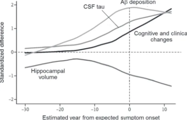
morphological changes have been detected 25 years before the onset of
symptoms. Bateman et al..
2)recently reported the deposition of A 15 years
before the onset of symptoms, followed by brain amyloidosis, tau protein in
the cerebrospinal fluid (CSF), hippocampal atrophy, and declines in cognitive
and clinical functions (Fig. 1). Thus, A, rather than tau protein, deposition
occurs at the early stages in the pathogenesis of AD.
Figure 1 Comparison of clinical, cognitive, and biochemical changes. Quoted
from Ref. 2 with modifications. The vertical axis shows normalized differences between AD mutation carriers and non-carriers.
2
Amyloid
-protein
The amyloid cascade hypothesis plays a central role in the pathogenesis
of AD.
3,4)This hypothesis proposes that A aggregates form senile plaques
and induce neuronal cell death. A mainly consists of 40- and 42-mer
proteins (A40 and A42)
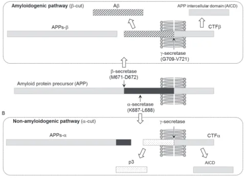
5,6)that are generated from the amyloid protein
precursor (APP) by - and -secretases, respectively (amyloidogenic pathway,
Fig. 2A). -Secretase has been identified as an aspartyl protease of the
pepsin family, called -site APP-cleaving enzyme (BACE-1).
7)BACE-1
cleavage (-cut) generates a membrane-bound APP C-terminal fragment
(CTF), which is a precursor for the cleavage of -secretase (Fig. 2A). Two
presenilin homologues, presenilin 1 (PS1) and presenilin 2 (PS2), have been
shown to play an important role in the activity of -secretase. However,
these presenilins require three other cofactors [nicastrin (Nct), anterior
pharynx-defective phenotype (APH-1), and presenilin-enhancer (PEN-2)] to
form a complex exhibiting -secretase activity.
7)The PS1 complex exhibits
stronger activity than the PS2 complex. Other A species (e.g. 37-, 38-, or
43-mer) are occasionally produced because the sites cleaved by -secretases
at the C-terminal region of APP vary. The potent amyloidogenicity and
pathogenicity of A43 have been rediscovered.
8)3
In contrast, APP is cleaved by -secretase between residues 16 and 17 to
produce APPs- and C83 (-cut), and A40 and A42 are not generated
from these cleaved precursors (non-amyloidogenic pathway, Fig. 2B). APP
processing of -secretase and -secretase produces a smaller fragment,
referred to as p3 (N-terminal of CTF) and APP intracellular domain (AICD).
However, the physiological roles of these APP metabolites have yet to be
elucidated in detail in spite of their ubiquitous expression in almost all human
organs.
9)Although the ratio of A40:A42 is approximately 1:10 under
physiological conditions, the aggregative ability and neurotoxicity of A42
are higher than those of A40. The ratio of A40:A42 has been shown to
increase in patients with familial AD, the onset of which is 10~20 years
earlier than sporadic AD, and approximately 24 mutations for APP and 199
mutations for presenilin have been identified to date.
10)These findings
suggest that A42 plays a more pivotal role in the pathogenesis of AD.
4)The mechanism of A42 fibril formation can be explained by a
nucleation-dependent polymerization model that consists of (1) a nucleation
phase and (2) extension phase.
11,12)In the nucleation phase,
thermodynamically unstable nuclei serve as a rate-limiting step. Binding of
monomers to such nuclei facilitates the formation of thermodynamically
stable oligomers and fibrils.
13)Amyloid oligomer hypothesis
Insoluble A fibrils had previously been considered to cause neuronal
death.
14)However, Walsh et al. demonstrated that the soluble A
oligomeric assembly, but not insoluble fibrils, was responsible for the
pathology of AD, such as memory loss and neurodegeneration.
13,15,16)A
oligomers are defined as soluble assemblies of A that can induce
synaptotoxicity and memory decline. Many types of A oligomers have
been identified, and at least two pathways of assembly
4,17)have been shown
to form insoluble protofibrils and fibrils (‘on-pathway’, Fig. 3) or
high-molecular weight oligomers, such as A*56 (12-mer), amylospheroids
(150-700 kDa), and A-derived diffusible ligands (ADDLs: ~90 kDa)
(‘off-pathway’, Fig. 3), which are stable and do not form fibrils.
13)These
high-molecular weight oligomers cause synaptic dysfunction and
excitotoxicity by blocking long-term potentiation (LTP) and facilitating
4
long-term depression (LTD), in spite of differences in their structure, stability,
and concentration.
4)Furthermore, A oligomers could accelerate the
phosphorylation of the tau protein to reduce microtubule-binding capacity
and form NFTs.
3)A oligomers have been shown to interact with membrane lipids and
proteins.
18)A is known to induce membrane-related toxicity by
influencing the fluidity of the lipid bilayer, perturbing the structure and
function of the plasma membrane, and promoting the release of lipids such as
cholesterol from neuronal membranes, which disrupts neuronal lipid
homeostasis. Furthermore, A can bind to some membrane proteins; the A
monomer was shown to bind to the insulin receptor expressed in neurons and
glial cells, thereby impairing the utilization of glucose, and A assemblies
interacted with N-methyl-
D-aspartate (NMDA)-receptors, which are
expressed in neurons, to induce internalization signals.
18)Figure 3 Scheme of A aggregation from oligomers to fibrils, showing both the
5
Oxidative stress via A
Oxidative stress is one of the major contributing factors to the
progression of AD.
19)Extensive oxidative damage, including protein
oxidation, lipid peroxidation, DNA damage, and mitochondrial dysfunction,
has been reported in the AD brain (Fig. 4A).
20)A-induced neurotoxicity
has been shown to play a role in this oxidative damage through protein
radicalization both in vitro
21,22)and in vivo.
23,24)Transition metals, such as Cu(II), Zn(II), and Fe(III), are known to be
enriched in senile plaques. Cu(II) has been shown to induce the
radicalization of A as well as generic proteins. The histidine residues at
positions 6, 13, and 14, tyrosine residue at position 10 (Tyr10), and aspartic
acid residues at positions 1 and 7 in A are involved in the formation of a
complex with Cu(II) (Fig. 4B, C).
25-27)Tyr10, in particular, is converted to a
phenoxy radical through the reduction of Cu(II) to Cu(I), leading to the
production of H
2O
2(Fig. 4A).
28,29)Figure 4 (A) Cu(II) induces Aβ radicalization, which leads to the production of
reactive oxygen species (ROS).29) (B,C) The proposed binding model
of Cu(II) with (B) His6,13,14 and Tyr10 in A1-2826) or (C) His13,14,
6
Met35 is another residue that plays a role in A-induced neurotoxicity
and oxidative stress.
21)The oxidative form (sulfoxide) of Met35 has been
detected in the brains of AD patients.
30)This finding indicates that the
S-oxidized radical cation can abstract an allylic hydrogen from the
phospholipid acyl chain to generate allyl radicals, which is followed by the
subsequent formation of lipid peroxides (Fig. 5A).
31)Pal et al.
32)reported
that neurodegeneration is aggravated in knockout mice deficient in a
methionine sulfoxide reductase isoform, suggesting the role of Met35
oxidation in the brain pathology of AD.
The S-oxidized radical cation is generally considered too unstable to
cause continuous oxidative damage. Murakami et al.
22)recently reported
the mechanism underlying A42-induced neurotoxicity based on how the
S-oxidized radical cation is stabilized for long-lasting oxidative stress. In
this mechanism, the turn structure at Glu22 and Asp23 in A42 moves the
phenoxy radical at Tyr10 closer to Met35, generating S-oxidized radical
cations
(Fig. 5B). Moreover, another C-terminal turn at Gly38 and Val39
could enable the carboxylate anion at Ala42 to interact with the S-oxidized
radical cation to form S-O bonding, which can stabilize the radical cation.
The resultant hydrophobic core in the C-terminal region may accelerate A
aggregation concomitantly with this stabilization.
Figure 5 (A) Methionine oxidation and subsequent radical formation. Quoted
from Ref. 31 with modifications. (B) The mechanism of stabilization of S-oxidized radical cation in Met35.
7
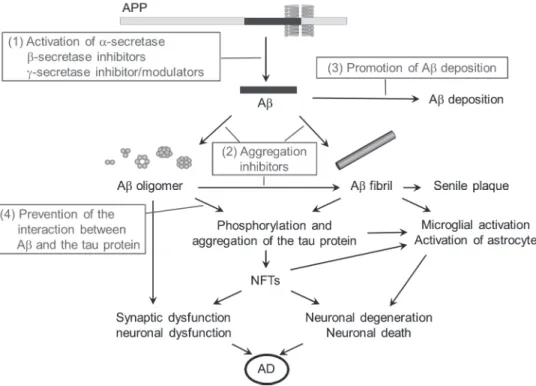
Therapeutic strategies targeting A
Several therapeutic agents against A have been developed based on the
amyloid oligomer hypothesis
33,34)to (1) decrease A production, (2) inhibit
A aggregation, (3) enhance A degradation, and (4) prevent the interaction
between A and the tau protein, and these have been referred to as
disease-modifying agents (Fig. 6). On the other hand, symptom-alleviating
agents are focused on improving neuronal function and protection. For
example, the administration of acetylcholinesterase (AChE) and NMDA
antagonists to improve cognitive function are widely accepted as the current
standard treatment for AD worldwide. Although the AChE inhibitor,
donepezil,
35)is available as a symptom-alleviating agent even in Japan, its
effectiveness is limited and is not considered to be disease-modifying.
35-37)Figure 6 A-associated AD pathogenesis (black letters) and various therapeutic
8
Several disease-modifying agents have been developed and advanced to
clinical trials in recent years.
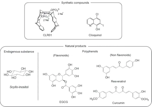
38)McLaurin et al.
39)demonstrated that
scyllo-inositol (Fig. 7), an endogenous stereoisomer of inositol in humans,
inhibited the aggregation of A42 by preventing its transformation into
-sheets. Fibril formation and cytotoxicity in differentiated-PC12 cells
were also prevented. The administration of scyllo-inositol to a transgenic
mouse model of AD (TgCRND8 line) decreased plaque deposition, amount
of amyloid in the brain, and synaptic dysfunction.
40)The presence of
scyllo-inositol was also detected in the brain using gas chromatography-mass
spectrometry.
41)Scyllo-inositol is currently undergoing clinical trials due to
its favorable pharmacokinetics, safety, and tolerability (Phase II).
Synthetic inhibitors that limit A42 aggregation have been reported.
Because Lys residues play an important role in the assembly and toxicity of
A42 due to their hydrophobicity and the formation of electrostatic
interactions, molecular tweezer CLR01 was designed to bind selectively to
Lys residues in A42 (Fig. 7).
42,43)CLR01 modified these properties of Lys
residues by pinching the whole side chain of Lys and forming the salt bridge
between the phosphonate of CLR01 and amino group of Lys residues.
CLR01 was also shown to protect primary neurons from A42-induced
synaptotoxicity and ameliorate the accumulation of A and the tau protein in
a transgenic mouse model of AD (3xTg AD line).
44)Metal ion-induced radical formation and aggregation are considered to be
targets for anti-A aggregation. PBT2, an 8-hydroxy quinolone (clioquinol,
Fig. 7) analog, was designed based on the criteria of easier chemical
synthesis, higher solubility, and blood-brain barrier permeability. PBT2
prevented the metal-induced aggregation of A42 and generation of reactive
oxygen species in vitro.
45)PBT2 was recently shown to reverse frontal lobe
dysfunction and reduced A42 levels in the CSF in a clinical trial (phase
IIa).
46)PBT2 may inhibit radical formation of A42 through the chelation
of extracellular Cu(II) and Zn(II), resulting in the clearance of these ions.
45)9
Polyphenols as anti-A
42 aggregative agents
AD is considered to be a lifestyle-related disease; therefore, the treatment
or prevention of AD with dietary components has received a lot of
attention.
12,47,48)Many studies have demonstrated the protective effects of
various polyphenols derived from food products (e.g. turmeric, red wine, and
green tea) against A aggregation and neurotoxicity (Fig. 7).
49-51)Curcumin is the main constituent of the spice turmeric, the daily intake of
which has been attributed to the lower incidence of AD in India.
52,53)Curcumin exhibited a potent anti-amyloidogenic effect against A in vitro,
such as fibril formation, the destabilization of preformed fibrils, and oligomer
formation in vitro,
54)and was also shown to preventively or therapeutically
reduce amyloid plaques in vivo.
55)A previous study demonstrated that
curcumin protected against A-induced neurotoxicity in PC12 cells.
56)The
binding of curcumin with A is due to hydrophobicity (- stacking) deduced
from the benzene ring.
57)10
Epidemiological studies have suggested that the moderate consumption of
red wine may protect against the development of AD.
58)Resveratrol, a
component in red wine, prevented A oligomer-induced neurotoxicity by
remodeling toxic oligomers into non-toxic conformers.
59,60)It also lowered
A levels in AD model mice by promoting the non-amyloidogenic processing
of APP through the enhanced activity of -secretase.
61)(–)-Epigallocatechin-3-gallate (EGCG), one of the main flavonoids in
green tea, inhibited A42 aggregation
49)by stabilizing A42 as a non-toxic
assembly.
62)Furthermore, EGCG protected hippocampal cells (primary
cultured neurons from Sprague-Dawley rats) from A-induced cell death,
63)and the in vivo administration of EGCG decreased A levels and plaque
formation by increasing the activity of -secretase.
64)Flavonoids are polyphenols that are commonly found in the human diet
and plants. Many studies have examined the ability of flavonoids to reduce
the production of A production either by enhancing the activity of
-secretase or inhibiting that of -secretase
51), to inhibit A aggregation
49,65),
and to enhance A clearance
51).
Although curcumin, resveratrol, and EGCG are currently undergoing
clinical or preclinical trials for the treatment of AD, most trials have reported
conflicting results.
47,66)No significant difference was observed in
Mini-Mental State Examination (MMSE; the most commonly used test for
complaints of memory problems) scores or in A plasma levels in patients
treated with 1 or 3 g curcumin per day for 6 months.
67)Clinical trials using
resveratrol and EGCG are currently being conducted. The above findings
for curcumin motivated us to verify the interaction between polyphenols and
A so that polyphenols could be developed as therapeutic agents for AD.
Regarding the molecular interaction between A and flavonoids, a docking
simulation by Keshet et al.
68)predicted the involvement of Lys28 and the
C-terminal region in the binding with myricetin. However, the precise
mode of binding with flavonoids has not been elucidated in detail, except for
limited studies using NMR spectroscopy [curcumin
69), EGCG
70,71), and
myricetin
72)].
11
Objectives
Silymarin, a seed extract of Silybum marianum that contains
flavonolignane diastereomers,
73)has been used as an anti-hepatotoxic
medicine without notable adverse effects,
74)and is particularly efficacious
against the damage induced by alcohol and disturbances in the function of the
gastrointestinal tract.
75)Murata et al.
76)recently demonstrated that
silymarin attenuated AD-like pathological features, such as senile plaques,
neuronal inflammation, behavioral dysfunction, and A oligomer formation
in a well-established AD mouse model (J20 line).
The purpose of this study is to clarify the inhibitory mechanism for A42
aggregation by polyphenols. Concretely speaking, (+)-taxifolin was
identified as one of the active components among six flavonoids isolated
from silymarin. Structure-activity relationship studies were performed
using methylated (+)-taxifolin and taxifolin enantiomers to clarify the
structure of (+)-taxifolin required for the anti-aggregative properties of A42
(Chapter 2). In Chapter 3, the involvement of autoxidation in the inhibitory
ability of (+)-taxifolin against A42 aggregation in the presence of an oxidant
or under anaerobic conditions was estimated using Th-T, UV spectrometry,
and CD spectrometry. Based on these results, together with other
catechol-type flavonoids, an inhibitory mechanism was proposed for A42
aggregation by catechol-type flavonoids. The mechanisms by which
(+)-taxifolin as a catechol-type polyphenol and curcumin as a
non-catechol-type one interacted with the -sheet region through / stacking
and hydrophobicity were compared using solid-state NMR experiments
(Chapter 4).
13
Chapter 2
Structure-activity relationship studies of (+)-taxifolin
isolated from silymarin as an inhibitor of
A42 aggregation*
Introduction
As mentioned in Chapter 1, Murata et al.
76)reported that silymarin
suppressed the aggregation and the neurotoxicity in PC12 cells of A42 in
vitro, and then they treated J20 mice with a powdered diet containing 0.1%
silymarin for 6 months. Silymarin rescued senile plaques and the amount of
A oligomers in the mouse brain as well as the behavioral dysfunction, but
almost no significant changes in BACE-1 activity was observed. These
results suggest that the protective effect of silymarin on A accumulation
could be originated from the inhibition of A aggregation, rather than the
attenuation of A production. Although silymarin is known as a mixture of
flavonolignan diastereomers,
73)such as silibinin, silydianin, and silychristin,
the effect of these components on A42 aggregation has not yet been
examined.
In this Chapter, the author screened the compounds focusing on the
flavonoids from silymarin with an anti-aggregative ability against A42, and
then examined the structure-activity relationships of the compound to
investigate its essential moiety required for the inhibition of A42
aggregation.
*The content described in this Chapter was originally published in Bioscience,
Biotechnology and Biochemistry. Mizuho Sato, Kazuma Murakami, Mayumi Uno,
Haruko Ikubo, Yu Nakagawa, Sumie Katayama, Ken-ichi Akagi and Kazuhiro Irie: Structure-activity relationship for (+)-taxifolin isolated from silymarin as an inhibitor of amyloid aggregation. Biosci. Biotechnol. Biochem. 2013; 77: 1100-1103 © The Japan Society for Bioscience, Biotechnology, and Agrochemistry.
14
Results
Isolation of (+)-taxifolin as an active component in silymarin
Silymarin was fractionated by column chromatography followed by high
performance liquid chromatography (HPLC) to yield six kind of flavonoids;
silibinin A (isolated yield 6.7%),
77)silibinin B (isolated yield 10%),
77)silydianin (isolated yield 8.4%),
78)(+)-taxifolin (isolated yield 2.2%),
79)isosilychristin (isolated yield 1.3%),
80)and silychristin (isolated yield
8.4%)
81)(Fig. 8A). Effects of these flavonoids on A42 aggregation were
examined using the assay of thioflavin-T (Th-T), a regent that fluoresces
when bound to A aggregates, and using transmission electron microscopy
(TEM), as previously described.
82,83)Only (+)-taxifolin, a catechol-type
flavonoid, among the isolated flavonoids significantly reduced the Th-T
fluorescence induced by A42, meaning the potent inhibition of A42
aggregation by (+)-taxifolin (Fig. 8B). In the analysis of TEM, A42
formed the typical fibril, but shorter or slighter fibrils were observed in the
case of A42 incubated with (+)-taxifolin (Fig. 9C). These inhibitory
effects of (+)-taxifolin on A42 aggregation were almost equal to that of
silymarin, indicating the (+)-taxifolin as one of the active components (Fig.
8B). Moreover, (+)-taxifolin disaggregated the preformed fibrils of A42
(Fig. 8C).
To test the effect on the neurotoxicity of A42, the author carried out the
MTT assay using PC12 cells, which are sensitive to A peptide and are
generally used for detecting cytotoxicity as a neurotoxicity model.
84)(+)-Taxifolin was neuroprotective in a dose-dependent manner (Fig. 8D).
Structure-activity relationship of (+)-taxifolin for anti-A
42 aggregation
To identify which hydroxyl groups of (+)-taxifolin are involved in the
inhibitory effect, four O-methyl derivatives of (+)-taxifolin were prepared by
diazomethane treatment (Scheme 1A). In the Th-T assay,
(+)-7-O-methyl-taxifolin prevented the aggregation of A42 in a manner
similar to (+)-taxifolin, whereas (+)-7,3'-di-O-methyl-, (+)-7,4'-di-O-methyl-,
and (+)-7,3',4'-tri-O-methyltaxifolin did not (Fig. 9A). In TEM images,
A42 fibrils treated with (+)-7-O-methyl-taxifolin, but not with
(+)-7,3'-di-O-methyl-taxifolin, were similar to those treated with (+)-taxifolin
(Fig. 9C). These results indicate 3',4'-dihydroxyl groups (catechol structure)
15
on the B-ring of (+)-taxifolin to be important to prevent A42 aggregation,
whereas the 7-hydroxyl group is less.
Figure 8 (A) Structure of the flavonoids isolated from silymarin. (B) The effect
of each flavonoid on A42 aggregation was estimated by the Th-T method. A42 (25 M) was incubated with or without each flavonoid (50 M) in PBS (50 mM sodium phosphate, 100 mM NaCl, pH 7.4) at 37
oC. ◆, A42 without flavonoid; ▲, A42 with (+)-taxifolin; ●,
A42 with silymarin; ◇, A42 with silibinin A; △, A42 with silibinin B; □, A42 with silydianin; ○, A42 with isosilychristin and ×, A42 with silychristin. The molecular weight of silymarin was defined as 482, which was that of the main components (silibinin A and B, silydianin, isosilychristin, and silychristin) in silymarin. Data are presented as the mean ± s.e.m. (n = 8). (C) The effect of (+)-taxifolin on the preformed fibrils of A42. ◆, A42 fibril (56.4 g/500 L) without flavonoid and ▲ , A42 fibril (56.4 g/500 L) with (+)-taxifolin (50 M); (D) The effect of (+)-taxifolin on A42-induced neurotoxicity on PC12 cells using MTT assay.
16
Scheme 1 (A) Methylation of (+)-taxifolin. (B) Synthesis (+)- and (–)-taxifolin
17
(+)-Taxifolin was not methylated at position 5 on the A-ring by
diazomethane, implying that the hydroxyl group at position 5 could not be
involved in the intermolecular interaction. Indeed, the hydroxyl group at
position 5 of (+)-taxifolin could participate in the intramolecular hydrogen
bond with the carbonyl oxygen on the C-ring, which was deduced from the
1H NMR chemical shift [11.7 ppm in (CD
3)
2CO]. The practical implication
Figure 9 (A) The inhibitory effects of methylated (+)-taxifolins on A42
aggregation determined by the Th-T assay. A42 (25 M) was
incubated with or without each flavonoid (50 M) in PBS at 37 oC.
◆, A42 without flavonoid; ▲, A42 with (+)-taxifolin; ■, A42 with (+)-7-O-methyl-taxifolin; ◇, A42 with (+)-7,3'-di-O-methyl- taxifolin; △, A42 with (+)-7,4'-di-O-methyl-taxifolin and □, A42 with (+)-7,3',4'-tri-O-methyl-taxifolin. Data are presented as the mean ± s.e.m. (n = 8). (B) The effects of the stereochemistry on the C-ring in (+)-taxifolin on A42 aggregation were evaluated by the Th-T test. ◆, A42 (25 M) without flavonoid; ▲, A42 (25 M) with (+)-taxifolin and △, A42 with (–)-taxifolin (50 M). (C) The TEM analysis of A42 fibrils treated with the (+)-taxifolin derivatives. A42 (25 M) was incubated with or without each flavonoid (50 M) in PBS at 37 oC for 48 h. Scale bar, 200 nm.
18
of this result is that the hydroxyl group at position 5 does not contribute to
the inhibition of A42 aggregation by (+)-taxifolin. Although the
methylated (+)-taxifolin at position 3 on the C-ring was not also obtained, the
report
86)that luteolin without a hydroxyl group at position 3 inhibited A42
aggregation means that the hydroxyl group at position 3 of (+)-taxifolin
would not participate in the inhibitory activity.
Finally, (
–)-taxifolin, 2,3-(S,S)-trans form, was synthesized to examine
the effect of the stereochemistry at position 2 and 3 on the C-ring of
(+)-taxifolin, 2,3-(R,R)-trans form, on the inhibition of A42 aggregation
(Scheme 1B). The inhibitory ability against A42 aggregation was almost
the same between these two enantiomers. Regarding the slight difference of
the inhibition of A42 aggregation by (+)-taxifolin between Figs. 8 and 9, the
fluorescence intensity in the Th-T assay are sometimes influenced by several
factors, for example, outside temperature, batch of A42, and so on.
However, similar results were obtained by another independent experiment.
Several groups reported no difference of the inhibitory activities against
A40 or A42 aggregation between (+)-catechin (2,3-trans form, Fig. 10)
and (–)-epicatechin (2,3-cis form, Fig. 10).
87)Furthermore, myricetin and
quercetin (Fig. 10) with a double bond between C2 and C3 on the C-ring
were previously reported to inhibit A42 aggregation.
12)These findings did
not contradict the results obtained in Fig. 9B; the stereochemistry at positions
2 and 3 not to play an important role in the inhibitory effects of (+)-taxifolin
against A42 aggregation.
19
Discussion
There were some reports on the preventive role of silymarin in the
inflammation and grail activation by inhibiting the production of
inflammatory factors such as NF-B, TNF-, iNOS and COX-2, leading to
neuroprotection.
88)The anti-neuroinflammation by silymarin observed in
the previous studies
76)might be associated with these benefits by silymarin.
We identified (+)-taxifolin as one of the active components with
anti-aggregative ability against A42 from silymarin, and this is the first
report that (+)-taxifolin has a preventive effect on A42 aggregation. The
quantification analysis using HPLC revealed that silymarin contained 1.2%
(+)-taxifolin, which was slightly less than the isolated yield (2.2%, purity
99.6%). The low content rate of (+)-taxifolin does not exclude the existence
of other active components in silymarin.
The structure-activity relationship studies of (+)-taxifolin clarified the
requisite moiety (catechol structure on B-ring) for inhibitory activity against
A42 aggregation, which is consistent with the findings that only
(+)-taxifolin has a catechol moiety among the flavonoids isolated from
silymarin in this study. These findings do not contradict the report by
Akaishi et al.
86)that the 3',4'-dihydroxyl group, not the 7-hydroxyl group, is
essential to the inhibitory effect of fisetin (quercetin analog without
5-hydroxyl group) on A42 fibril formation. A detailed inhibitory
mechanism for A42 aggregation by such a catechol-type flavonoid will be
discussed in the following Chapters.
20
Experimental procedure
General remarks
The following spectroscopic and analytical instruments were used: 1H NMR,
AVANCE III 500 and AVANCE III 400 (Bruker, Germany); EI-MS, JMS-600H (70 eV, 300 A; JEOL, Tokyo, Japan); polarimeter, P-2200 (Jasco, Tokyo, Japan); matrix-assisted laser desorption/ionization time-of-flight mass spectrometry (MALDI-TOF-MS), AXIMA-CFR (Shimadzu, Kyoto, Japan); high performance liquid chromatography (HPLC), Waters 600E multisolvent delivery system with 2487 UV dual absorbance detector (Milford, MA); transmission electron microscopy (TEM), H-7650 electron microscope (Hitachi, Ibaraki, Japan); micro-plate reader, MPR-A4II, (TOSOH, Tokyo, Japan), or Fluoroskan Ascent
(Thermo Scientific, Rockford, IL); peptide synthesizer, PioneerTM peptide
synthesizer (Applied Biosystems, Foster City, CA).
Following N--(9-fluorenylmethoxycarbonyl) (Fmoc) amino acid were used for peptide synthesis: Ala-OH, Asn(Trt)-OH, Asp(OtBu)-OH, Arg(Pbf)-OH, Gln(Trt)-OH, Glu(OtBu)-OH, Gly-OH, His(Trt)-OH, Ile-OH, Leu-OH, Lys(Boc)-OH, Met-OH, Phe-OH, Ser(tBu)-OH, Tyr(tBu)-OH, Val-OH (Applied Biosystems). 1-[bis(dimethylamino)methylene]-1H-1,2,3-triazolo[4,5-b]
pyridinium 3-oxid hexafluorophosphate (HATU), Fmoc-L-Ala-polyethylene
glycol-polystylene support (PEG-PS) resin, and N,N-diisopropylethylamine (DIPEA) were purchased from Applied biosystems. N,N-Dimethylformamide (DMF), trifluoroacetic acid (TFA), 1,2-ethanedithiol, thioanisole, m-cresol, and diethyl ether (peroxide free) were obtained from Nacalai tesque (Kyoto, Japan).
Separation of silymarin
Silymarin (lot no. 228-216, LKT Laboratories, St. Paul, MN) was fractionated
by column chromatography eluted with 5.0% MeOH/CHCl3 on Wakogel C-200
(Wako, Osaka, Japan) to give two major fractions containing flavonoids. The first fraction was chromatographed by HPLC on a YMC-Pack ODS-A column (20 mm
i.d. x 150 mm; YMC, Kyoto, Japan) using 50% MeOH/H2O to yield silibinin A (16
mg from 240 mg of silymarin)77) and silibinin B (25 mg from 240 mg of
silymarin),77) and using 40% MeOH/H
2O to yield silydianin (31 mg from 370 mg of
silymarin),78) respectively. The second fraction was separated on a YMC-Pack
ODS-AL column (20 mm i.d. x 150 mm; YMC) using 40% MeOH/H2O to give
(+)-taxifolin (6.7 mg from 310 mg of silymarin),79) isosilychristin (3.9 mg from 310
mg of silymarin),80) and silychristin (26 mg from 310 mg of silymarin)81) (Fig. 8A).
The structures of these compounds were confirmed by 1H NMR77-81), EI-MS, and
specific optical rotation (P-2200).
Silibinin A: 1H NMR (500 MHz, 295.0 K, acetone-d 6, 31.9 mM, ref. tetramethylsilane) 3.52 (1H, dd, J = 12.4, 4.3 Hz), 3.75 (1H, dd, J = 12.4, 2.5 Hz), 3.87 (3H, s), 4.16 (1H, ddd, J = 7.0, 4.3, 2.5 Hz), 4.64 (1H, d, J = 11.5 Hz), 5.00 (1H, d, J = 8.0 Hz), 5.07 (1H, d, J = 11.5 Hz), 5.94 (1H, d, J = 2.1 Hz), 5.97 (1H, d, J = 2.1 Hz), 6.89 (1H, d, J = 8.1 Hz), 6.96 (1H, d, J = 8.3 Hz), 6.99 (1H, dd, J = 8.1, 1.9
21
Hz), 7.09 (1H, dd, J = 8.3, 2.0 Hz), 7.15 (1H, d, J = 1.9 Hz), 7.16 (1H, d, J = 2.0 Hz).
EI-MS: m/z, calcd: 482, found: 482 [M]+). []
D +26.0o (c 0.25, MeOH, 25.6 oC). Silibinin B: 1H NMR (500 MHz, 295.0 K, acetone-d 6, 26.9 mM, ref. tetramethylsilane) 3.52 (1H, dd, J = 12.4, 4.2 Hz), 3.75 (1H, dd, J = 12.4, 2.5 Hz), 3.88 (3H, s), 4.15 (1H, ddd, J = 8.0, 4.2, 2.5 Hz), 4.65 (1H, d, J = 11.5 Hz), 5.00 (1H, d, J = 8.0 Hz), 5.09 (1H, d, J = 11.5 Hz), 5.96 (1H, d, J = 2.0 Hz), 5.99 (1H, d, J = 2.0 Hz), 6.88 (1H, d, J = 8.1 Hz), 6.97 (1H, d, J = 8.3 Hz), 6.99 (1H, dd, J = 8.1, 1.9 Hz), 7.10 (1H, dd, J = 8.3, 2.0 Hz), 7.15 (2H, t, J = 2.0 Hz). EI-MS: m/z, calcd: 482, found: 482 [M]+). [] D +12.0o (c 0.19, MeOH, 25.9 oC). Silydianin: 1H NMR (500 MHz, 295.0 K, acetone-d 6, 41.5 mM, ref. tetramethylsilane) 2.93 (1H, brs), 3.28 (1H, dd, J = 6.8, 2.9 Hz), 3.41 (1H, brs), 3.67 (1H, dd, J = 4.1, 2.1 Hz), 3.88 (1H, d, J = 7.8 Hz), 4.29 (1H, dd, J = 7.8, 3.2 Hz), 4.65 (1H, d, J = 11.3 Hz), 4.92 (1H, d, J = 11.3 Hz), 6.00 (2H, s), 6.20 (1H, dd,
J = 6.9, 1.4 Hz), 6.76 (2H, s), 6.89 (1H, s). EI-MS: m/z, calcd: 482, found: 482
[M]+). [] D +230o (c 0.005, MeOH, 26.8 oC). (+)-Taxifolin: 1H NMR (400 MHz, 296.9 K, acetone-d 6, 3.94 mM, ref. tetramethylsilane) 4.58 (1H, d, J = 11.5 Hz), 4.99 (1H, d, J = 11.5 Hz), 5.92 (1H, d, J = 2.1 Hz), 5.96 (1H, d, J = 2.1 Hz), 6.84 (1H, d, J = 8.1 Hz), 6.90 (1H, dd, J = 8.1,
1.9 Hz), 7.05 (1H, d, J = 1.9 Hz), 11.7 (1H, brs). EI-MS: m/z, calcd: 304, found: 304 [M]+). [] D +22.2o (c 0.12, MeOH, 29.1 oC). Silychristin: 1H NMR (500 MHz, 295.0 K, acetone-d 6, 41.5 mM, ref. tetramethylsilane) 3.61 (1H, ddd, J = 6.9, 6.8, 5.5 Hz), 3.84 (3H, s), 3.86 (1H, dd, J = 10.8, 6.8 Hz), 3.92 (1H, dd, J = 10.8, 5.5 Hz), 4.63 (1H, d, J = 11.6 Hz), 5.04 (1H, d, J = 11.6 Hz), 5.58 (1H, d, J = 6.9 Hz), 5.95 (1H, d, J = 2.0 Hz), 5.99 (1H, d, J = 2.0 Hz), 6.83 (d, 1H, J = 8.1 Hz), 6.93 (1H, dd, J = 8.1, 1.9 Hz), 6.99 (1H, brd, J
= 1.1 Hz), 7.02 (1H, brs), 7.10 (1H, d, J = 1.9 Hz). EI-MS: m/z, calcd: 482, found: 482 [M]+). [] D +112o (c 0.30, MeOH, 26.3 oC). Isosilychristin: 1H NMR (500 MHz, 295.0 K, acetone-d 6, 7.05 mM, ref. tetramethylsilane) 3.66 (1H, dd, J = 10.8, 8.9 Hz), 3.76 (3H, s), 3.86 (1H, ddd, J = 8.8, 4.5, 3.0 Hz) 3.96 (1H, dd, J = 10.8, 4.5 Hz), 4.64 (1H, d, J = 11.7 Hz), 5.24 (1H, d, J = 11.7 Hz), 5.68 (1H, d, J = 3.0 Hz), 5.90 (1H, d, J = 2.1 Hz), 5.93 (1H, d, J = 2.1 Hz), 6.75 (1H, d, J = 8.1 Hz), 6.84 (1H, d, J = 8.4 Hz), 6.89 (1H, dd, J = 8.1, 2.0 Hz), 6.99 (1H, d, J = 2.0 Hz), 7.08 (2H, d, J = 8.4 Hz). EI-MS: m/z, calcd: 482, found: 482 [M]+). [] D +206o (c 0.005, MeOH, 26.4 oC). Methylation of (+)-taxifolin
N-Methyl–N-nitroso-p-toluenesulfonamide dissolved in ether/EtOH (35 mL/15
mL, 0.20 mM) was heated at 70 oC. To the solution was added a potassium
hydroxide solution (one gram of potassium hydroxide in 15 mL of water) to yield diazomethane, which was condensed in a cold tube as a yellow ether solution. (+)-Taxifolin (65 mg, 0.21 mmol) was dispersed in benzene (1.0 mL) and diethylether (3.0 mL), to which an aliquot of the diazomethane solution (12 mL) was
added at 0 oC, and the mixture was stood at the same temperature for 1.5 h. The
solution was evaporated in vacuo, and the residue was separated by preparative thin layer chromatography, followed by HPLC on a YMC-Pack ODS-A column (20 mm
22
i.d. x 150 mm; YMC) with a linear gradient of 50-100% CH3CN/H2O for 30 min to
yield (+)-7-O-methyl- (18 mg, 28% yield), (+)-7,3'-di-O-methyl- (6.3 mg, 9.7% yield), (+)-7,4’-di-O-methyl- (7.3 mg, 11% yield), and (+)-7,3',4'-tri-O-methyl-taxifolin (2.1 mg, 3.2% yield, Scheme 1A). Their
structures were confirmed by 1H NMR79) and EI-MS to be identical to those reported
previously.
(+)-7-O-Methyl-taxifolin: 1H NMR (400 MHz, 296.8 K, acetone-d
6, 16.3 mM,
ref. tetramethylsilane) 3.85 (3H, s),4.64 (1H, d, J = 11.5 Hz), 5.04 (1H, d, J = 11.4 Hz), 6.04 (1H, d, J = 2.2 Hz), 6.07 (1H, d, J = 2.3 Hz), 6.85 (1H, d, J = 8.1 Hz), 6.90 (1H, d, J = 8.0 Hz), 7.05 (1H, brs). EI-MS: m/z, calcd: 318, found: 318 [M]+.
(+)-7,3'-O-Methyl-taxifolin: 1H-NMR (400 MHz, 296.8 K, acetone-d 6, 16.3 mM, ref. tetramethylsilane) 3.85 (3H, s),3.87 (3H, s), 4.72 (1H, d, J = 11.8 Hz), 5.10 (1H, d, J = 11.8 Hz), 6.04 (1H, d, J = 2.3 Hz), 6.07 (1H, d, J = 2.3 Hz), 6.86 (1H, d, J = 8.1 Hz), 7.03 (1H, dd, J = 8.2 , 2.0 Hz), 7.21 (1H, d, J = 2.0 Hz). EI-MS: m/z, calcd: 332, found: 332 [M]+. (+)-7,4'-O-Methyl-taxifolin: 1H NMR (400 MHz, 296.8 K, acetone-d 6, 9.94 mM, ref. tetramethylsilane) 3.85 (3H, s),3.87 (3H, s), 4.66 (1H, dd, J = 11.5, 3.1 Hz) , 4.75 (1H, brd, J = 3.7 Hz), 5.09 (1H, d, J = 11.5 Hz), 6.05 (1H, d, J = 2.2 Hz), 6.07 (1H, d, J = 2.2 Hz), 6.97 (1H, d, J = 8.2 Hz), 7.00 (1H, dd, J = 8.3, 1.8 Hz),
7.08 (1H, d, J = 1.9 Hz). EI-MS: m/z, calcd: 332, found: 332 [M]+.
(+)-7,3',4'-O-Methyl-taxifolin: 1H NMR (400 MHz, 296.8 K, acetone-d
6, 4.04 mM, ref. tetramethylsilane) 3.83 (3H, s), 3.84 (3H, s),3.86 (3H, s), 4.73-4.76 (1H, m), 5.13 (1H, d, J = 11.1 Hz), 6.05 (1H, d, J = 2.2 Hz), 6.08 (1H, d, J = 2.2 Hz), 6.98 (1H, d, J = 8.3 Hz), 7.10 (1H, dd, J = 8.3, 2.0 Hz), 7.22 (1H, d, J = 2.0 Hz). EI-MS: m/z, calcd: 346, found: 346 [M]+.
Synthesis of (–)-taxifolin
(–)-Taxifolin, 2,3-(S,S)-trans form, was synthesized basically according to the
method of Roschek et al.,85) except for the use of 3,4-trihydroxybenzaldehyde as a
substrate. Briefly, vanillin (0.10 g, 0.63 mmol) dissolved in CH2Cl2 was
demethylated by treatment with 1 M boron tribromide in dichloromethane (2.6 mL,
2.6 mmol) at 4 oC for 1 h to give quantitatively 3,4-dihydroxybenzaldehyde. The
phenolic hydroxyl groups were protected with methoxymethyl groups (73% yield). On the other hand, the phenolic hydroxyl groups of 2,4,6-trihydroxyacetophenone were also protected with methoxymethyl groups (24% yield). A cross-aldol reaction of these two products in KOH/MeOH (83% yield), followed by treatment
with H2O2 under alkaline condition, yielding quantitatively the epoxide, which was
cyclized and deprotected under HCl/MeOH to give (±)-taxifolin (59% yield). Enantiomers were separated by HPLC on a CHIRALCEL OJ-RH column (10 mm
i.d. x 150 mm; Daicel Corporation, Osaka, Japan) using 15% CH3CN/H2O
containing 0.1% acetic acid.89) The ratio of enantiomers was almost 1 : 1, based on the isolated yield of each enantiomer:
(+)-taxifolin: 1H NMR (400 MHz, 297.1 K, acetone-d
6, 19.7 mM, ref.
tetramethylsilane) 4.60 (1H, d, J = 11.6 Hz), 5.01 (1H, d, J = 11.6 Hz), 5.94 (1H, d,
23 2.0 Hz), 7.06 (1H, d, J = 2.0 Hz). []D +17.3o (c 0.10, MeOH, 16 oC, lit.80) []D +19.0o, c 0.1, MeOH). (–)-taxifolin: 1H NMR (400 MHz, 297.1 K, acetone-d 6, 19.7 mM, ref. tetramethylsilane) 4.61 (1H, d, J = 11.2 Hz), 5.02 (1H, d, J = 11.6 Hz), 5.95 (1H, s), 5.98 (1H, s), 6.86 (d, 1H, J = 8.0 Hz), 6.91 (1H, d, J = 8.0 Hz), 7.07 (1H, s). []D –16.2o (c 0.10, MeOH, 16 oC). Synthesis of A42
The A42 were synthesized in a stepwise fashion on 0.1 mmol of preloaded
Fmoc-L-Ala-PEG-PS resin using a PioneerTM Peptide Synthesizer as reported
previously.90) The Fmoc group was deblocked with 20% piperidine in DMF for 5
min (flow rate; 5.0 mL/min). The coupling reaction was carried out using Fmoc amino acid (0.4 mmol), HATU (0.4 mmo), and DIEPA (0.8 mmol) in DMF for 30 min (flow rate; 30 mL/min). After each coupling reaction, the N-terminal Fmoc group was removed with 20% piperidine in DMF. After the chain elongation was completed, the peptide-resin was treated with a cocktail containing trifluoroacetic acid, m-cresol, thioanisol, and 1,2-ethanedithiol for final deprotection and cleavage from the resin. The crude peptide was precipitated by diethylether and purified by HPLC on Develosil ODS UG-5 column (20 mm i.d. x 150 mm; Nomura Chemical
Co., Seto, Aichi, Japan) using 80 min liner gradient of 10-50% CH3CN/H2O
containing 0.1% NH4OH, described previously.82) After lyophilization, the
corresponding pure A42 peptide was obtained, the purity of which was confirmed by HPLC (>98%). The molecular weight of A42 was confirmed by
MALDI-TOF-MS; A42 (m/z, calcd: 4515.14, found: 4515.25 [M+H]+)
Thioflavin-T fluorescence assay
The aggregative ability of A42 was evaluated at 37 oC by the thioflavin-T
(Th-T) method developed by Naiki et al.91) The procedure was described
elsewhere.82) In brief, A42 was dissolved in 0.1% NH
4OH at 250 M, and each
flavonoid was dissolved in EtOH at 5.0 mM, followed by dilution with sodium phosphate-buffered saline (PBS: 50 mM sodium phosphate and 100 mM NaCl, pH 7.4) at the desired concentration (A42, 25 M; flavonoids, 50 M). After
incubation at 37 oC, 2.5 L of each A42 solution was added to 250 L of 5.0 M
Th-T in 5.0 mM Gly-NaOH (pH 8.5). Fluorescence intensity was measured at 420 nm excitation and 485 nm emission using a micro-plate reader (MPR-A4II or Fluoroskan Ascent).
Transmission electron microscopy (TEM)
The aggregates of A42 after a 48-h incubation were examined under a H-7650
electron microscope. The experimental procedure was described elsewhere.82) In
brief, each Aβ42 solution was prepared as described in Th-T assay and incubated at
37 oC for 48 h. After centrifugation at 4 oC for 10 min, the supernatant was
removed from the pellets, and the aggregates were suspended in distilled water by gentle vortex mixing. The suspensions were applied to a 200-mesh Formvar-coated copper grid (Nissin EM, Tokyo, Japan) and dried in air before being
24
negatively stained for a few seconds with 2% uranyl acetate. The aggregates were examined with the H-7650 transmission electron microscope (Hitachi).
Estimation of cell survival
The reduction of 3-(4,5-dimethylthiazol-2-yl)-2,5-diphenyltetrazolium bromide
(MTT) by mitochondrial reductase was carried out according to Shearman et al.84)
PC12 cells were cultured in Dulbecco's modified Eagle's medium (Sigma) containing 5% fetal calf serum, 10% horse serum, and 1% penicillin/streptomycin. Before using for the experiment, near-confluent culture of the cells were plated at
approximately 104 cells/100 L/well in 96-well tissue culture plate coated with
collagen, and incubated at 37 oC under 5.0% CO
2 overnight. A42 was dissolved
in 0.1% NH4OH at 10 M, and (+)-taxifolin was dissolved in DMSO at 1 mM,
followed by dilution with flesh medium at the desired concentration (A42, 1 M; flavonoids, 10 M). One hundred microlitters of the medium was exchanged with
old one, and then the 96-well plate was incubated at 37 oC under 5.0% CO
2. After 48-h incubation, 10 L of 5 mg/mL MTT in PBS (pH 7.4) was added to the cell
culture, which was incubated at 37 oC under 5.0% CO
2 for 4 h. The culture
medium was removed, and then 10% SDS/0.1 M NH4Cl (100 L) was added to the
cells. After further incubation overnight in the dark at room temperature, the absorbance at 595 nm was measured. The absorbance obtained by addition of vehicle was taken as 100%.
25
Chapter 3
Inhibitory mechanism for A42 aggregation
by catechol-type flavonoids*
Introduction
Many researchers have reported protective effects of various polyphenols
from green tea, turmeric, and red wine etc., against A aggregation and
neurotoxicity.
49-51)Several compounds [e.g. (–)-epigallocatechin-3-gallate
(EGCG), curcumin, and resveratrol] are in clinical or preclinical trials for AD
treatment.
47,66)However, the recent failures of some trials
33)motivated the
author to clarify the mechanism by which polyphenols inhibit the aggregation
of A42 to develop promising leads for clinical use.
As mentioned in Chapter 2, the author isolated (+)-taxifolin,
80)a
flavanonol which has a catechol moiety on the B-ring, as one of the active
components in silymarin against A42 aggregation (Fig. 8 in Chapter 2).
Altough the catechol moiety on B-ring was suggested to be important for the
anti-aggregative ability, the mechanism how the catechol moiety contributes
to the activity is not fully understood. This Chapter describes a
comprehensive study on the role of autoxidation of (+)-taxifolin in the
prevention of aggregation and -sheet transformation of A42, and the
effects of various flavanonols and flavonols on the aggregation of A42
mutants substituted at Arg5, Lys16, and/or Lys28 with norleucine (Nle).
The experiments using liquid chromatography-mass spectrometry (LC-MS)
were also carried out to propose a site-specific inhibitory mechanism for
A42 aggregation by catechol-type flavonoids.
*The content described in this Chapter was originally published in Journal of
Biological Chemistry. Mizuho Sato, Kazuma Murakami, Mayumi Uno, Yu
Nakagawa, Sumie Katayama, Ken-ichi Akagi, Yuichi Masuda, Kiyonori Takegoshi and Kazuhiro Irie: Site-specific inhibitory mechanisms for amyloid-42 aggregation by catechol-type flavonoids targeting the Lys residues. J. Biol. Chem. 2013; 288: 23212-23224. © The American Society for Biochemistry and Molecular Biology.
26
Results
Effects of autoxidation of (+)-taxifolin on its ability to prevent the
aggregation of A
42
A catechol moiety is easily oxidized to form an o-quinone.
92,93)To
investigate the contribution of autoxidation to the inhibitory ability, the
author examined the aggregative ability of A42 in the presence of
(+)-taxifolin treated with sodium periodate (NaIO
4), which is known as a
mild oxidant of catechol but affecting little effect on peptide bond.
94)As
shown in Fig. 11A, NaIO
4extensively promoted the suppressive ability of
(+)-taxifolin compared with (+)-taxifolin alone. These observations were
also confirmed by the TEM experiment (Fig. 12A). NaIO
4treatment in the
presence of (+)-taxifolin formed only shorter and thinner fibrils compared
with (+)-taxifolin alone. A42 formed the typical fibrils even in the
presence of NaIO
4alone, and almost no differences (e.g. length, thickness)
were observed between the morphology in the presence and absence of
NaIO
4(Fig. 12A).
NaIO
4alone slightly affected the Th-T fluorescence of A42 aggregates
(Fig. 11A) possibly because NaIO
4can oxidize Met35 in A42 to its
sulfoxide, the formation of which was confirmed by HPLC (Fig. 11E) and
MALDI-TOF-MS (A42-M35
ox; m/z: calcd: 4531.14, found: 4531.55
[M+H]
+). This is in good agreement with a report that oxidation using
hydrogen peroxide, a strong oxidant, reduced A42 aggregation.
95)However, in the presence of both (+)-taxifolin (50 M) and NaIO
4(100 M),
Met35 was not oxidized by NaIO
4; this was confirmed by HPLC (Fig. 11E)
and MALDI-TOF-MS (A42-M35
red; m/z: calcd: 4515.14, found: 4516.26
[M+H]
+). This indicates that NaIO
4preferred to oxidize (+)-taxifolin more
than the sulfur atom of the Met35 of A42.
In addition, the author tested whether the treatment of NaIO
4leads to the
oxidation of Met35 in the preformed A42 fibrils. The fibrils (ca. 28 g)
treated with NaIO
4for 4 h were dissolved in formic acid (10 L), and were
sonicated for 1 h. After volatilization, the resultant pellets were resolved in
50% acetonitrile containing 0.1% trifluoroacetic acid, followed by subjection
to MALDI-TOF-MS analysis. NaIO
4did not oxidize Met35 in the
preformed A42 fibril (A42-M35
red; m/z: calcd: 4515.14, found: 4515.12
[M+H]
+). Also in Th-T assay, A42 fibril was not disassembled by NaIO
427
(data not shown). These mean that NaIO
4could partially oxidize Met35 in
the monomeric A42, but not the fibrils.
Furthermore, in order to investigate the role of autoxidation, the effect by
(+)-taxifolin on A42 aggregation was tested in vacuo. Notably,
(+)-taxifolin little suppressed the aggregation of A42 under an anaerobic
condition (Fig. 11B). In TEM images, typical fibril formation was observed
even in the presence of (+)-taxifolin under the anaerobic condition (Fig. 12A).
A42 also aggregated in the presence of (+)-taxifolin and
tris(2-carboxy-ethyl)phosphine (TCEP), a reductant (Fig. 11C). These
results strongly suggest the autoxidation of (+)-taxifolin to be required for
inhibitory activity against A42 aggregation.
As mentioned in Chapter 1, the mechanism of A42 fibril formation is
well explained by a nucleation-dependent polymerization model mainly
consisted of nucleation phase and extension phase.
11,12)To determine which
stage (nucleation phase or extension phase) is affected by (+)-taxifolin, the
author examined the effect of (+)-taxifolin on A42 aggregation in the
presence of the fibril seed as a template, according to the protocol developed
by Naiki et al.
91)There was a nucleation phase (~1 h) when A42 was
incubated alone, whereas addition of the seeds skipped the nucleation phase,
resulting in the rapid formation of A42 fibrils (Fig. 11D). In the case of
co-incubation of (+)-taxifolin with the seeds, the nucleation phase of A42
did not drastically change, but the fluorescence gradually decreased after
incubation for 4 h, suggesting that (+)-taxifolin could prevent the elongation
phase (~2 or 4 h) in A42 aggregation, rather than the nucleation phase (~1
h) (Fig. 11A, D). Although the slight difference of the length of elongation
phase between Fig. 11A and 11D might be deduced from several factors, for
example, outside temperature, batch (lot) of A42, 2~4 h as an averaged time
for elongation phase were observed in another independent experiments.
Moreover, (+)-taxifolin destabilized the preformed A42 fibril. The
disappearance of nucleation phase in the presence of seed and NaIO
4(Fig.
11D) implied the ability of oxidized taxifolin to disassemble even the seed.
Indeed, the fluorescence of preformed A42 fibrils immediately disappeared
after addition of (+)-taxifolin treated with NaIO
4(data not shown).
28
Figure 11 (A) The effect of NaIO4, an oxidant, on A42 aggregation estimated by
Th-T tests. A42 (25 M) was incubated with or without (+)-taxifolin
(50 M) and/or NaIO4 (100 M) in PBS (50 mM sodium phosphate,
100 mM NaCl, pH 7.4) at 37 oC. (B) The ability of (+)-taxifolin to
suppress the aggregation of A42 under an anaerobic condition. A42 (25 M) was incubated with or without (+)-taxifolin (50 M) in PBS in
vacuo at room temperature. (C) The effect of TCEP, a reductant, on
A42 aggregation. A42 (25 M) was incubated with or without
(+)-taxioflin (50 M) and/or TCEP (100 M) in PBS at 37 oC. (D)
The aggregative ability of A42 in the presence of A42 seed and/or
(+)-taxifolin, NaIO4. A42 (25 M) was incubated with or without the
seed (10 g/mL) and/or (+)-taxifolin (50 M), NaIO4 (100 M) in PBS
at 37 oC. The data are presented as the mean ± s.e.m. (n = 8). Th-T
relative fluorescence was expressed as a percentage of the fluorescence for wild-type A42 alone, whose maximum value was taken as 100%. (E) HPLC analysis of A42 solution with the indicated treatment. A42 (25 M) was incubated with or without (+)-taxifolin (50 M)
and/or NaIO4 (100 M) in PBS at 37 oC for 4 h. An aliquot was
centrifuged by 20,130 g at 4 oC for 10 min, and the supernatant was
subjected to HPLC on a Develosil ODS UG-5 column under a gradient
of 10-50% CH3CN containing 0.1% NH4OH for 40 min. (F) Detection
29
Next, the author measured UV spectra of (+)-taxifolin incubated with
A42 to evaluate the effects of NaIO
4or the anaerobic condition on the
autoxidation of (+)-taxifolin. When A42 was incubated with (+)-taxifolin
under air, the intensity of the peak at 260 nm and 400 nm gradually increased,
and that of the peak at 320 nm decreased during 48 h of incubation (Fig. 12B).
These spectral changes are characteristic of the oxidation of catechol-type
flavonoids to form the o-quinone structure.
96)The addition of NaIO
4accelerated these UV changes (Fig. 12B). In contrast, there was almost no
changes in the UV spectra when (+)-taxifolin and A42 were co-incubated in
vacuo or with TCEP (Fig. 12B). These results indicate that the o-quinone
formation in (+)-taxifolin through autoxidation plays a critical role in the
inhibition of A42 aggregation. The UV spectra of A42 alone remained
almost constant during the incubation (data not shown), meaning that the
spectra of A42 itself did not affect those of (+)-taxifolin.
Conversion to the o-quinone from (±)-taxifolin in the presence of NaIO
4was also verified by reacting with o-phenylenediamine to yield phenazine
(Fig. 11F), whose structure was confirmed by
1H NMR and HR-EI-MS.
These data confirm the presence of o-quinone in (±)-taxifolin.
Effects of autoxidation of (+)-taxifolin on its ability to inhibit transformation
of a random structure into a
-sheet in A
42
The effects of autoxidation of (+)-taxifolin on the secondary structure of
A42 was investigated by using CD spectroscopy. Shown in Fig. 13 is the
data for A42; the positive peak at 195 nm and negative peak at 215 nm
drastically increased even after 4 h of incubation, and remained until 48 h of
incubation, suggesting that a random structure transformed into a -sheet in
A42 (Fig. 13A). On the other hand, (+)-taxifolin strongly delayed the
transformation of A42 (Fig. 13B). Furthermore, addition of NaIO
4decelerated the transformation process during 0~8 h (Fig. 13C).
The author also measured the CD spectra under an anaerobic condition
(Fig. 13D, E). A spectrum related to the -sheet formation was found only
after 24 h of incubation, but its peak intensity was weaker than that of A42
under air in Fig. 13A. Since radicalization of A42 induced by reactive
oxygen species is indispensable to its aggregation,
22)these results seem to be
reasonable. The transformation into a -sheet was not suppressed either by
(+)-taxifolin in vacuo. The findings suggest that the effects of autoxidation
30
Figure 12 (A) TEM images of A42 aggregates after 48 h of incubation. A42
(25 M) was treated with or without (+)-taxifolin (50 M) and/or
NaIO4 (100 M) in PBS under an aerobic or anaerobic condition.
Scale bar, 200 nm. (B) UV-visible spectra of (+)-taxifolin (50 M)
treated with A42 (25 M) in the presence of NaIO4 or TCEP (100 M)
31
of (+)-taxifolin on its ability to inhibit A42 aggregation are closely
associated with prevention of the transformation into a -sheet.
LC-MS analysis of A
42 treated with oxidized taxifolin
The o-quinone of flavonoids can form covalent bonds with nucleophilic
residues in proteins (e.g. Cys, Arg, Lys) to modulate their activity.
97)Because A42 has three basic amino acid residues (Arg5, Lys16, and Lys28),
the author asked if these residues bound to oxidized taxifolin covalently.
The o-quinone derived from (+)-taxifolin can react with Lys or Arg residues
in A42 through a Michael addition or Schiff base formation (Fig. 14A).
Figure 13 Effects of autoxidation of (+)-taxifolin on its ability to prevent the
transformation into the -sheet structure of A42 using CD spectrometry. (A-C) A42 (25 M) was incubated (A) without, with
(+)-taxifolin (50 M) in the (B) absence or (C) presence of NaIO4 (100
M) at 37 oC for the period indicated. (D and E) A42 (25 M) was
incubated (D) without or (E) with (+)-taxifolin (50 M) in vacuo at room temperature for the period indicated.
32
An A42 solution incubated with (+)-taxifolin and NaIO
4for 4 h was
analyzed using a highly sensitive ion trap type LC-MS equipped with a TOF
mass analyzer (LCMS-IT-TOF). As shown in Fig. 14B, LC-MS
measurements gave the mass envelop at +7, +6, and +5 charge distribution
(deconvoluted mass: 4817.12, calcd: 4816.38), corresponding to the A42–
oxidized taxifolin adduct resulted from Michael addition. These results
imply that the basic amino acid residues of A42 might be involved in the
covalent bonding with the oxidized taxifolin.
Figure 14 (A) The proposed structures of adducts of A42 with oxidized taxifolin.
(upper) Lys16, Lys28, and (lower) Arg5 in A42 could attack the
o-quinone of (+)-taxifolin, resulting in (left) Michael addition or (right)
Schiff base formation with the indicated calculated mass. (B) LCMS-IT-TOF analysis of the A42 solution treated with oxidized taxifolin. After A42 (25 M) was incubated with (+)-taxifolin (50
M) in the presence of NaIO4 (100 M) in PBS at 37 oC for 4 h, an
33
Inhibitory effect of (+)-taxifolin on aggregation of A
42 mutants substituted
at Arg5, Lys16, and/or Lys28
Although formation of Michael adducts between the o-quinone of
(+)-taxifolin and the Lys residues of A42 was suggested in LC-MS (Fig.
14B) together with the verification of the o-quinone formation (Fig. 11F), an
attempt to determine the Lys residues involved in the adduct formation by
LC-MS/MS analysis was disappointing, possibly because of the extremely
low amount and/or instability of the adduct. To obtain further insight into
the mechanism by which (+)-taxifolin inhibits the aggregation of A42, we
prepared five A42 mutants [R5Nle-, K16Nle-, K28Nle-, K16,K28(Nle)
2-,
and R5,K16,K28(Nle)
3-A42], where the basic amino acid residues of A42
were substituted with norleucine (Nle). The aggregative ability in the
presence or absence of (+)-taxifolin was also estimated (Fig. 15A-E). These
mutants retained substantial aggregative abilities to form fibrils (70~80%)
compared with wild-type A42 in Th-T test (Fig. 15F). (+)-Taxifolin did
not suppress the aggregative ability of K16Nle-A42 (Fig. 15B).
K28Nle-A42 also aggregated in the presence of (+)-taxifolin, though
intensity of the Th-T fluorescence slightly decreased than that for
K28Nle-A42 alone (Fig. 15C). Moreover, (+)-taxifolin did not prevent the
aggregation of K16,K28(Nle)
2-A42 and R5,K16,K28(Nle)
3-A42 (Fig. 15D,
E). On the other hand, (+)-taxifolin largely suppressed the aggregation of
R5Nle-A42 (Fig. 15A). These results indicate that adduct formation with
lysine residues at positions 16 and 28 have an important role in prevention of
the A42 aggregation. More correctly, since the aggregative ability of
K28Nle-A42 was slightly suppressed by (+)-taxifolin compared with that of
K16Nle-A42 (Fig. 15B, C), Lys16 would be more effective target than
Lys28 in inhibition of A42 aggregation.
Inhibition of A
42 aggregation by non-catechol-type flavonoids
Almost the flavonoids (myricetin, quercetin, morin, and kaempferol),
which were previously reported to inhibit A42 aggregation,
12)belong to the
flavonols. Flavonols contain a double bond between C2 and C3 on the
C-ring, whereas flavanonols like (+)-taxifolin do not (Fig. 16). To obtain
the insight into the inhibitory mechanism for A42 aggregation by flavonols
as well as flavanonols, the author evaluated the effects of these compounds
on A42 aggregation. The IC
50was calculated for A42 aggregation from
34
the inhibitory rate (%) of each flavonoid [10, 25, 50, 100 M for a
strong-class inhibitor (dihydromyricetin, (+)-taxifolin, myricetin, quercetin);
25, 50, 100, 250 M for a middle-class inhibitor (morin, kaempferol,
datiscetin); 50, 100, 250, 500 M for a weak-class inhibitor
(dihydrokaempferol, pinobanksin, galangin)] on the aggregation of A42 (25
M) after a 24-h incubation (Fig. 16).
Figure 15 The aggregative ability of (A) R5Nle-A42, (B) K16Nle-A42, (C)
K28Nle-A42, (D) K16,K28(Nle)2-A42, and (E)
R5,K16,K28(Nle)3-A42 in the presence of (+)-taxifolin was examined
by Th-T assay. Each A42 mutant (25 M) was incubated with or
without (+)-taxifolin (50 M) in PBS at 37 oC. ◆, A42mutant
without flavonoid; ◇, A42 mutant with (+)-taxifolin. The data are presented as the mean ± s.e.m. (n = 8). Th-T relative fluorescence was expressed as a percentage of the fluorescence for A42 mutant alone, whose maximum value was taken as 100%. *p < 0.05 compared with A42 mutant alone. The time point without asterisk means no significant difference between A42 mutant treated and untreated with (+)-taxifolin. (F) The comparison of aggregative ability of A42 mutants. Th-T relative fluorescence of each mutant after incubation for 24 h was expressed as a percentage of the fluorescence for wild-type A42 alone, whose maximum value was taken as 100%. The data are presented as the mean ± s.e.m. (n = 8).
35
Among flavanonols, dihydromyricetin (IC
50= 25.3 M) as well as
(+)-taxifolin (IC
50= 33.0 M) with contiguous hydroxyl groups on the B-ring
suppressed the aggregation of A42, whereas dihydrokaempferol and
pinobanksin (IC
50>500 M) with one or no hydroxyl group did not (Fig. 16),
suggesting vicinal hydroxyl groups on the B-ring to be essential for the
inhibitory activity of flavanonols. Similarly, among flavonols, the author
compared the ability to inhibit A42 aggregation of myricetin, quercetin,
morin, kaempferol, datiscetin, and galangin. The aggregation of A42 was
strongly suppressed by myricetin (IC
50= 15.1 M) and quercetin (IC
50= 15.3
M) with vicinal hydroxyl groups on the B-ring, while galangin (IC
50>500
M) without a hydroxyl group on the B-ring did not show any inhibitory
activity (Fig. 16). Interestingly, morin (IC
50= 30.3 M), kaempferol (IC
50=
75.1
M), and datiscetin (IC
50= 55.4 M) without a catechol moiety
moderately suppressed the aggregation of A42.
Figure 16 The structures and IC50 values of (upper) flavanonols and (lower)
flavonols examined in this study. The IC50 value was calculated from the inhibitory rate (%) of each flavonoid on A42 (25 M) aggregation after 24 h incubation using Th-T assay.