The Effects of Onset-Age and Exposure Duration on the L2 as
Observed in Brain Activation: an fNIRS Study
TAURA, Hideyuki∗ and NASU, Aoi∗∗
Abstract
This study aims to examine how the age of exposure to L2 English (onset-age or OA) and exposure duration (length of residence or LOR) affect brain activation using the fNIRS machine. Thirty-two Japanese-English bilinguals were divided into four groups according to the level of English proficiency based on their OA and LOR. While wearing the brain cap to monitor brain waves, the participants were individually presented with two tasks - a verbal fluency task and a numeral task in two languages, English and Japanese. Behavioural data showed no significant differences among four groups, which proved the task difficulty was equal among the groups. The results firstly demonstrated that the earlier one's initial intensive and extensive exposure to an L2 is, the less the brain activation in both hemispheres is observed, even in those who were exposed to their L2 before the critical period. The second finding revealed that brain activation decreases in both hemispheres as one's L2 proficiency improves.
∗ Professor, Graduate School of Language Education and Information Science
1. Introduction
1.1 LateralizationThe specialization of various functions into the two cerebral hemispheres is called lateralization, seen for example in language processing which is predominantly carried out in the left hemisphere in over 90% of right-handed people (e.g., Knecht, 2000). This is only a tendency, however, as the right hemisphere does become involved in certain aspects of language use such as prosody. The regions of the brain used for language include Broca's area for language production, which is located in the inferior third frontal gyrus in the dominant cerebral hemisphere (Brodmann areas 44 and 45). Another brain region is Wernicke's area supporting language comprehension and it is found in the posterior section of the superior temporal gyrus in the dominant cerebral hemisphere (Brodmann area 22).
The age at which language lateralization takes place is controversial to date. Recent development of neuroimaging technologies allows us to investigate brain activation in small children in a non-invasive way. Wartenburger et al. (2007), for instance, found that by using fNIRS (functional near-infrared imaging), four-year-old children already showed signs ofL1 lateralization in the left hemisphere. Other researchers report on lateralization beginning at an even earlier age. In Dahaene-Lambertz's (2000) study, 4-month-old infants showed left hemisphere involvement when presented with auditory stimuli and in Peña et al's (2003) research, infants as young as two to five days old displayed more activation in the left hemisphere while listening to children’s stories. These findings suggest that L1 lateralization in the left hemisphere seems to occur quite early on.
Some researchers have explored whether the brain processes L1 or L2 differently. Perani
et al. (1997) found that there are no cortical response differences observed between early
bilinguals (onset age of bilingualism, zero to four years of age) and late bilinguals (onset age of ten years of age or older) when listening to stories. Quaresima et al. (2002) discovered that early bilinguals (onset age of zero to five years of age) showed similar activation patterns in the left lateral frontal lobe whether a translation task was from L1 (Dutch) to L2 (English) or vice versa. Weber-Fox and Neville (1996) argued that for early and late bilinguals there were no differences in brain activation during a semantic judgment task, but there were differences in the activated regions on an L1 and L2 grammatical judgment task. This issue of task-dependency is also reported by Yokoyama et al. (2010) who researched late Japanese-English bilinguals and found that different brain regions are used to process more complex structures (passive form) while the same brain areas are activated on easier tasks in both Japanese and English. In contrast, Suh et al. (2007) who examined Korean-English bilinguals found that they used a totally different cerebral network for their L1 compared to their L2, in comprehending syntactically complex sentences. Thus, the research into which areas of the brain are activated for L1 or L2 is still inconclusive.
The Effects of Onset-Age and Exposure Duration on the L2 as Observed in Brain Activation: an fNIRS Study
1.2 The Critical Period and L2 proficiency
The critical period hypothesis for language acquisition was first proposed by Lenneberg (1967 and 1975) who argued that it is difficult to achieve native-like proficiency once one passes the critical period (puberty). Data supportive of this hypothesis have been presented by some researchers such as Johnson & Newport (1989). They examined immigrants to the U.S.A. whose age of arrival varied from 3 to 39 years old, using a grammatical judgment test. They found that age 16 marked the dividing line: those who arrived before 16 were able to produce more accurate L2 in comparison to those whose arrival was after that age. Luk and Shirai's (2009) research produced similar results, though the dividing age was found to be a lot earlier age than 16. On the other hand, Hakuta et al. (2003) rejected the critical period for L2 acquisition.
There are some researchers who attempted to identify whether L1 and L2 processing takes place in the same or different brain regions, depending upon one's onset age for L2. Kim et al, (1997) found early bilinguals (onset age in infancy) show that both the L1 and L2 activate the same region in Broca’s area whereas late bilinguals (OA in early adulthood) showed activation in a different region of Broca’s area. Dehaene et al., (1997) reported their bilingual participants (onset age before seven) as showing a left-hemispheric dominance for comprehending L1, but during the L2 listening task, the right hemisphere showed signs of activation. Sakai (2005) argues cortical activation is influenced not only by the L2 age of acquisition, but also by the proficiency level. Oishi (2006) found that Japanese EFL learners showed a positive correlation between their proficiency and the level of cerebral activation until they had reached a certain level of proficiency with TOEFL and TOEIC tests - little activation in novice learners, followed by hyper-activation in advanced learners and reduced activation in very proficient learners.
Thus, the onset age of L2 acquisition and proficiency level appear to be key variables when examining if language processing occurs in the same cerebral region for the production of the L1 or L2.
1.3 Research Questions
Research Question 1: Do simultaneous bilinguals (OA at birth) show different brain activation in
their L2 use when compared to sequential bilinguals (OA before the age of seven)?
Research Question 2: Is the length of exposure to L2 important in brain activation when the L2
2. Methodology
2.1 ParticipantsA total of 32 subjects (14 male and 18 female; M=17.41, SD=7.75, age range of 8.03 to 45.09) participated in this study. All of them were right-handed as assessed with the Edinburgh Inventory (Oldfield, 1971). The participants were divided into four groups. Group 1 (G1, hereafter) consisted of eight subjects (six male and two female) whose English exposure began at birth and who continued to be exposed to both Japanese and English languages up until this experiment took place. Group 2 (G2) consisted of nine subjects (three male and six female) who were first exposed to English before the age of seven years old and thus prior to the critical period for language acquisition. They had a mean length of residence in English-speaking countries of 8.78 years. Group 3 (G3) consisted of seven subjects (three male and four female) who were exposed to English intensively for varying periods of from one to six years after the age of 16. Group 4 (G4) consisted of eight subjects (six male and two female) who were in seventh grade at a public junior high school. Thus, G1 participants are labeled as simultaneous bilinguals. G2 became bilingual sequentially. G3 learned English as a foreign language both in Japan and in English speaking countries. In contrast, G4 had just begun to learn English in a formal classroom setting in Japan six months prior to this experiment. The participants' demographic data are summarized in Table 1.
Table 1. Participants' demographic data
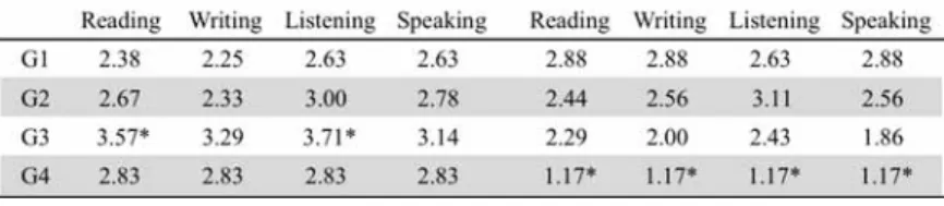
Participants self-evaluated their own language level in the four skills (speaking, listening, reading, and writing) in Japanese and English on a five-point Likert scale with '5' being superior, '3' equal, and '1' inferior to native speakers of the same age (Table 2).
Table 2. Self-evaluation of four skills in Japanese and English
The Effects of Onset-Age and Exposure Duration on the L2 as Observed in Brain Activation: an fNIRS Study
Levene's test for homogeneity of variances was checked and it was found that we did not violate the homogeneity of variance assumption. T-tests revealed some statistically major features among the groups: (1) G3 rated their Japanese proficiency in two skills (reading and listening) better than G1, and (2) G4 self-evaluated themselves statistically inferior to G1 in all four skills of English. This reveals that G3, with their overseas experiences, may have a greater linguistic confidence and that G4, with only six months of English learning, find themselves equipped with very limited skills in English.
Prior to the experiment, a signed consent form was obtained from all the participants or from their parents, if they were under 18 years of age. This study was approved by the Ritsumeikan University Institutional Ethics Committee in Japan.
2.2 Procedure
Participants were individually shown into a laboratory and seated in a chair with their eyes approximately 30 cm away from the monitor (Panasonic Let's Note CF-F9). While a flexible cap with 27 fiber probes was being placed on their head, information was gathered on their language background, right or left-handedness, and self-assessment of their four skills (speaking, listening, writing, and reading) in the two languages - Japanese and English. Once the cap was appropriately placed and connected, photos were taken to record the position of the cap from three angles—from the left, right and front. Then, a video clip was shown to inform the participants of what the experiment involved. When participants were made fully aware of the task, they were told that it was their right to stop it at any time if they felt uncomfortable, once the experiment had started. Upon completion of the experiment, each participant was given an appropriate token of gratitude—a book voucher.
2.3 Tasks
Using TOEFL or TOEIC items as tasks in brain research has been harshly criticized as the tasks involve too many intervening variables to differentiate (Ishikawa, 2009; Taura et al., 2010). Verbal fluency tasks are frequently used to elicit brain activation data from bilingual subjects because of the relative ease to make comparable tasks between two languages (e.g., Arai
et al., 2006; Ehlis et al., 2007; Fallgatter et al., 2004; Kameyama et al., 2004; Schecklmann et al.
2008; Taura et al., 2010).
Bilingual children are reported to possess better metalinguistic awareness (Bialystok, 2012) and be more capable of handling cognitively demanding tasks such as the bilingual Stroop test (Taura, 2008). More cognitively demanding tasks trigger greater brain activation in the pre-frontal lobe as disclosed by a brain-imaging study by Izzetoglu et al. (2004). Stephan et al. (2003), using cognitively demanding tasks, have also found that letter decision tasks induce
activation in the left anterior cingulate cortex and left inferior frontal gyrus whereas visuo-spatial decision tasks activate right anterior cingulate cortex and right parietal areas. Thus, employing a mathematical task, where participants need to attend to both letter and colour information (further explained below), is expected to activate both hemispheres more since the task is cognitively more demanding than a simple verbal task.
Thus, two types of tasks were devised for this study. The first task was a simple verbal fluency task while the second verbal task was cognitively more demanding as the participants needed to control their answers depending on the colour and nature of the one-digit numbers presented on the PC screen.
2.3.1 Verbal Fluency Task
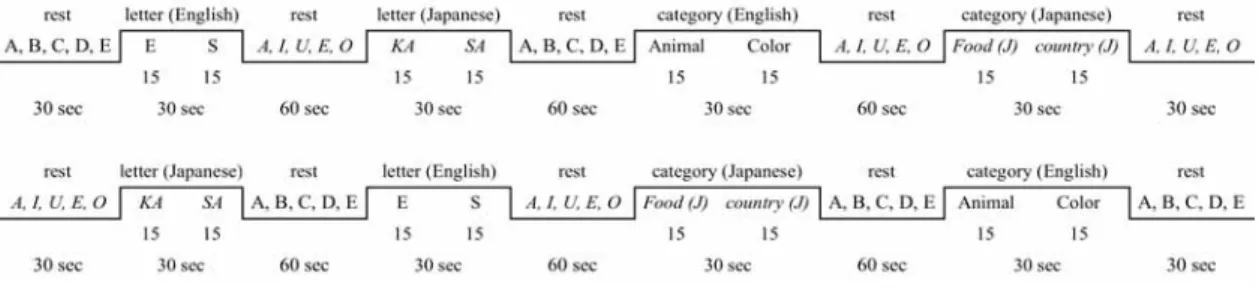
The Verbal Fluency Task (VFT) included four sub-tasks as shown in the blocked design in
Figure 1 that also shows the task stimuli and the timing of the tasks. The experiment began with a 30-second rest. Then the first letter task was given for 30 seconds followed by a 60-second rest, before the second letter task began. This pattern repeated itself again with category tasks in each language. At the end of the fourth task, there was another rest lasting for 30 seconds. To offset the language sequence effects, a counter-balanced version was used for every second participant.
Figure 1. Blocked design for Verbal Fluency Task
Two kinds of tasks were given to the participants: the letter task and the category task in Japanese and English. For the Japanese letter task, hiragana prompts were projected onto the computer screen of the letters 'ka' or 'sa', and the participants were then expected to say aloud as many Japanese words as possible that started with 'ka' in the first 15 seconds and 'sa' in the second 15 seconds. The English letter task used the letters 'E' and 'S' in the same way. Meanwhile, in the Japanese category task, participants were required to produce as many words as they could that related to cue words such as 'country', where for example they may have said 'Japan' or 'France'. Both Japanese and English category tasks had two category words that were shown on the PC screen for 15 seconds each. The actual category words shown in English were 'animal' and 'colour' while the words 'food' and 'country' were projected onto the screen in Japanese. Our decision to
The Effects of Oneet-Age and Exposure Duration on the L2 as Observed in Brain Activation: an fNIRS Study
use such English letters and categories are based on previous studies by Schecklmann et al. (2008) and Ehlis et al. (2007) while the hiragana examples were selected from Japanese studies (e.g., Arai et al., 2006; Kameyama et al., 2004; Murai et al., 2004).
2.3.2 Numeral Task
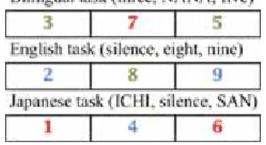
There were three kinds of sub-tasks that made up The Numeral Task—the bilingual task, the English task, and the Japanese task (Figure 2).
Figure 2. Numeral Task
In each task, ten different slides were presented for three seconds each. The Numeral Task was comprised of each task (30 seconds in length) followed by a 60-second rest, except for the first and last rests, which were 30 seconds long. The ten slides each presenting a one-digit number, were shown on the PC monitor one by one (Figure 3). Each stimulus was shown for 3 seconds. The bilingual task involved both English and Japanese, with a number projected on each slide in either red or green. A number in red required the participants to say it aloud in Japanese while a green number had to be identified aloud in English. For the English task using only the English language, a number was shown on the slide in either green or blue. A green number had to be named aloud in English but when it was in blue, only the number 3 or a multiple of 3, was to be said aloud. For example, when the slide ‘3’ in blue was shown, the correct answer was ‘three’ in English but when a slide with a blue ‘7’ was presented, they had to remain silent as 7 is not a multiple of 3. The Japanese proceeded likewise, with the only difference being the red color of the Japanese numbers instead of green. The sequence of the three tasks was counter-balanced for every second participant.
2.4 Brain Imaging Technique
Among the variety of brain imaging techniques, the non-invasive functional
neuro-imaging method known as fNIRS is said to be easier to use and sensitive to detecting small substance concentrations, with a high temporal resolution (Toga and Mazziotta, 2002). In principle, fNIRS measures brain activation using changes in the intensity of light detected by source and detector probes that are attached to the head with a harmless light that penetrates the brain. Other brain imaging techniques including fMRI (functional magnetic resonance imaging), PET (positron emission tomography), and MEG (magnetoencephalography) require large and bulky instruments which make it difficult for small children to be examined. In comparison, fNIRS uses light via fibre optics, which can even allow babies to be examined (Miyai et al., 2001). In addition, fNIRS is sensitive to a very low substance concentration using a fluorescence method and the result is similar to a PET scan without the disadvantage of radioactive tracers. Temporal and spatial resolution differ vastly from one brain imaging method to another: high temporal resolution is obtained using the MEG eletrophysiological method (starting from 1 m.s.) while
hemodynamics-based techniques such as fMRI (starting from 1 mm) provide a greater spatial resolution. fNIRS offers a smaller, easier-to-use option to monitor vascular, metabolic-cellular, and neuronal responses with a high temporal resolution of about 1,000 m.s., despite a low spatial resolution of about 3 cm. It is also recognized that fNIRS data are consistent with fMIR data in predicting hemispheric dominance on linguistic tasks (e.g., Kennan et al., 2002). fNIRS has been widely used and proved to be useful in various disciplines (see Kovelman et al., 2008 for
summary). Therefore, for this study where younger children were involved, we decided to use an fNIRS - a multichannel continuous wave optical imager (Shimadzu FOIRE-3000).
The Effects of Onset-Age and Exposure Duration on the L2 as Observed in Brain Activation: an fNIRS Study
Shimadzu FOIRE-3000 has 13 emitters and 14 receivers, 3 centimetres apart from each other to detect 42 areas (channels). The fNIRS machine uses three wavelengths of near-infrared light (780±5nm, 805±5nm, 830±5nm). According to the International 10/20 system (Jasper, 1958), emitters and receivers of fNIRS were placed bilaterally over the frontal lobe, including Broca's area in the left inferior frontal cortex, with the lowest probes being positioned along the
T3-Fp1-Fz-Fp2-T4 line (Figures 4 and 5). The length between the nasion (right above the nose) and the inion (the most prominent projection point of the occipital bone at the base of the skull) was measured to pinpoint the one-tenth distance from the nasion where the eighth receiver hole is aligned. Next, the distance between the midpoints of the two ears was measured to make sure that the one-tenth above each mid-ear point was aligned with the T3-Fp1-Fz-Fp2-T4 line. These two steps were taken for all the participants.
Our decision to examine the frontal lobe in both hemispheres was based on findings from Herrmann et al. (2003 and 2005), Just et al. (1996), Kameyama et al. (2004), and Yamadori (1998) that in a language task, not only the left laterosuperior temporal cortex (Wernicke's area) and the left inferior frontal gyrus (Broca's area) but also their homologous right-hemisphere areas are found to be activated, though the right areas had much smaller volumes of activation.
2.5 Data analysis
Data from the fNIRS were processed using the Shimadzu FOIRE (a functional optical imagery software for research) and PASW SPSS software. The first stage involved identifying in the individual participants, the two distinct areas of the brain for this study, namely, Broca's area and its homologous area in the right hemisphere of the prefrontal lobe. After checking that all the probes were aligned properly, we visually identified Broca's area and its right homologous area and wrote down their corresponding probe numbers. Diagrams displaying changes in oxygenated hemoglobin (oxy-Hb), deoxygenated hemoglobin (deoxy-Hb), and total hemoglobin (total-Hb) using the FOIRE software were produced, in order to discard any data at particular channels that showed the involvement of artefacts which may have been due to a jerking movement of the participant or probes popping out of the holder sockets. Despite its excellent temporal resolution, fNIRS lacks spatial resolution (Toga and Marital, 2002), therefore we needed to include several channels of data to cover the designated two regions of the brain. Broca's area generally included channels 25, 33, and 34 while its right hemispheric counterpart included channels 18, 26, and 27 in most participants.
FOIRE-3000 shows three different fNIRS parameters (oxy-Hb as a red line, deoxy-Hb as a blue line, and total-Hb as a green line on the monitor). Based on the findings that fNIRS oxy-Hb exhibits a high correlation with the blood oxygenation level dependent (BOLD) signal used in fMRI (e.g., Ryo et al., 2008; Shimoyama et al., 2006; Toronov et al., 2007), and the data produced
may therefore be used to represent all three parameters (Fukuda, 2009; Malonek et al., 1997; Strangman et al., 2002), this study focused solely on oxy-Hb.
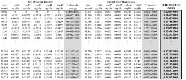
fNIRS raw data (mM-mm) reflect the comparison between the change in concentration of hemoglobin and wavelengths of near-infrared light (fNIRS value) from 130 milliseconds before. Therefore, the raw data cannot be used for intra- and inter-subject analysis and need to be
subtracted from the baseline data (e.g. Yokoyama, 2010; Peterson et al., 1988). In other words, the raw fNIRS data during each task need to be subtracted from the fNIRS data in the preceding rest task. This subtraction method was originally devised by Peterson et al. (1988) using PET scanning and has since been widely used in neuroimaging studies. The subtraction process takes place in the following manner: (1) fNIRS (oxy-Hb) data in the channels (24, 25, 33, 41, and 42, for instance) that were identified as Broca's area are averaged out for a particular 30-sec task, (2) the same calculation is conducted for the preceding 30-sec rest time, and (3) the rest average fNIRS is subtracted from the task average fNIRS (Table 3). The same procedure is repeated with the fNIRS data in the right hemisphere. For a group comparison, these individual subtraction figures are averaged out within each group as representative figure.
Table 3. Subtraction of the average rest-time fNIRS data from the average task fNRIS data
3. Results
The results of the two experiments– the Verbal Fluency Task and Numeral Task–are provided here. Each task is dealt with separately showing oxy-H in mM-mm.
3.1 Verbal Fluency Task 3.1.1 VFT behavioural data
The Effects of Onset-Age and Exposure Duration on the L2 as Observed in Brain Activation: an fNIRS Study
English letter task, 4.0 in the Japanese letter task, 6.6 in the English category task, and 7.0 in the Japanese category task (for individual group averages, see Figure 6). The chi-square test revealed that there was no significant difference among groups in the number of output words uttered in each of the four VFT: the English letter task (χ²=33.492, df=33, p>.05), the Japanese letter task (χ²=33.168, df=33, p>.05), the English category task (χ²=49.782, df=45, p>.05), and the Japanese category task (χ²=51.468, df=45, p>.05). There was no significant difference when four tasks were compared and all groups combined (χ²=70.608, df=63, p>.05). These results showed that there were no differences in the difficulty of each task among the four groups, and that difficulty was not a factor that affected the fNIRS data.
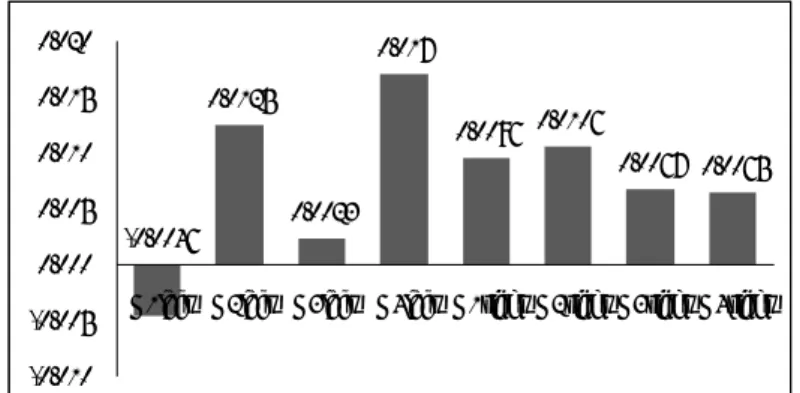
Figure 6. Number of words produced Figure 7. English letter VFT in mM-mm
3.1.2. VFT: English letter task
Descriptive data are provided in Figure 7. We carried out an analysis of variance (ANOVA) on fNIRS data in both hemispheres among four groups (F(7,139)=411.322, p<.001, eta
squared=.954). The results of a post hoc Bonferroni are as follows: G1<G2<G3<G4 in Broca's
area while G1<G2<G3=G4 in its right homologous area. Thus, G1 showed the least amount of brain activation in the English letter task in both hemispheres. G2 revealed the second least amount of brain activation in both hemispheres. Meanwhile, both G3 and G4 needed more brain activation than G1 and G2, irrespective of whether it was in the left or right hemisphere.
3.1.3. VFT: Japanese letter task
Figure 8 summarizes the fNIRS data on which we carried out an ANOVA (F(7,139)=50.220, p<.001, eta squared=.717). The results of a post hoc Bonferroni are as follows: G1=G3<G4<G2 in Broca's area and G1<G3<G2<G4 in its right homologous area. Thus, G1 showed the least amount of brain activation on the Japanese letter task in both hemispheres. G3 demonstrated the second least amount of brain activation in the right hemisphere and as minimal in
0.00 1.00 2.00 3.00 4.00 5.00 6.00 7.00 8.00 9.00 G1 3.81 3.13 7.12 5.00 G2 5.06 5.00 7.61 8.83 G3 5.07 4.57 7.14 8.14 G4 2.50 3.42 4.58 6.33 English Letter Japanese Letter English Category Japanese Category 0.0031 0.0069 0.0128 0.0186 0.001 0.0137 0.0126 -0.0033 -0.005 0.000 0.005 0.010 0.015 0.020
G1 left G2 left G3 left G4 left G1 right G2 right G3 right G4 right
the amount of activation as G1 in the left hemisphere. Meanwhile, both G2 and G4 needed more brain activation than G1 and G3, irrespective of whether in the left or right hemisphere.
0.0035 0.0107 0.0035 0.0083 0.0054 0.0008 0.0077 -0.0044 -0.006 -0.004 -0.002 0.000 0.002 0.004 0.006 0.008 0.010 0.012
G1left G2left G3left G4left G1right G2right G3right G4right
Figure 8. Japanese letter VFT in mM-mm
3.1.4 VFT: English category task
Figure 9 presents the summary of fNIRS data on which we conducted an ANOVA (F(7,139)=92.782, p<.001, eta squared=.824). The results of a post hoc Bonferroni are as follows: G1=G2, G1=G4, G3=G4, G1<G3, G2<G3/4 in Broca's area while G3<G1<G2<G4 in its right homologous area. Thus, there were few statistically significant differences in the Broca's area among the four groups while each group differed from other in the right hemisphere in the order of G3 being the least activated, then G1, G2, and G4 being the most activated.
0.0112 0.0106 0.0141 0.0126 0.0063 0.0089 0.0134 -0.0036 -0.006 -0.004 -0.002 0.000 0.002 0.004 0.006 0.008 0.010 0.012 0.014 0.016
G1left G2left G3left G4left G1right G2right G3right G4right
Figure 9. English category VFT in mM-mm
3.1.5. VFT: Japanese category task
The summary of the fNIRS data is presented in Figure 10. We carried out an ANOVA on the fNIRS data in both hemispheres in all the four groups (F(7,139)=148.025, p<.001, eta
The Effects of Onset-Age and Exposure Duration on the L2 as Observed in Brain Activation: an fNIRS Study
area while G1=G2=G4, G1>G3, G2>G3/4, G3<G1/2, G4=G1/3 in its right homologous area. There is little difference among the four groups in the right hemisphere, which signifies that, regardless of OA for L2 and LOR in the L2 environment, the Japanese category task seems to activate the right hemisphere to the same degree for everyone.
0.0125 0.0023 0.017 0.0096 0.0106 0.0067 0.0065 -0.0046 -0.010 -0.005 0.000 0.005 0.010 0.015 0.020
G1left G2left G3left G4left G1right G2right G3right G4right
Figure 10. Japanese category VFT in mM-mm
3.1.6. VFT: summary
The results from the four tasks, particularly the L2 English tasks, are combined here in an attempt to seek the answers to our research questions.
The results of the English letter task disclosed that the earlier the onset age of exposure to English and the longer that exposure lasts, the less the brain activation is observed in Broca's area in the left hemisphere. Onset age (OA) to English was important when distinguishing G1 (OA at birth) from G2 (OA before age 7) and G3/4 (OA after 12). Furthermore, English learning duration differentiated G3 with the experience of studying in an English-speaking country for from one to six years, from G4 who had received a formal English education for only six months in Japan. The second finding concerned the brain activation level in the right hemisphere which was identical to the left hemisphere except there was no statistical difference seen between G3 and G4.This can be interpreted that one to six years of study in an English-speaking country is not as sufficient for dominant use of the left hemisphere for L2 (letter fluency) processing. Thirdly, G1 and G2 (OA before 7) use Broca's area during the English letter task significantly more than the right counterpart, whereas G3 uses both hemispheres to the same degree and G4 dominantly use their left hemisphere.
The English category task showed both similar and different pictures in the results. The similar results can be seen in the right hemisphere where brain activation increased in the order of G1 (least), G2, and G3 (most). Meanwhile, a totally different tendency was observed in Broca's area where no statistical differences were found among the four groups. Previous research has found that the letter fluency task is generally more difficult and requires more brain activation than
the category fluency task (e.g., Kubota et al., 2005; Ehlis et al., 2007). This helps us to interpret that the category task is easy enough for most L2 English learners to carry out using Broca's area (automatization possibly leads to no group differences). On the other hand, in the right hemisphere where language function is indirectly involved, brain activation is still affected by L2 proficiency in terms of OA and LOR as shown by more brain activation for G4 and the least amount for G1.
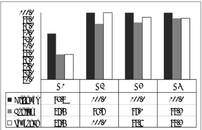
3.2. Numeral Task 3.2.1. Accuracy rates
The summary of the accuracy rates for each task in each group is provided in Figure 11. The average accuracy rates for each task were 98.4% for the Bilingual Numeral Task, 94.9% for the English Numeral Task, and 96.1% for the Japanese Numeral Task. A chi-square test revealed that there was no significant difference among the groups in the accuracy rates for the three tasks:
Bilingual Numeral Task (χ²=2.845, df=3, p>.05), English Numeral Task (χ²=7.214, df=9, p>.05),
and Japanese Numeral Task (χ²=7.567, df=9, p>.05). The average accuracy rates for each group were 89.5% for G1, 98.8% for G2, 98.6% for G3, and 98.8% for G4, which revealed no group differences as well (χ²=14.049, df=8, p>.05). This indicates no impact from the task difficulty on the fNIRS data among the groups.
80.0 82.0 84.0 86.0 88.0 90.0 92.0 94.0 96.0 98.0 100.0 Bilingual 93.8 100.0 100.0 100.0 English 87.5 96.7 97.1 98.3 Japanese 87.5 100.0 98.6 98.3 G1 G2 G3 G4
Figure 11. Accuracy rate (%) for each task in each group
3.2.2. Bilingual Numeral Task
fNIRS data of the bilingual Numeral Task are summarized in Figure 12. An ANOVA showed statistical differences among the four groups (F(7,139)=972.150, p<.01, eta squared=.784). A post hoc Bonferroni revealed group differences of G4<G1<G2=G3 in Broca's area and G1=G3<G2=G4 in its right homologous site. At the same time, within-group analyses revealed that significantly more right hemispheric activation was seen in G1 and G4 whereas the trend was reversed in G3 and both hemispheres became activated at the same level in G2.
The Effects of Onset-Age and Exposure Duration on the L2 as Observed in Brain Activation: an fNIRS Study 0.0003 0.0066 0.006 0.004 0.0062 0.0032 0.0066 -0.0019 -0.003 -0.002 -0.001 0 0.001 0.002 0.003 0.004 0.005 0.006 0.007 0.008
G1left G2left G3left G4left G1right G2right G3right G4right
Figure 12. Bilingual Numeral Task
3.2.3. English Numeral Task
fNIRS data of the English Numeral Task are summarized in Figure 13. An ANOVA showed statistical differences among the four groups (F(7,139)=80.744, p<.01, eta squared=.803). A post hoc Bonferroni revealed group differences, G3<G1=G2<G4 in Broca's area and G3<G1=G2<G4 in the right homologous site. At the same time, within-group analyses revealed that significantly more left hemispheric activation was seen in G1, G2, and G3 whereas both hemispheres became activated at the same level in G4.
0.0082 0.0084 0.0055 0.0125 0.0069 0.0058 0.0033 0.014 0.000 0.002 0.004 0.006 0.008 0.010 0.012 0.014 0.016
G1left G2left G3left G4left G1right G2right G3right G4right
Figure 13. English Numeral Task
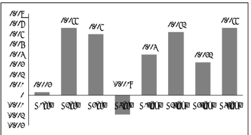
3.2.4. Japanese Numeral Task
fNIRS data of the Japanese Numeral Task are summarized in Figure 14. An ANOVA showed statistical differences among the four groups (F(7,139)=116.920, p<.01, eta squared=.855). A post hoc Bonferroni revealed group differences of G1=G3<G4<G2 in Broca's area and G3<G1<G2<G4 in the right homologous site. At the same time, within-group analyses revealed
that significantly more left hemispheric activation was seen in G2 and G3 whereas the trend was reversed in G1 and G4. 0.0044 0.0214 0.006 0.0164 0.0084 0.0138 0.0013 0.0276 0.000 0.005 0.010 0.015 0.020 0.025 0.030
G1left G2left G3left G4left G1right G2right G3right G4right Figure 14. Japanese Numeral Task
3.2.5. Numeral Task summary
Our focus is placed on the results of the bilingual and English numeral tasks since this study mainly explores the critical period for L2 acquisition. The most striking finding relates to G4's fNIRS data for the bilingual and English numeral tasks. G4 needed the most amount of cerebral blood flow on the English task in both hemispheres along with the right hemisphere on the bilingual task whereas G4 displayed the least activation in Broca's area for the bilingual task. According to Stephan et al. (2003), the left hemisphere of the brain is activated on letter decisions (identifying the number in the task) and the right hemisphere attends to visuo-spatial decisions (recognizing the ink colour that each number is written in). Considering the fact that L1 processing takes place in the left part of the brain, G4 must have had a hard time processing the English numeral task concurrently in both the left side of the brain (where recognition of each stimulus number and verbalizing the answer in L2 English are executed) and in the right brain hemisphere (where discrimination of the colour of each number is made, in addition to L2 processing). This difficulty revealed itself in their fNIRS data on the English numeral task. On the bilingual numeral task, however, G4 automatically processed the letter information in L1 with ease (which is seen in the least brain activation fNIRS data on the left side of the brain) and were able to process both colour identification and L2 English easily too.
Other findings include clear differences between groups in some fNIRS data and no differences for other data. The first tendency is seen (1) in the difference between G1 and G2 and between G3 and G4 on the bilingual numeral task in both hemispheres, and (2) in the difference between G3 and G4 on the English numeral task in both hemispheres. Similarities, on the other hand, are seen between G1 and G2 for the English numeral task in both hemispheres.
The Effects of Onset-Age and Exposure Duration on the L2 as Observed in Brain Activation: an fNIRS Study
4. Discussion
The fNIRS data relevant to our query into the critical period for L2 acquisition are presented in Figure 15 to seek the answers to our two research questions.
VFT: English letter VFT: English category
Numeral: Bilingual Numeral: English
Figure 15. Summary of English related fNIRS data
4.1 Research Question 1:
Do simultaneous bilinguals (OA at birth) show different brain activation in their L2 use when compared to sequential bilinguals (OA before the age of seven)?
The English VFT results show that G2 (OA before seven) need significantly more brain activation than G1 (OA at birth) in both hemispheres on the letter task and in the right hemisphere for the category task. The same tendency holds true for the bilingual Numeral Task in both
hemispheres. Meanwhile, no such differences were observed in the left hemisphere on the English category task, or both hemispheres on the English Numeral Task.
0.0112 0.0106 0.0141 0.0126 0.0063 0.0089 0.0134 -0.0036 -0.006 -0.004 -0.002 0.000 0.002 0.004 0.006 0.008 0.010 0.012 0.014 0.016
G1left G2left G3left G4left G1right G2right G3right G4right 0.0031 0.0069 0.0128 0.0186 0.001 0.0137 0.0126 -0.0033 -0.005 0.000 0.005 0.010 0.015 0.020
G1 left G2 left G3 left G4 left G1 right G2 right G3 right G4 right 0.0003 0.0066 0.006 0.004 0.0062 0.0032 0.0066 -0.0019 -0.003 -0.002 -0.001 0.000 0.001 0.002 0.003 0.004 0.005 0.006 0.007 0.008
G1left G2left G3left G4left G1right G2right G3right G4right
0.0082 0.0084 0.0055 0.0125 0.0069 0.0058 0.0033 0.014 0.000 0.002 0.004 0.006 0.008 0.010 0.012 0.014 0.016
Table 4. Comparison between G1 and G2
(= indicates no statistical differences)
The overall picture reveals that G2 needed to activate their brain areas as much as or more than G1 for all four tasks. A more accurate picture, however, manifests itself with a closer look at the data considering the cognitive demand perspective of the tasks. Izzetoglu et al. (2004) and Stephan et al. (2003), who used VFT tasks in their brain-imaging experiments, reported that the letter task was more demanding than the category task. In addition, for the numeral task, the bilingual version is obviously more demanding than the English task in that participants must function in the dual language mode while they only needed to attend to a single language in the latter task. In the more cognitively demanding (VFT letter and bilingual Numeral) tasks, the G1 activation level was significantly lower than their counterparts (Figure 16). In the relatively less demanding (the VFT category and the English Numeral) tasks, the G2 activation level was similar to G1's. Thus, it appears that we can validly conclude that the onset age of L2 exposure at birth results in less brain activation in both hemispheres than participants with the onset age of L2 exposure before age seven, which is observable particularly on cognitively demanding tasks.
G2 (English letter task in Broca's area) G1 (English letter task in Broca's area)
G2 (Bilingual Numeral task in Broca's area) G1 (Bilingual Numeral task in Broca's area)
The Effects of Onset-Age and Exposure Duration on the L2 as Observed in Brain Activation: an fNIRS Study
4.2 Research Question 2:
Is the length of exposure to L2 important in brain activation when the L2 is learned after the critical period?
This research question is an attempt to seek an answer to our query as to whether or not brain activation changes as L2 proficiency improves in those who are first exposed to an L2 at age 12 - after the critical period for language learning. Table 5 summarizes the fNIRS data comparing G3 to G4. G3 has the experience of one to six years of intensive and extensive exposure to English by studying in an English-speaking country. G4 had only a six-month formal English education in Japan with no overseas experience before they participated in this experiment.
Table 5. Comparison between G3 and G4
(= indicates no statistical differences)
A similar picture can be seen, as in RQ1, although not in terms of the L2 onset age but connected to the L2 learning duration: The longer the study time, the less the brain activation that occurred as was seen in 63% of the tasks. In addition, G4 showed the same degree of brain activation as G3 in 25% of the tasks. Both ratios were calculated including both cognitively non-demanding and demanding tasks. Thus, 88% of the tasks were accounted for in this manner.
However, there is one instance when comparing G3 to G4 when the data did not follow this argument. Here, when examining the fNIRS data on the left side of the brain on the bilingual
Numeral task, G3 exhibited more blood flow than G4 (highlighted in Table 5). In this task, the
participants were instructed to say the numbers aloud either in Japanese or English, depending on the ink colour of the numbers displayed on the computer monitor. One possible interpretation for the low fNIRS signals in G4 could be that they had to pay so much attention to the ink colour, activating the visuo-spatial faculty in the right anterior cingulate cortex and right parietal areas, as well as their L2 in the right hemisphere, that they had little resources left for L1 processing in the left side of the brain.
Another interpretation of the fNIRS data can be made by examining the low brain activation in the left hemisphere, in G1 and G4 on the bilingual Numeral task. As far as English
proficiency is concerned, the participants' language background information based on OA and L2 exposure duration predicts that G1 would be the most proficient followed by G2, G3, and G4. The fNIRS data, however, do not reflect the prediction of G1 having the least brain activation and G4 the highest, but the data show more activation in G2 (OA<7) and G3 (OA=12, L2 sojourn for one to six years) than G1 (OA=0) and G4 (OA=12, duration=6 months). G1 aside, this tendency of more proficient L2 learners in G2 and G3showing more activation in Broca's area, than the novice learners in G4 could be due to a combination of the timing of exposure to L2 and the foundation of their L1. G1 participants developed both their languages simultaneously from birth and G4 had a solid L1 foundation before they began to learn English at the age of twelve. Meanwhile, G2's first intensive and extensive exposure to English began before seven, when their L1 had not yet been fully developed, and G3 underwent such experiences in their teens or twenties when their L2 needed fine-tuning. Thus, the significantly higher brain activation in G2 and G3 than G1 and G2 could be caused by an interruption to their L1 development in their teens and twenties.
Regardless of how valid our interpretation may be, the general tendency observed here leads us to conclude that, the longer the exposure to L2, the less is the brain activation in both hemispheres, irrespective of whether the tasks involved are cognitively demanding or not.
5. Conclusion
The present study used a linguistic Verbal Fluency Task as well as a more cognitively demanding Numeral Task to examine brain activation in relation to the critical period for language acquisition and L2 learning duration. The results first demonstrated that the earlier one's first intensive and extensive exposure to an L2 is, the less brain activation is shown in both hemispheres, even in those exposed to L2 before the critical period. This suggests that L2 processing requires less energy in the brain when exposure to the language takes place earlier on in life. The second finding revealed that brain activation lessens in both hemispheres as L2 proficiency improves, implying that one needs less energy in L2 processing as L2 learning extends in time. In addition, an incidental finding hinted at a need to firmly solidify L1 without the interruption of L2 acquisition or learning while still fine-tuning the L1 in the teens and twenties.
This study is not without its limitations. To confirm our conclusion, further research is needed to see if the results are due to a longer language exposure or the interplay of the longer duration and the impact of being immersed in an L2 community (G3, in particular, included only those who have studied for some time in an English-speaking country). Secondly, more participants of a similar age in the different groups are needed to eliminate an intervening variable such as maturation, which could have affected the results. In addition, a longitudinal study to further examine the L2 novice learners, could lend support to our tentative conclusion in this study.
The Effects of Onset-Age and Exposure Duration on the L2 as Observed in Brain Activation: an fNIRS Study
Acknowledgements: We are grateful to the participants in this study and Mr. Yomota (Shimadzu Corporation) for his invaluable suggestions. This research is partly subsidized by a Ritsumeikan University grant promoting academic research.
References
Arai, H., Takano, M., Miyakawa, K., Ota, T., Takahashi, T., Asaka, H., Kawaguchi, T. (2006). A quantitative near-infrared spectroscopy study: A decrease in cerebral hemoglobin
oxygenation in Alzheimer’s disease and mild cognitive impairment. Brain and Cognition,
6, 2, 189–194.
Bialystok, E. (2012). Emerging bilingualism: Dissociating advantages for metalinguistic awareness and executive control. Cognition, 122, 1, 67-73.
Dahaene-Lambertz, G. (2000). Cerebral specialization for speech and non-speech stimuli in infants.
Journal of Cognitive Neuroscience, 12, 3, 449-460.
Dehaene, S., Dupoux, E., Mehler, J., Cohen, L., Paulesu, E., Perani, D., van de Moortele, P.F., Lehericy, S., and Le Bihan, D. (1997). Anatomical variability in the cortical
representation of first and second language. NeuroReport,8, 17, 3809-3815. Ehlis, A. C., Herrmann, M. J., Plichta, M. M., and Fallgatter, A. J. (2007). Cortical activation
during two verbal fluency tasks in schizophrenic patients and healthy controls as assessed by multi-channel near-infrared spectroscopy. Psychiatry Research, 156, 11-13.
Fallgatter, A.J., Ehlis, A., Wagener, A., Michel, T., & Herrmann, M.J. (2004). Near-infrared spectroscopy in psychiatry. Nervenarzt,75, 911-916.
Fukuda, M. (2009). Seishinshikkan to NIRS [Mental illness and near-infrared spectroscopy] Tokyo: Nakayama shoten.
Hakuta, K., Bialystok, E., and Wiley, E. (2003). A test of the critical-period hypothesis for second-languageb acquisition. Psychological Science, 14, 1, 31-38.
Herrmann, M. J., Ehlis, A.C., & Fallgatter, A. J. (2003). Frontal activation during a verbal-fluency task as measured by near-infrared spectroscopy. Brain Research Bulletin, 61, 1, 51.56. Ishikawa, S. (2009) Dainigengoshutokukenkyu to nokagaku [Research of second language acquisition and neuroscience] システム制御情報学会誌53, 4, 143-148.
Izzetoglue, K., Bunce, S., Onaral, B., Pourrezaei, K., and Chance, B. (2004). Functional Optical Brain Imaging Using Near-Infrared During Cognitive Tasks. International Journal of
Human-Computer Interaction, 17, 2, 211-227.
Jasper, H. H. (1958). The ten-twenty electrode system of the International Federation.
Electroencephalography and Clinical Neurophysiology, 10, 371–375.
Just, M.A., Carpenter, P.A., Keller, T.A., Eddy. W.F., and Thulborn, K.R. (1996). Brain Activation Modulated by Sentence Comprehension. Science, 274, 5284, 114-116.
Johnson J. and Newport, E. (1989). Critical period effects in second language learning: The influence of maturational state on the acquisition of English as a second language.
Cognitive Psychology, 21, 60-99.
Kameyama, M., Fukuda, M., Uehara, T., and Mikuni, M. (2004). Sex and age dependencies of cerebral blood volume changes during cognitive activation: A multichannel near-infrared spectroscopy study. Neuroimage, 22, 4, 1715-1721.
Kennan, R.P., Kim, D., Maki, A., Koizumi, H., and Constable, R.T. (2002). Non-Invasive Assessment of Language Lateralization by Transcranial Near Infrared Optical Topography and Functional MRI. Human Brain Mapping, 16, 183–189.
Kim, K., Relkin, N. R., Lee, K., and Hirsch, J. (1997). Distinct cortical areas associated with native and second languages. Nature, 388, 171-174.
Knecht S, Dräger B, Deppe M, Bobe L, Lohmann H, Flöel A, Ringelstein EB, Henningsen H. (2000). Handedness and hemispheric language dominance in healthy humans. Brain,
123, 12, 2512-2518.
Kovelman, I., Shalinsky, M. H., Barens, M. S., and Petitto, L. (2008). Shining new light on the brain’s “bilingual signature”: A functional Near Infrared Spectroscopy investigation of semantic processing. Neuroimage, 39, 1457- 1471.
Kubota Yasutaka, MotomiToichi, Mitsue Shimizu, Richard A. Mason, Christinel M. Coconcea, Robert L. Findling, Kokichi Yamamoto & Joseph R. Calabrese. (2005). Prefrontal activation during verbal fluency tests in schizophrenia- a near-infrared spectroscopy (NIRS) study. Schizophrenia Research, 77, 65-73.
Lenneberg, E. (1967). Biological Foundations of Language. New York: J Wiley and Sons. Lenneberg, E. (1975). In search of a dynamic theory of aphasia. In Lenneberg E.L. and
Lenneberg E. (Eds.), Foundation of Language Development. A multidisciplinary
Approach. San Diego, CA: Academia Press.
Luk, Z.P., and Shirai, Y. (2009). Is the acquisition order of grammatical morphemes impervious to L1 knowledge? Evidence from the acquisition of plural -s, articles, and possessive's.
Language Learning, 59, 4, 721-754.
Malonek, D., Dirnagl, U., Lindauer, U., Yamada, K., Kanno, I., Grinvald, A., (1997). Vascular imprints of neuronal activity: relationships between the dynamics of cortical blood flow, oxygenation, and volume changes following sensory stimulation. Proc. Natl. Acad. Sci.
U.S.A. 95, 14826-14831.
Miyai, I., Tanabe, H., Sase, I., Eda, H., Oda, I., Konishi, I., Tsunazawa, Y., Suzuki, T., Yanagida, T., and Kubota, K. (2001). Cortical mapping of gait in humans: A near-infrared
spectroscopic topography study. NeuroImage, 14, 1186-1192.
The Effects of Onset-Age and Exposure Duration on the L2 as Observed in Brain Activation: an fNIRS Study
C. (2004). Shoniyogosokikadai no kokoromi – shogakuseihenojissenrei [Attempts of
productive vocabulary for children: practical research on elementary school children]. Osaka: Osaka University of Education. 53, 1, 83-89.
Oishi, H. (2006). Nokagakukara no dainigengoshutokuron [Second language acquisition theory from neuroscience]. Kyoto: Showado.
Oldfield, R.C. (1971). The assessment and analysis of handedness: The Edinburgh inventory.
Neuropscychologia, 9, 1, 97-113.
Peña, M., Maki, A., Kovačc, D., Dehaene-Lambertz, G., Koizumi, H., Bouquest, F., and Mehler, J. (2003). Sounds and silence: An optical topography study of language recognition at birth.
Proceedings of National Academy of Science, USA 100: 11702-11705.
Perani, E., Paulesu, E., Galles, N. S., Dupoux, E., Dehaene, E., Bettinardi, V., Cappa, S.F., Fazio, F., and Mehler, J. (1997). The bilingual brain. Proficiency and age of acquisition of the second language. Brain. 121, 10, 1841-1852.
Petersen, S. E., Fox, P. T., Posner, M. I., Mintun, M., and Raichle, M. E. (1988). Positron emission tomographic studies of cortical anatomy of single-word processing. Nature, 331,
585-589.
Quaresima, V., Giosue, P., Roncone, R., Casacchia, M., Ferrari, M. (2009). Prefrontal cortex dysfunction during cognitive tests evidenced by functional near-infrared spectroscopy.
Brain Research Bulletin, 59, 30, 235-243.
Ryo, S., Matsuno, K., and Sugiura, T. (2008). Korokeshonno shorikateiniokeru nonaikasseibuito
shintekifuka [Brain activation areas and psychological stress in processing collocations].
Summary report on Grants-in-Aid for Scientific Research by Japanese government
(#17320084), 197-220.
Sakai, K., (2005). Language acquisition and brain development. Science, 310, 5749, 815-819. Schecklmann, M., Ehlis, A.-C., Plichta, M. M., and Fallgatter, A. J. (2008). Functional
near-infrared spectroscopy: A long-term reliable tool for measuring brain activity during verbal fluency. Neuroimage, 43, 1, 147- 155.
Schecklmann, M., Ehlis, A. C., Plichta, M. M., and Fallgatter, A. J. (2010). Influence of muscle activity on brain oxygenation during verbal fluency assessed with functional
near-infrared spectroscopy. Neuroscience, 171, 2, 434-442.
Shimoyama, I., Sato, F., Nakazawa, K., and Ono, K. (2006). Single measurement of oxy- and deoxyhemoglobin for a functional near infra-red spectroscopy. Abstracts for the 29th
Annual Meeting of the Japan Neuroscience Society (Neuroscience 2006), 232-235.
Stephan, K.E., J.C. Marshall, K.J.Friston, J.B.Rowe, A.Ritzl, K.Zilles, and G.R.Fink. (2003). Lateralized Cognitive Processes and Lateralized Task Control in the Human Brain.
Strangman, G., Boas, D.A., Sutton, J.P. (2002). Non-invasive neuroimaging with near-infrared light. Biological Psychiatry, 52, 679-693.
Suh, S., Yoon, H. W., Lee, S., Chung, J-Y., Cho, Z., and Park, H. (2007). Effects of syntactic complexity in L1 and L2; An fMRI study of Korean-English bilinguals. Brain Research,
1136, 178-189.
Taura, H. (2008). Bilinguality and Bilingualism in Japanese School-aged children. Tokyo: Akashi shoten.
Taura, H., Nasu, A., Abe, D., Hirai, S., Inoue, M., Nakagawa, A., Nakamura, T., Oga, M., and Takamura, R. (2010). Effects of L2 immersion experiences on translation task
performance through a brain-imaging technique of functional near-infrared spectroscopy.
Studies in Language Science, 1, 31-53.
Toga, A.W. and Mazziotta, J.C. (2002). Brain Mapping: The Methods. California: Academic Press.
Toronov, V.Y., Zhang, X., and Webbb, A.G. (2007). A spatial and temporal comparison of hemodynamic signals measured using optical and functional magnetic resonance imaging during activation in the human primary visual cortex. NeuroImage, 34, 1136-1148.
Wartenburger, I., Steinbrink, J., Telkemeyer, S., Friedrich, M., Friederici, A. D., and Obrig, H. (2007). The processing of prosody: Evidence of interhemispheric specialization at the age of four. NeuroImage, 34, 416 – 425.
Weber-Fox, C.M. and Neville, H.J. (1996). Maturational constraints of functional specialization for language processing: ERP and behavioral evidence in bilingual speakers. Journal of
Cognitive Neurolinguistic, 17, 79-91.
Yamadori, S. (1998). Hitoha naze kotobawo tsukaeruka [Why can people use language?] Tokyo: Kodansha.
Yokoyama, S. (2010). Nokarano gengokyouiku nyumon: saishinno chikenkara kenkyuhouhou
made [Introduction to language research on brain: the latest information and procedure