Title
Hemagglutinin/Adhesin domains of Porphyromonas gingivalis play key roles in coaggregation with Treponema denticola
Author(s) Alternative
Ito, R; Ishihara, K; Shoji, M; Nakayama, K; Okuda, K
Journal FEMS immunology and medical microbiology, 60(3): 251-260
URL http://hdl.handle.net/10130/2681
Right
This is the pre-peer-reviewed version of the
following article: FEMS Immunol Med Microbiol. 2010 Dec;60(3):251-60, which has been published in final form at
Hemagglutinin/Adhesin Domains of Porphyromonas gingivalis Play
Key Roles in Coaggregation with Treponema denticola
Rieko Ito1, 2, Kazuyuki Ishihara1, 2*, Mikio Shoji3, Koji Nakayama3 and Katsuji Okuda1,2
1
Department of Microbiology and 2Oral Health Science Center, Tokyo Dental College
3
Division of Microbiology and Oral Infection, Department of Molecular Microbiology and Immunology, Nagasaki University Graduate School of Biomedical Science
Running title: P. gingivalis-T. denticola coaggregation
Key words: colonization, protease, gingipain, biofilm, periodontitis
*Corresponding author: Kazuyuki Ishihara
Department of Microbiology Tokyo Dental College 1-2-2 Masago, Mihama-ku, Chiba 261-8502, Japan Phone: +81-43-270-3742
Fax: +81-43-270-3744 e-mail: [email protected]
Abstract
Porphyromonas gingivalis and Treponema denticola are major pathogens of periodontal
disease. Coaggregation between microorganisms plays a key role in the colonization of the gingival crevice and the organization of periodontopathic biofilms. We
investigated the involvement of surface ligands of P. gingivalis in coaggregation. Two triple mutants of P. gingivalis lacking Arg-gingipain A (RgpA), Lys-gingipain (Kgp) and Hemagglutinin A (HagA) or RgpA, Arg-gingipain B (RgpB) and Kgp showed
significantly decreased coaggregation with T. denticola, whereas coaggregation with a major fimbriae (FimA)-deficient mutant was the same as that with the P. gingivalis wild-type parent strain. rgpA, kgp and hagA code for proteins that contain 44 kDa Hgp44 adhesin domains. The coaggregation activity of a rgpA kgp mutant was significantly higher than that of the rgpA kgp hagA mutant. Furthermore, anti-Hgp44 IgG reduced coaggregation between P. gingivalis wild-type and T. denticola. T.
denticola sonicates adhered to recombinant Rgp domains. Coaggregation following
co-culture of the rgpA kgp hagA mutant expressing the RgpB protease with the rgpA
rgpB kgp mutant expressing the unprocessed HagA protein was enhanced compared to
that of each triple mutant with T. denticola. These results indicate that the processed P.
gingivalis surface Hgp44 domains are key adhesion factors for coaggregation with T.
denticola.
Introduction
Chronic periodontitis is induced by anaerobic Gram-negative rods and spirochetes in dental plaque biofilms (Genco, et al., 1988, Ellen & Galimanas, 2005).
Porphyromonas gingivalis and Treponema denticola have been shown to be major
pathogens of chronic periodontitis (Lamont & Jenkinson, 1998, Ishihara & Okuda, 1999). The coaggregation of these periodontopathic bacteria to antecedent
microorganisms on the tooth surface appears to play a pivotal role in the formation of periodontopathic biofilms (Kolenbrander, et al., 1993). A large number of
microorganisms colonize periodontal biofilms by coaggregation (Kolenbrander, et al., 1993), with P. gingivalis being shown to coaggregate with T. denticola (Grenier, 1992, Onagawa, et al., 1994).
Interbacterial communication between the consortia of microorganisms is an essential factor in the organization of periodontopathic biofilms (Park, et al., 2006, Saito, et al., 2008). Socransky et al. (1998) designated the combination of P. gingivalis, T.
denticola and Tannerella forsythia as the “red complex” and these three bacteria have
been shown to increase proportionally in chronic periodontitis lesions. T. denticola and P. gingivalis are commonly detected together in chronic periodontitis lesions
(Kigure, et al., 1995) and synergistic effects between these microorganisms have been demonstrated (Simonson, et al., 1992, Yamada, et al., 2005). In addition, Grenier
(1992) suggested that coaggregation between P. gingivalis and T. denticola was bimodal. Several cell surface components of P. gingivalis contribute to its adhesive properties, including fimbriae, vesicles and hemagglutinins (Okuda & Takazoe, 1974, Okuda, et al., 1986, Lee, et al., 1993, Duchesne, et al., 1995). The proteases Arg-gingipain A (RgpA),
arg-gingipain B (RgpB) and lys-gingipain (Kgp) are also involved in hemagglutination (Shi, et al., 1999). RgpA and Kgp consist of propeptide, catalytic and C-terminal hemagglutinin/adhesion domains (Hgp44, Hgp15, Hgp17 and Hgp27), whereas RgpB contains a propeptide and a catalytic domain (Potempa, et al., 2003). The Hgp44 and Hgp15 domains of RgpA and Kgp mediate hemagglutination and hemoglobin-binding activity (Shi, et al., 1999). The Hgp44 domain is also encoded by the hemagglutinin gene (hagA) of P. gingivalis (Han, et al., 1996). Several motifs in the Hgp domains were also reported to adhere to intercellular matrix components such as collagen and fibronectin (O'Brien-Simpson, et al., 2005). Furthermore, P. gingivalis fimbriae mediate coaggregation with various plaque-forming bacteria such as Actinomyces
viscosus, Streptococcus gordonii and Streptococcus oralis (Goulbourne & Ellen, 1991,
Lamont, et al., 1993, Amano, et al., 1997). Hashimoto et al. (2003) showed that P.
gingivalis fimbriae and T. denticola dentilisin were involved in coaggregation between
these two bacteria. However, the molecular basis for the mechanism of this coaggregation still remains to be fully elucidated.
The aim of this study was to further clarify periodontopathic biofilm formation by identifying the P. gingivalis ligands involved in coaggregation with T. denticola.
Materials and Methods
Bacterial strains and culture conditions
and its mutants were grown in Tryptic soy broth (Becton Dickinson and Co., Sparks, MD) supplemented with hemin (5 μg/ml) and menadione (0.5 μg/ml) at 37 °C under anaerobic conditions (N2: 80%, H2: 10%, CO2: 10%). T. denticola ATCC 35405 was
grown in TYGVS medium (Ohta, et al., 1986) or modified TYGVS medium (E-TYGV) in which 1% EX-CYTE (Millipore, Billerica, MA) was used instead of 10% rabbit serum at 37 °C under anaerobic conditions. Growth rates in E-TYGV medium were found to be similar to those in TYGVS medium. Luria-Bertani (LB) broth and LB agar plates (Becton Dickinson and Co.) were used for growth of Escherichia coli strains. Antibiotics were used at the following concentrations: ampicillin (Am; 100 μg/ml),
erythromycin (Em; 10 μg/ml), chloramphenicol (Cm, 20 μg/ml) and tetracycline (Tet; 0.7 μg/ml).
Coaggregation assay
Coaggregation between T. denticola and P. gingivalis was evaluated by the modified turbidimetric assay method of Nagata et al. (1990). Three-day cultures of T. denticola and P. gingivalis were harvested by centrifugation at 10,000 g for 20 min. Cells were washed twice with phosphate-buffered saline (PBS, pH 7.2) and resuspended in
coaggregation buffer, which consisted of 1 mM phosphate buffer (pH 6.0) containing CaCl2 (0.1 mM), MgCl2 (0.1 mM) and 150 mM NaCl. Each cell suspension was
adjusted to an optical density of 0.5 at 660 nm using a UV-visible spectrophotometer UV-2550 (Shimadzu, Kyoto, Japan). Fifteen hundred µl of P. gingivalis ATCC 33277 suspension and 1,000 µl of T. denticola were mixed in the cuvette of the
spectrophotometer and the mixture then incubated at room temperature.
spectrophotometer for 60 min. When 20% or more autoaggregation of P. gingivlis or T.
denticola was observed, the results were excluded. The optimal conditions for
coaggregation (buffer pH, NaCl concentration and ratio of P. gingivalis to T. denticola) were determined in preliminary experiments. Coaggregation percentage was
calculated according to the following equation: Coaggregation = [(pre-incubation value [OD660] – post-incubation value [OD660])/(pre-incubation value [OD660])] x 100. The effects of each amino acid and inactivation of specific genes were expressed as a percentage of the coaggregation achieved with wild-type strains of P. gingivalis ATCC 33277 and T. denticola ATCC 35405. The effects of specific amino acids (L-arginine, L-lysine, L-cysteine, L-histidine, L-asparagine, L-proline, L-serine, L-glutamate and L-α-alanine) as well as the sugars (D-galactosamine, D-galactose,
N-acetylgalactosamine, D-sorbitol and D-fructose) in addition to specific protease inhibitors (Na-p-tosyl-L-lysine chloromethyl ketone (TLCK) and leupeptin) were also evaluated. Coaggregation assays were performed in triplicate or more and repeated three times on separate days.
Overlay assay and immunoblotting
To determine the size of the P. gingivalis ligand involved in coaggregation, an overlay assay was performed. Three-day cultures of T. denticola were harvested by
centrifugation at 10,000 g for 20 min. Cells were then washed twice with PBS and resuspended in the same buffer. Cell suspensions were disrupted by sonication on ice at 100 W for 5 min using a sonicator (SONIFIER 250, Branson, Danbury, CT). Unbroken cells were removed by centrifugation at 10,000 g for 20 min. Three-day cultures of P. gingivalis ATCC 33277 cells were washed twice with PBS, followed by
resuspension in SDS-solubilizing buffer containing 0.25 mM TLCK and boiling at 100 °C for 5 min, after which they were fractionated by SDS-polyacrylamide gel electrophoresis using 12.5% or 10-20% gradient gels. For immunoblotting, the proteins on the gels were electrophoretically transferred to a PVDF membrane
(Immobiline, Millipore, Billerica, MA) by using a semi-dry blotting system (BIO-RAD, Hercules, CA) according to the manufacturer’s instructions. After blocking with Milk Diluent Blocking Solution Concentrate (KPL, Gaithersburg, MD) for 1 h, the membrane was washed with PBS containing 0.05% Tween 20 (PBST), incubated with sonic
extracts of T. denticola ATCC 35405 (1 mg/ml) at room temperature for 1 h and washed with PBST. The membrane was immunostained using 1:1000 diluted rabbit antiserum against whole cells of T. denticola ATCC 35405 (Ishihara, et al., 1998) for 1 h. The membrane was then washed thoroughly with PBST and reacted with
peroxidase-conjugated anti-rabbit IgG (Bio-Rad). After washing with PBST, the membranes were developed with TMB Membrane Peroxidase Substrate (KPL). Immunoblotting was performed as described previously (Ishihara, et al., 1998) using anti-Hgp44 serum (Yasaki-Inagaki, et al., 2006). To perform immunoblot and overlay assays on the same membrane, the membrane was separately reacted with different solutions using Screenerblotter (SANPLATEC, Osaka, Japan).
Construction of P. gingivalis mutant strains
P. gingivalis genome sequence data were obtained from The Institute for Genomic
Research website (http://www.tigr.org) to construct mutants. The P. gingivalis rgpA mutant (RK103) was constructed as follows: a DNA fragment corresponding to a 1.4-kb region within rgpA was generated by PCR using P. gingivalis ATCC 33277
chromosomal DNA as the template with a forward primer,
5'-GGGGGGAATTCCGGAGAGATCGCCACGCTTGATGATCCTTTT, containing an
EcoRI site (underlined) and a reverse primer,
5'-GGGGGGGATCCTCAGACCTGTCAGATTGATTGTAGCTGTTC, containing a
BamHI site (underlined). The resulting fragment was cloned into the pCR4 vector
(Invitrogen, Carlsbad, CA) and the plasmid designated as pRK1. An ermF-ermAM DNA fragment (Fletcher, et al., 1995) was inserted into an EcoRV site of the rgpA fragment in pRK1, resulting in pRK2. pRK2 plasmid DNA was then linearized by
NotI digestion and introduced into the P. gingivalis kgp mutant (KDP129) cells by
electroporation (Kikuchi, et al., 2005).
The transformants were spread on Tryptic soy agar plates containing 10 µg/ml Em, 20 µg/ml Cm, 5 µg/ml hemin and 0.5 µg/ml menadione, and incubated anaerobically for 7 days. Insertion of the Em-resistant cassette following transformation (RK103) was verified by Southern and Western blot analyses.
Inter-bacterial complementation.
The RgpAB/Kgp-null mutant (P. gingivalis KDP 136) was co-cultured with the HagA/RgpA/Kgp-null mutant (P. gingivalis KDP 137) in Tryptic soy broth (Becton, Dickinson and Co.) supplemented with hemin (5 µg/ml) and menadione (0.5 µg/ml) under anaerobic conditions according to the method of Naito et al. (2006).
Coaggregation of cells co-cultured with T. denticola ATCC 35405 was analyzed as described above.
To determine which Rgp domains were involved in coaggregation with T. denticola, activity was measured using recombinant Rgp domains. Recombinant proteins of the catalytic domain, as well as the Hgp44 and Hgp15-27 domains, were purified as described previously (Inagaki, et al., 2003). One hundred µl of each
domain-containing coating buffer (10 µg/ml) was applied to 96-well EIA plates
(Corning Incorporated, Corning, NY) at 37 °C for 2 h. Control wells were coated with 2% BSA using the same procedure. The wells were then washed with PBST and blocked with 2% BSA-PBS for 1 h. The wells were washed again with PBST and 100-µl sonicates of T. denticola (10 µg/ml) added and incubated for 1 h. After washing the plate with PBST, 1:500 diluted anti-T. denticola antibody (Ishihara, et al., 1998) in 0.5% BSA-PBST was added and incubated for 1 h. Following washing of the plates, 1:2,000 diluted peroxidase-labeled goat anti-rabbit antibody in 0.5% BSA-PBST was added and incubated for 1 h. Finally, after washing the plate with PBST, color development was performed using the ABTS Peroxidase Substrate (KPL), and optical densities at 410 nm were measured with a microplate reader (BIO-RAD).
Effects of anti-Hgp44 antibody on coaggregation
Normal rabbit serum was obtained from Pel-Freez Biologicals (Rogers, AR, USA). Rabbit IgG of each serum was purified by protein G Sepharose Fast Flow
chromatography (GE Healthcare UK Ltd, Little Chalfont, Buckinghamshire, UK) according to the supplier's instructions. T. denticola and P. gingivalis were then adjusted to an absorbance of 0.5 at 660 nm. P. gingivalis (750 µl) suspensions in the 24-well microtiter plates were incubated with 0.002-0.046 mg/ml protein G-purified normal rabbit IgG, anti-P. gingivalis ATCC 33277 whole cell IgG, or anti-Hgp44 IgG for
10 min, followed by addition of T. denticola ATCC 35405 (500 µl). The plates were then shaken gently at room temperature for 30 min. Coaggregation was evaluated visually based on the formation of large clumps of microorganisms.
Statistical analysis
Coaggregation activity among various P. gingivalis strains and attachment of T.
denticola sonicates to P. gingivalis Rgp domains were subjected to a one-way analysis
of variance and Tukey’s multiple comparison at a 5% level of significance. Reduction in coaggregation activity by treatment with amino acid, protease inhibitor and heat treatment were assessed with Student t-test.
Results
Effects of amino acids and protease inhibitors on coaggregation reactions
P. gingivalis ATCC 33277 strongly coaggregated with T. denticola ATCC 35405
between 15 min to 40 min, with no significant autoaggregation of either organism (data not shown). Heating of P. gingivalis cells at 75 °C for 30 min resulted in significant reduction in coaggregation activity (p < 0.01), whereas the same treatment for T.
denticola cells showed no effects on coaggregation (Table 2). None of the tested
sugars affected the coaggregation reactions at 100 or 200 mM (data not shown). However, L-arginine, L-lysine, L-histidine, leupeptin and TLCK strongly inhibited coaggregation in a dose-dependent manner (Table 2).
The apparent sizes of the P. gingivalis ligands involved in coaggregation were determined by overlay assays. Sonicates of T. denticola adhered primarily to approximately 61-, 53-, 43-, 40 and 38.9-kDa P. gingivalis ATCC 33277 proteins separated by SDS-PAGE (Fig. 1A lane 1). As the 43 kDa protein was similar to the Hgp44 protein, overlay assays and immunoblotting were performed at the same time. The two bands obtained from the overlay assay were almost the same size as that detected by anti-Hgp44 antibody (Fig. 1B).
Coaggregation activity of P. gingivalis mutants lacking cell surface components Inhibition of coaggregation by cysteine protease inhibitors, arginine or lysine suggested the involvement of gingipains in coaggregation between P. gingivalis and T. denticola. Therefore, to verify the involvement of gingipains in the coaggregation reaction,
interactions between P. gingivalis mutants defective in these components and T.
denticola ATCC 35405 were performed. Coaggregation with T. denticola was strongly
reduced in P. gingivalis KDP137, which lacked intact rgpA, kgp and hagA genes, and in
P. gingivalis KDP136, which was defective in the rgpA, rgpB and kgp genes (Fig. 2).
The mutant defective in only kgp and the doubly-deficient mutant lacking rgpA and
rgpB exhibited only weak attenuation of coaggregation activities. On the other hand,
the coaggregation reaction was not reduced by mutation of the fimA gene.
Adherence of recombinant Rgp domains to T. denticola
Adherence of T. denticola sonicates was significant for all three Rgp domains compared with the control (Fig. 3A). This suggested an interaction between Rgp domains and T.
components.
Inhibition of coaggregation by anti-Hgp44 IgG
To clarify the role of Hgp44 in coaggregation, we evaluated the effects of antibody against Hgp44 on these interactions. Anti-Hgp44 antibody at 0.012-0.046 mg/ml and anti-P. gingivalis whole cell IgG at 0.006-0.046 mg/ml significantly inhibited
coaggregation, whereas normal rabbit IgG at these concentrations did not (Fig. 3B). These results provide additional support for an important role for the Hgp44 domain in coaggregation.
Inter-bacterial complementation
The reduction in coaggregation with T. denticola in the rgpA kgp hagA mutant KDP137 suggested the involvement of mature Hgp domains. To confirm this, expression of the Hgp domains on the surface of the P. gingivalis gingipain-null mutant KDP136 by co-culture of the HagA-deficient and RgpB-positive mutant KDP137 was evaluated. We hypothesized that, as the Rgp proteases are secreted by P. gingivalis and involved in the processing of gingipains and HagA (Naito, et al., 2006), extracellular RgpB
liberated by KDP137 might process the HagA proprotein on the surface of KDP136, leading to complementation of the weak coaggregation exhibited by the latter mutant. Indeed, coaggregation following co-culture of KDP136 and KDP137 was restored to 58.9% of that of the wild-type parent strain, whereas coaggregation with KDP136 and KDP137 alone were 0% and 20.1% of that with the wild-type parent strain, respectively (Fig. 4) (P <0.05).
Effect of Hgp44 on coaggregation reactions
Several serum components have been reported to adhere to T. denticola and P. gingivalis, suggesting their possible involvement in coaggregation between these microorganisms. To assess the effects of these components, coaggregation reactions were also evaluated using T. denticola grown in serum-free E-TYGV medium. Coaggregation between wild-type P. gingivalis and T. denticola was observed at almost the same level as in serum containing media (Fig. 5). We next examined the contribution of the Hgp44 domains to coaggregation. Coaggregation reactions decreased according to the copy number of the Hgp44 coding region. Coaggregation of the rgpA kgp mutant (RK103) was significantly stronger than that of the rgpA kgp hagA mutant KDP137, further suggesting the involvement of the Hgp44 domains in coaggregation (Fig. 5).
Discussion
The results of the present study indicate that gingipain Hgp domains such as Hgp44 also act as ligands in coaggregation between P. gingivalis and T. denticola. Biofilm formation is important in the colonization of the oral cavity by bacteria as this is a hostile environment in which saliva continuously flows across tissue surfaces and contains anti-microbial agents secreted from mucosal membranes (Marsh, 1989, Scannapieco, 1994). Therefore, oral bacteria colonizing the oral cavity must adhere tightly to the surfaces of oral tissue and teeth, as well as to microorganisms already present. Colonization of dental plaque by P. gingivalis may, therefore, be facilitated by its adhesion to other bacteria such as Fusobacterium nucleatum (Kolenbrander &
Andersen, 1989, Bradshaw, et al., 1998), T. denticola (Grenier, 1992, Onagawa, et al., 1994) and S. gordonii (Lamont, et al., 2002). Indeed, T. denticola is commonly isolated together with P. gingivalis (Kigure, et al., 1995, Socransky, et al., 1998). These two species were also reported to exhibit synergistic effects during biofilm formation in vitro (Yamada, et al., 2005).
P. gingivalis-T. denticola coaggregation activity was inhibited in a
dose-dependent manner by cysteine protease inhibitors, as well as by arginine or lysine, which is consistent with previous reports (Grenier, 1992, Onagawa, et al., 1994). Loss of coaggregation activity in P. gingivalis with heating suggests that the adhesion
molecule on the surface of this organism is heat labile. The inhibition profile also suggests the involvement of gingipains in the coaggregation reaction, since all
gingipains are inhibited by TLCK and Rgps by leupeptin (Kadowaki, et al., 1994, Pike,
et al., 1994). The low coaggregation activity exhibited by the gingipain-null mutant
KDP136 observed in this study is consistent with earlier results (Grenier, 1992,
Onagawa, et al., 1994). However, the interaction between hydrophobic tosyl groups of TLCK and cell surface components of T. denticola may act independently of gingipain inhibition. Such interactions may affect coaggregation reactions. Further study is required to clarify the mechanism of inhibition by protease inhibitors.
The binding ability of the major outer sheath protein (Msp) to fibrinogen and fibronectin and of a factor H-like protein 1 to complement has been reported in T.
denticola (Edwards, et al., 2005, McDowell, et al., 2005). As T. denticola is usually
grown in serum-containing medium, it was possible that serum components attach to T.
denticola and are involved in its coaggregation activity. Therefore, in this study, the
medium containing 1% EX-CYTE, which contains bovine lipoprotein and cholesterol in a liquid concentrate, or in serum-free TYGVS medium (data not shown). The results suggest that the coaggregation between P. gingivalis and T. denticola seen here was serum-independent.
Coaggregation was also significantly reduced in the rgpA kgp hagA triple mutant (adhesin-null mutant). The involvement of these genes in coaggregation between P. gingivalis and A. viscosus was also reported (Abe, et al., 2004). HagA has been shown to be a hemagglutinin of P. gingivalis (Han, et al., 1996). HagA contains domains which are highly homologous to some of the Hgp domains of RgpA and Kgp (Potempa, et al., 2003). These Hgp domains were reported to play a role in P.
gingivalis adherence to type-V collagen, fibronectin and fibrinogen (O'Brien-Simpson,
et al., 2005). The weak coaggregation displayed by the KDP137 triple mutant (rgpA
kgp haA) in this study suggests that the Hgp domains also play an important role in
coaggregation between P. gingivalis and T. denticola. In the rgpA rgpB double mutant (KDP133), coaggregation showed no significant decrease in comparison with the parental strain. It is possible that Hgp domains from other molecules such as Kgp and HagA mediated the coaggregation reaction.
Overlay assays revealed that P. gingivalis 38.9- and 43- kDa proteins reacted with T. denticola. The molecular size of the proteins was very similar to that detected by anti-Hgp44 antibody. RgpA and Kgp have hemagglutinin/adhesion domains downstream from their catalytic domains, and these domains exhibit extensive homology with one another (Potempa, et al., 2003). Among these, the catalytic
domains and Hgp44 of RgpA are approximately the same size. This latter domain also shares homology with the hemagglutinin (HagA) of P. gingivalis. These results
suggest that the Hgp44 domain is involved as a ligand in coaggregation with T.
denticola.
Complementation assays indicated that coaggregation was restored 58.9% by inter-bacterial complementation with the adhesin-null but RgpB-positive mutant KDP137 and the HagA-positive but gingipain-null mutant KDP136. Naito et al. (2006) reported that complementation with these mutants resulted in maturation of Hgp44. Taken together, these data suggest that RgpB protease produced by KDP137 partially restores coaggregation activity of the gingipain-null mutant KDP136 by processing the proprotein of HagA.
Among the recombinant Rgp domains, recombinant Hgp44 showed the strongest adherence to T. denticola sonicates, and, furthermore, anti-Hgp44 antibody inhibited coaggregation. These results suggest that the Hgp44 domain is largely responsible for coaggregation with T. denticola and that the maturation of Hgp44 is important in this interaction. Maturation appears to be primarily dependent upon Rgp protease activity. We obtained evidence to support this following complementation of the rgpA rgpB kgp triple mutant with the rgpA kgp hagA triple mutant. The results suggest a role for Rgp proteases in such maturation and are compatible with the effects of protease inhibitors. A similar phenomenon was also observed for hemagglutination by P. gingivalis (Shah, et al., 1992). In addition, coaggregation activity decreased according to the number of the Hgp44 coding genes. A comparison of the rgpA kgp double mutant (RK 103) with the rgpA kgp and hagA mutant (KDP137) also suggested the involvement of Hgp44 in coaggregation.
Several other surface components of P. gingivalis and T. denticola have been reported. FimA has been shown to be a structural component of the major fimbriae of
P. gingivalis and appeared to be involved in its coaggregation reactions (Hashimoto, et
al., 2003). These reports indicated that FimA binds to the dentilisin protease of T.
denticola according to overlay assays and noted that coaggregation was inhibited in the
presence of a synthetic FimA peptide. Previously, we observed that a
dentilisin-deficient mutant coaggregated normally with P. gingivalis (Ishihara, et al., 1998). The fimA mutant also coaggregated with T. denticola in the present study. Rosen et al. (2008) further reported that T. denticola mutants lacking the Msp, dentilisin, or a leucine-rich repeat protein adhered strongly to P. gingivalis cells. These results taken together suggest that FimA is not a major ligand for coaggregation. Grenier (1992) earlier suggested a bimodal coaggregation reaction between T. denticola and P.
gingivalis. The adherence of Msp to P. gingivalis has also been reported (Rosen, et al.,
2008). It is possible that several surface components are involved in this reaction. However, further analysis is required to fully clarify how these ligands interact during the coaggregation process.
Taken together, the present findings indicate that the Hgp44 domains of RgpA, Kgp and HagA are the adhesins involved in the coaggregation between P. gingivalis and
T. denticola. Such interactions may play an important role in the incorporation of
these periodontopathic bacteria into biofilms present in the gingival crevice, which may induce the formation of peridontitis lesions.
Acknowledgements
This study was partially supported by Grant 16591837 from the Ministry of Education, Science, Sport, Culture and Technology of Japan and a grant from the
Waksman Foundation of Japan. We would like to thank Prof. . H. K. Kuramitsu for his critical advice in the preparation of this manuscript and Associate Professor Jeremy Williams, Tokyo Dental College, for editorial assistance.
References
Abe N, Baba A, Takii R, Nakayama K, Kamaguchi A, Shibata Y, Abiko Y, Okamoto K, Kadowaki T & Yamamoto K (2004) Roles of Arg- and Lys-gingipains in
coaggregation of Porphyromonas gingivalis: identification of its responsible molecules in translation products of rgpA, kgp, and hagA genes. Biol. Chem. 385: 1041-1047.
Amano A, Fujiwara T, Nagata H, Kuboniwa M, Sharma A, Sojar HT, Genco RJ, Hamada S & Shizukuishi S (1997) Prophyromonas gingivalis fimbriae mediate coaggregation with Streptococcus oralis through specific domains. J. Dent. Res. 76: 852-857.
Bradshaw DJ, Marsh PD, Watson GK & Allison C (1998) Role of Fusobacterium
nucleatum and coaggregation in anaerobe survival in planktonic and biofilm oral
microbial communities during aeration. Infect. Immun. 66: 4729-4732.
Duchesne P, Grenier D & Mayrand D (1995) Demonstration of adherence properties of
Porphyromonas gingivalis outer membrane vesicles using a new microassay. Oral
Microbiol. Immunol. 10: 76-80.
Edwards AM, Jenkinson HF, Woodward MJ & Dymock D (2005) Binding properties and adhesion-mediating regions of the major sheath protein of Treponema denticola ATCC 35405. Infect Immun 73: 2891-2898.
Ellen RP & Galimanas VB (2005) Spirochetes at the forefront of periodontal infections.
Periodontol. 2000 38: 13-32.
Fletcher HM, Schenkein HA, Morgan RM, Bailey KA, Berry CR & Macrina FL (1995) Virulence of a Porphyromonas gingivalis W83 mutant defective in the prtH gene.
Imfect. Immun. 63: 1521-1528.
Genco RJ, Zambon JJ & Christersson LA (1988) The role of specific bacteria in
periodontal disease: The origin of periodontal infections. Adv. Dent. Res. 2: 245-259. Goulbourne PA & Ellen RP (1991) Evidence that Porphyromonas (Bacteroides)
gingivalis fimbriae function in adhesion to Actinomyces viscosus. J. Bacteriol. 173:
5266-5274.
Grenier D (1992) Demonstration of a bimodal coaggregation reaction between
Porphyromonas gingivalis and Treponema denticola. Oral Microbiol. Immunol. 7:
280-284.
Han N, Whitlock J & Progulske-Fox A (1996) The hemagglutinin gene A (hagA) of
Porphyromonas gingivalis 381 contains four large, contiguous, direct repeats. Infect.
Immun. 64: 4000-4007.
Hashimoto M, Ogawa S, Asai Y, Takai Y & Ogawa T (2003) Binding of Porphyromonas
gingivalis fimbriae to Treponema denticola dentilisin. FEMS Microbiol. Lett. 226:
267-271.
Inagaki S, Ishihara K, Yasaki Y, Yamada S & Okuda K (2003) Antibody responses of periodontitis patients to gingipains of Porphyromonas gingivalis. J. Periodontol. 74: 1432-1439.
Ishihara K & Okuda K (1999) Molecular pathogenesis of the cell surface proteins and lipids from Treponema denticola. FEMS Microbiol. Lett. 181: 199-204.
Ishihara K, Kuramitsu HK, Miura T & Okuda K (1998) Dentilisin activity affects the organization of the outer sheath of Treponema denticola. J. Bacteriol. 180:
3837-3844.
characterization of a novel arginine-specific cysteine proteinase (argingipain) involved in the pathogenesis of periodontal disease from the culture supernatant of
Porphyromonas gingivalis. J. Biol. Chem. 269: 21371-21378.
Kigure T, Saito A, Seida K, Yamada S, Ishihara K & Okuda K (1995) Distribution of
Porphyromonas gingivalis and Treponema denticola in human subgingival plaque at
different periodontal pocket depths examined by immunohistochemical methods. J.
Periodontal Res. 30: 332-341.
Kikuchi Y, Ohara N, Sato K, Yoshimura M, Yukitake H, Sakai E, Shoji M, Naito M & Nakayama K (2005) Novel stationary-phase-upregulated protein of Porphyromonas
gingivalis influences production of superoxide dismutase, thiol peroxidase and
thioredoxin. Microbiology 151: 841-853.
Kolenbrander PE & Andersen RN (1989) Inhibition of coaggregation between
Fusobacterium nucleatum and Porphyromonas (Bacteroides) gingivalis by lactose
and related sugars. Infect. Immun. 57: 3204-3209.
Kolenbrander PE, Ganeshkumar N, Cassels FJ & Hughes CV (1993) Coaggregation: specific adherence among human oral plaque bacteria. FASEB J. 7: 406-413.
Lamont RJ & Jenkinson HF (1998) Life below the gum line: pathogenic mechanisms of
Porphyromonas gingivalis. Microbiol. Mol. Biol. Rev. 62: 1244-1263.
Lamont RJ, Bevan CA, Gil S, Persson RE & Rosan B (1993) Involvement of
Porphyromonas gingivalis fimbriae in adherence to Streptococcus gordonii. Oral
Microbiol. Immunol. 8: 272-276.
Lamont RJ, El-Sabaeny A, Park Y, Cook GS, Costerton JW & Demuth DR (2002) Role of the Streptococcus gordonii SspB protein in the development of Porphyromonas
Lee JY, Sojar HT, Sharma A, Bedi GS & Genco RJ (1993) Active domains of fimbrillin involved in adherence of Porphyromonas gingivalis. J. Periodontal Res. 28:
470-472.
Marsh PD (1989) Host defenses and microbial homeostasis: role of microbial interactions. J. Dent. Res. 68: 1567-1575.
McDowell JV, Lankford J, Stamm L, Sadlon T, Gordon DL & Marconi RT (2005) Demonstration of factor H-like protein 1 binding to Treponema denticola, a pathogen associated with periodontal disease in humans. Infect. Immun. 73: 7126-7132.
Nagata H, Murakami Y, Inoshita E, Shizukuishi S & Tsunemitsu A (1990) Inhibitory effect of human plasma and saliva on co-aggregation between Bacteroides gingivalis and Streptococcus mitis. J. Dent. Res. 69: 1476-1479.
Naito M, Sakai E, Shi Y, Ideguchi H, Shoji M, Ohara N, Yamamoto K & Nakayama K (2006) Porphyromonas gingivalis-induced platelet aggregation in plasma depends on Hgp44 adhesin but not Rgp proteinase. Mol. Microbiol. 59: 152-167.
O'Brien-Simpson NM, Pathirana RD, Paolini RA, Chen YY, Veith PD, Tam V, Ally N, Pike RN & Reynolds EC (2005) An immune response directed to proteinase and adhesin functional epitopes protects against Porphyromonas gingivalis-induced periodontal bone loss. J. Immunol. 175: 3980-3989.
Ohta K, Makinen KK & Loesche WJ (1986) Purification and characterization of an enzyme produced by Treponema denticola capable of hydrolyzing synthetic trypsin substrates. Infect. Immun. 53: 213-220.
Okuda K & Takazoe I (1974) Haemagglutinating activity of Bacteroides
melaninogenicus. Arch. Oral Biol. 19: 415-416.
properties of hemagglutinin from culture supernatant of Bacteroides gingivalis. Infect.
Immun. 54: 659-665.
Onagawa M, Ishihara K & Okuda K (1994) Coaggregation between Porphyromonas
gingivalis and Treponema denticola. Bull. Tokyo dent. Coll. 35: 171-181.
Park Y, James CE, Yoshimura F & Lamont RJ (2006) Expression of the short fimbriae of Porphyromonas gingivalis is regulated in oral bacterial consortia. FEMS
Microbiol. Lett. 262: 65-71.
Pike R, McGraw W, Potempa J & Travis J (1994) Lysine- and arginine-specific
proteinases from Porphyromonas gingivalis. Isolation, characterization, and evidence for the existence of complexes with hemagglutinins. J. Biol. Chem. 269: 406-411. Potempa J, Sroka A, Imamura T & Travis J (2003) Gingipains, the major cysteine
proteinases and virulence factors of Porphyromonas gingivalis: structure, function and assembly of multidomain protein complexes. Curr. Protein Pept. Sci. 4: 397-407. Rosen G, Genzler T & Sela MN (2008) Coaggregation of Treponema denticola with
Porphyromonas gingivalis and Fusobacterium nucleatum is mediated by the major
outer sheath protein of Treponema denticola. FEMS Microbiol. Lett. 289: 59-66. Saito Y, Fujii R, Nakagawa KI, Kuramitsu HK, Okuda K & Ishihara K (2008)
Stimulation of Fusobacterium nucleatum biofilm formation by Porphyromonas
gingivalis. Oral Microbiol. Immunol. 23: 1-6.
Scannapieco FA (1994) Saliva-bacterium interactions in oral microbial ecology. Crit.
Rev. Oral Biol. Med. 5: 203-248.
Shah HN, Gharbia SE, Progulske-Fox A & Brocklehurst K (1992) Evidence for
independent molecular identity and functional interaction of the haemagglutinin and cysteine proteinase (gingivain) of Porphyromonas gingivalis. J. Med. Microbiol. 36:
239-244.
Shi Y, Ratnayake DB, Okamoto K, Abe N, Yamamoto K & Nakayama K (1999) Genetic analyses of proteolysis, hemoglobin binding, and hemagglutination of
Porphyromonas gingivalis. Construction of mutants with a combination of rgpA,
rgpB, kgp, and hagA. J. Biol. Chem. 274: 17955-17960.
Shoji M, Naito M, Yukitake H, Sato K, Sakai E, Ohara N & Nakayama K (2004) The major structural components of two cell surface filaments of Porphyromonas
gingivalis are matured through lipoprotein precursors. Mol. Microbiol. 52:
1513-1525.
Simonson LG, McMahon KT, Childers DW & Morton HE (1992) Bacterial synergy of
Treponema denticola and Porphyromonas gingivalis in a multinational population.
Oral Microbiol. Immunol. 7: 111-112.
Socransky SS, Haffajee AD, Cugini MA, Smith C & Kent RLJ (1998) Microbial complexes in subgingival plaque. J. Clin. Periodontol. 25: 134-144.
Yamada M, Ikegami A & Kuramitsu HK (2005) Synergistic biofilm formation by
Treponema denticola and Porphyromonas gingivalis. FEMS Microbiol. Lett. 250:
271-277.
Yasaki-Inagaki Y, Inagaki S, Yamada S, Okuda K & Ishihara K (2006) Production of protective antibodies against Porphyromonas gingivalis strains by immunization with recombinant gingipain domains. FEMS Immunol. Med. Microbiol. 47: 287-295.
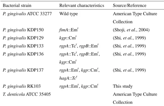
Table 1. Bacterial strains used in this study
Bacterial strain Relevant characteristics Source/Reference
P. gingivalis ATCC 33277 Wild type American Type Culture
Collection
P. gingivalis KDP150 fimA::Emr (Shoji, et al., 2004)
P. gingivalis KDP129 kgp::Cmr (Shi, et al., 1999)
P. gingivalis KDP133 rgpA::Tcr, rgpB::Emr (Shi, et al., 1999)
P. gingivalis KDP136 rgpA::Tcr, rgpB::Emr,
kgp::Cmr
(Shi, et al., 1999)
P. gingivalis KDP137 rgpA::Emr, kgp::Cmr,
hagA::Tcr
(Shi, et al., 1999)
P. gingivalis RK103 rgpA::Emr, kgp::Cmr This study
T. denticola ATCC 35405 American Type Culture
Table 2. Inhibition of coaggregation by addition of amino acids or protease inhibitors
Tested chemicals Concentration (mM) Inhibition (%)
Amino acids L-arginine 100 98.1 + 1.7* 50 94.0 + 1.0* 25 92.0 + 1.0* L-lysine 100 98.5 + 1.8* 50 93.1 + 1.1* 25 82.5 + 5.5* L-histidine 100 87.9 + 5.4* 50 92.2 + 0.5* 25 97.6 + 3.6* Inhibitor TLCK 10 99.6 + 2.7* 5 97.6 + 2.3* 0.5 69.6 + 7.2* Leupeptin 1 50.4 + 10.6* 0.5 43.0 + 7.9* 0.1 33.0 + 8.6* Heat treatment 75 °C 30 min P. gingivalis - 90.8 + 36.6* T. denticola - -17.7 + 1.38* *P < 0.05
Figure legends
Figure 1.
A. Evaluation of P. gingivalis ligands involved in coaggregation by overlay assay and immunoblotting. P. gingivalis proteins were separated following electrophoresis and blotted membranes incubated with sonic extracts of T. denticola ATCC 35405. Membranes were then immunostained with rabbit anti-serum against whole cells of T.
denticola ATCC 35405.
Lanes 1: P. gingivalis after reaction with T. denticola sonicate; 2: Control, P.
gingivalis not incubated with T. denticola sonicate. Arrows indicate the 61-, 53-,
43-, 40 and 38.9-kDa bands.
B. Comparison of bands obtained with overlay assay and immunoblotting with anti-Hgp44 serum. Lane 1: Membrane transferred with P. gingivalis antigen was blocked with 3% BSA containing PBS, incubated with 3% BSA containing PBS, reacted with anti-Hgp44 rabbit antibody and the reacted antibody detected with peroxidase conjugated anti-rabbit antibody. Lane 2: Membrane transferred with P.
gingivalis antigen was blocked with 3% BSA containing PBS, reacted with sonic
extracts of T. denticola ATCC 35405 (1 mg/ml), reacted with anti-T. denticola rabbit antibody and the reacted antibody detected with peroxidase conjugated anti-rabbit antibody.
Figure 2.
Coaggregation of T. denticola with P. gingivalis gingipain- and adhesin-deficient
and coaggregation activities evaluated after 60 min incubation. Autoaggregation (%) of P. gingivalis ATCC 33277, KDP150, KDP129, KDP133, KDP136 KDP137 and T.
denticola ATCC35405 alone were 2.76 ± 1.52, 2.49 ± 1.19, 3.46 ± 0.67, 19.1 ± 1.93,
1.63 ± 0.81, 2.39 ± 0.46 and 14.98 ± 25.16, respectively. *: P<0.05
Fig. 3
A. Adherence of recombinant Rgp domains to T. denticola. Each domain (10 µg/ml) of Rgp was coated onto 96-well EIA plates and sonicates of T. denticola (10 µg/ml) reacted for 1 h. Levels of adherent T. denticola proteins were evaluated using anti-T. denticola antibody.
Each experiment was performed 4 times using 5 samples. CAT: Catalytic subunit of RgpA, Hgp44: Hgp15-27, Non: Control *: P<0.05
B. Inhibition of coaggregation between T. denticola and P. gingivalis by P. gingivalis Hgp44 antibody. P. gingivalis cell suspensions (OD660=0.5) in 24-well
microtiter plates were incubated with anti-P. gingivalis ATCC 33277 whole cell IgG, anti-Hgp44 IgG or normal rabbit IgG (control) for 10 min, followed by addition of T.
denticola ATCC 35405 (OD660=0.5). After 30 min, coaggregation was evaluated
visually.
1. Anti-P. gingivalis ATCC 33277 whole cell IgG 2. Anti-Hgp44 IgG
3. Normal rabbit IgG (control) PBS: P. gingivalis treated with PBS.
Figure 4
Coaggregation increased by coculture with P. gingivalis mutants KDP136 and KDP137.
P. gingivalis KDP 136 was cocultured with P. gingivalis KDP 137 at 37°C for 1-2 days
and coaggregation activity with T. denticola ATCC 35405 evaluated. *: P<0.05
Figure 5
Coaggregation of T. denticola with P. gingivalis HGP-44 domain-deficient mutants. Cells of each P. gingivalis HGP44 domain-deficient mutant strain were mixed with T.
denticola ATCC 35405 grown in E-TYGV medium and coaggregation activities
evaluated after 60 min incubation. Autoaggregation (%) of P. gingivalis ATCC 33277, KDP129, RK103, KDP137 and T. denticola 35404 alone was 6.0 ± 3.37, 3.6 ± 1.8, 1.40 ± 0.62, 4.2 ± 1.6 and -5.6 ± 3.28, respectively.