Title
First isolation and genetic characterization of pseudocowpox virus
from cattle in Japan( 本文(Fulltext) )
Author(s)
OHTANI, Akifumi; YOKOYAMA, Akihiro; NARUSHIGE,
Hisato; INOSHIMA, Yasuo
Citation
[Virology Journal] vol.[14] no.[1] p.[172]
Issue Date
2017
Rights
© The Author(s). This article is distributed under the terms of the
Creative Commons Attribution 4.0 International License
(http://creativecommons.org/licenses/by/4.0/), which permits
unrestricted use, distribution, and reproduction in any medium,
provided you give appropriate credit to the original author(s) and
the source, provide a link to the Creative Commons license, and
indicate if changes were made. The Creative Commons Public
Domain Dedication waiver
(http://creativecommons.org/publicdomain/zero/1.0/) applies to
the data made available in this article, unless otherwise stated.
Version
出版社版 (publisher version) postprint
URL
http://hdl.handle.net/20.500.12099/74380
S H O R T R E P O R T
Open Access
First isolation and genetic characterization
of pseudocowpox virus from cattle in Japan
Akifumi Ohtani
1, Akihiro Yokoyama
1, Hisato Narushige
1and Yasuo Inoshima
2,3,4*Abstract
Background: Pseudocowpox virus (PCPV) infects cattle worldwide with zoonotic potential but has not been isolated in Japan. Thus, the epidemiological status of PCPV infection in cattle is undetermined.
Results: In May 2016, a cattle in a farm in Yamaguchi Prefecture showed white vesicles and hyperemia in the mucosa under the tongue surface, but not on the teats and coronary cushions. A parapoxvirus was isolated from the oral lesion swab and was genetically characterized based on the full-length sequence ofB2L gene encoding viral envelope. Phylogenetic analysis showed that the isolated virus was classified into PCPV.
Conclusion: This case indicates its potential spread in Japan. This is the first report of isolation of PCPV in Japan. Keywords: Pseudocowpox virus, Cattle, Oral lesions, Isolation,B2L gene
Background
Pseudocowpox virus (PCPV) is a member of the genus Parapoxvirusin the family Poxviridae, which includes bo-vine papular stomatitis virus (BPSV) and orf virus (ORFV) [1]. Parapoxviruses are commonly known as causative agents of dermal diseases in ruminants worldwide, leading to papular stomatitis and contagious pustular dermatitis, especially in the regions of the lips, nostrils, oral mucosa, and teats. The importance of PCPV is increasingly recog-nized, primarily because of economic losses to farmers in connection with disease outbreaks and because of their zoonotic potential [2].
In Japan, although serological surveys have revealed that seroprevalence of parapoxvirus is very high in cattle and sheep [3, 4] and multiple BPSVs have been isolated [5], no PCPV has yet been isolated; thus, the epidemiological sta-tus of PCPV infection in cattle is undetermined.
We here report the first case of the isolation of PCPV in Japan. We determined the full-length sequence of the B2L gene encoding viral envelope of this isolate, and evaluated its phylogenetic relation to known members of this virus group.
Methods
Clinical and epidemiological investigations
In May 2016, a breeding cow (Japanese Black, female, 13-month old) in a farm in Yamaguchi Prefecture, in the western part of Japan showed anorexia, mild fever, frothy salivation, and hyperemia in the mucosa under the tongue surface. No lesions were observed on the teats or coronary cushions. A few days after the onset of clinical signs, the cattle showed white vesicles on the tongue surface (Fig. 1). These signs were convalescent in about 1 week. No other cattle in the herd showed clinical signs.
Sample collection
An oral swab sample was collected from the mucosal le-sions of the affected cattle and was homogenized with Eagle’s minimum essential medium (MEM). The sample was centrifuged at 800×g for 10 min at 4 °C. The super-natant was filtered through a 450-nm membrane (Merck Millipore, Cork, Ireland) and used for virus isolation and DNA extraction.
Virus isolation
Virus isolation was performed through inoculation in primary bovine testis (BT) cells; two cell lines, hamster lung (HmLu-1) and Madin-Darby bovine kidney (MDBK) cells were also tested for comparison. The cells were grown in Eagle’s MEM with kanamycin (Nissui Phar-maceutical, Tokyo, Japan) supplemented with 0.295%
* Correspondence:[email protected]
2
Laboratory of Food and Environmental Hygiene, Cooperative Department of Veterinary Medicine, Gifu University, 1-1 Yanagido, Gifu, Gifu 501-1193, Japan
3The United Graduate School of Veterinary Sciences, Gifu University, 1-1
Yanagido, Gifu, Gifu 501-1193, Japan
Full list of author information is available at the end of the article
© The Author(s). 2017 Open Access This article is distributed under the terms of the Creative Commons Attribution 4.0 International License (http://creativecommons.org/licenses/by/4.0/), which permits unrestricted use, distribution, and reproduction in any medium, provided you give appropriate credit to the original author(s) and the source, provide a link to the Creative Commons license, and indicate if changes were made. The Creative Commons Public Domain Dedication waiver (http://creativecommons.org/publicdomain/zero/1.0/) applies to the data made available in this article, unless otherwise stated.
tryptose phosphate broth (TPB), 0.015% sodium bicarbon-ate, 0.03% L-glutamine, and 5–10% fetal bovine serum at
37 °C. The cells were cultured in rolling tubes and 24-well plates, washed with maintenance medium (MEM contain-ing 0.295% TPB, 0.015% sodium bicarbonate, 0.03%L -glu-tamine, and 0.1% bovine serum albumin), and inoculated with 0.1 ml of the processed samples. After adsorption for 60 min at 37 °C, the inocula were replaced with 0.5 ml of maintenance medium. The rolling tube-cultured and 24-well plates-cultured cells were incubated by rotary cultures and stationary cultures, respectively, and observed daily for cytopathic effects (CPE) for at least 7 days. Cultures without CPE were passaged twice in a blinded manner.
Genetic analysis
DNA was extracted both from the oral swab sample and from the BT cells showing a CPE using magLEAD 12gC (Precision System Science, Chiba, Japan). Polymerase chain reaction (PCR) amplifications were carried out with TaKaRa Ex Taq Hot Start Version (TaKaRa Bio, Shiga, Japan) using a SimpliAmp Thermal Cycler (Thermo Fisher Scientific, Kanagawa, Japan) for detection of the full-length (1137 bp) and partial-length (554 bp) B2L gene encoding envelope of parapoxvirus with the primer sets OVB2LF1/OVB2LR1 [6] and PPV1/PPV4 [5], respectively. PCR products were purified using NucleoSpin Gel and PCR Clean-up (Macherey-Nagel, Düren, Germany), and the nucleotide sequence was determined by direct sequen-cing using a BigDye Terminator Cycle Sequensequen-cing Kit v3.1 (Applied Biosystems, Austin, TX, U.S.A.). Sequence data were aligned using the ClustalW method [7]. Phylogenetic analysis was performed using MEGA 6 software [8]. Phylogenetic trees were constructed using maximum-likelihood methods, and the reliability of the branches was evaluated by bootstrapping with 1000 replicates. Nucleo-tide and deduced amino acid sequences were compared
with those of available corresponding parapoxviruses (Table 1). Bovine viral diarrhea virus [9], epizootic hemorrhagic disease virus [10], bluetongue virus [10], ovine herpesvirus 2 [11], and bovine herpesvirus 1 [12], were not detected by PCR using specific primers for de-tection of each viruses (data not shown).
Results
At the third passage, a distinct CPE was observed from 2 to 3 days after inoculation in the rolling tube-cultured BT cells, characterized by a rounded morphology and cell de-tachment (Fig. 2). We designated the isolate as strain YG2828. However, at the third passage, no CPE appeared in the other cell lines or in stationary cultures. For histo-logical observations, confluent monolayers of BT cells in the chamber slide system were inoculated with the isolate. Twenty-four hours after inoculation, the BT cells were fixed with acetone and stained with hematoxylin-eosin, which revealed pyknosis, and eosinophilic and basophilic cytoplasmic inclusion bodies (data not shown).
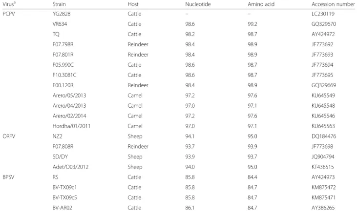
Fragments of expected size were amplified by PCR using both primer sets. Neither deletions nor insertions in the nucleotide sequence of the YG2828 strain were found (Additional file 1: Figure S1). Based on the nu-cleotide/amino acid identities and phylogenetic analysis of the full-length B2L gene, the YG2828 strain was clas-sified as PCPV (Fig. 3). The nucleotide identities against published parapoxviruses ranged from 85.8 to 98.6% (Table 1), and showed the highest identity (98.6%) to three PCPV strains: F05.990C and F10.3081C isolated from cattle in 2005 and 2010 in Finland, respectively, and VR634 isolated from a human in the USA in 1963 with “milker’s nodules” on the hands. The deduced amino acid identities ranged from 84.4 to 99.2%, and showed the highest identity (99.2%) to the VR634 strain (Table 1), even though these strains were isolated inde-pendently, chronologically, and geographically.
Discussion
As noted above, seroprevalence of parapoxvirus is very high in cattle in Japan and multiple BPSVs have been isolated [5], but no PCPV has yet been isolated. In this study, a PCPV was firstly isolated in Japan by rotary cul-tures, but not stationary cultures. Similarly, Mavromousta-kis et al. [13] reported that significantly (P < 0.01) less herpes simplex virus was produced in stationary than in rotary cultures. Although the procedures of rotary cul-tures are more burdensome than those of static culcul-tures, we suggest that rolling of inoculated cultures should be conventionally applied in clinical virology laboratories to aid in the isolation of PCPV. In the affected cattle in this study, there was no evidence of infection on the teats and udder, which are the more common lesion sites of pseudo-cowpox infection. The classification of parapoxviruses was
Fig. 1 Clinical presentation of an affected cattle with white vesicles in the mucosa under the tongue surface
formerly based on the natural host range, clinical signs, and serology [14], however this does not always reflect the classification revealed by molecular analysis [5] as evidenced by the present study.
Previously, there has only been one report describing the PCR detection of PCPV DNA in Japan, in which PCPV DNA was detected from oral lesions in a calf in Iwate Pre-fecture, in the northern part of Japan, but virus isolation was unsuccessful [15]. Notably, the partial-length sequence of the B2L gene determined from the PCR product (acces-sion no. AB921003) was identical to that of the present strain YG2828 (data not shown). Thus, our results con-firmed that PCPV can be isolated from atypical sites besides
the teats and udder, and suggest that YG2828-like PCPV may cause oral lesions in cattle. Moreover, since parapox-viruses cross-react antigenically and two similar strains in-fected cattle in different locations in separate years, YG2828-like PCPV might be spreading among the cattle population in Japan. It is known that cattle are frequently infected with parapoxvirus subclinically [15] and PCPV has zoonotic potential [2]. Therefore, we recommend to wear gloves for people with regular exposure to cattle mucosa.
Conclusion
A PCPV was firstly isolated in Japan from the oral lesion swab of cattle showing white vesicles and hyperemia in
Table 1 Nucleotide and deduced amino acid sequence identities (%) of the full-lengthB2L gene
Virusa Strain Host Nucleotide Amino acid Accession number PCPV YG2828 Cattle – – LC230119 VR634 Cattle 98.6 99.2 GQ329670 TQ Cattle 98.2 98.7 AY424972 F07.798R Reindeer 98.4 98.9 JF773692 F07.801R Reindeer 98.4 98.9 JF773693 F05.990C Cattle 98.6 98.7 JF773694 F10.3081C Cattle 98.6 98.7 JF773695 F00.120R Reindeer 98.4 98.9 GQ329669 Arero/05/2013 Camel 97.2 97.6 KU645549 Arero/04/2013 Camel 97.0 97.1 KU645548 Arero/02/2014 Camel 97.2 97.6 KU645546 Hordha/01/2011 Camel 97.0 97.1 KU645563 ORFV NZ2 Sheep 94.1 95.0 DQ184476 F07.808R Reindeer 93.7 93.9 JF773698 SD/DY Sheep 93.9 93.7 JQ904794 Adet/O03/2012 Sheep 94.0 95.0 KT438515 BPSV RS Cattle 85.8 84.4 AY424973 BV-TX09c1 Cattle 85.8 84.7 KM875472 BV-TX09c5 Cattle 85.8 84.7 KM875471 BV-AR02 Cattle 86.1 84.7 AY386265
a
PCPV, pseudocowpox virus; ORFV, orf virus; BPSV, bovine papular stomatitis virus
Fig. 2 Cytopathic effect observed in BT cells at passage 3. The cells were tested at day 3 after inoculation. Non-infected control (a) and infected (b) cells are shown
the mucosa under the tongue surface, but not on the teats and coronary cushions, by rotary cultures. Genetic characterization based on the full-length sequence of B2Lgene revealed that the isolated virus was genetically close to strains isolated from cattle in the USA and Finland. PCPV is responsible for significant economic losses in the cattle production. Further virological and epidemiological studies to characterize this strain and the possibility of its spread in Japan are highly required.
Additional file
Additional file 1: Figure S1. Alignment of the deduced amino acid sequences of the full-lengthB2L gene. Amino acids identical to the pseudocowpox virus strain YG2828 at given positions are represented by dots. (PPTX 73 kb)
Abbreviations
BPSV:Bovine papular stomatitis virus; BT: Bovine testis; CPE: Cytopathic effects; HmLu-1: Hamster lung; MDBK: Madin-Darby bovine kidney; MEM: Minimum essential medium; ORFV: Orf virus; PCPV: Pseudocowpox virus; PCR: Polymerase chain reaction; TPB: Tryptose phosphate broth Acknowledgements
We acknowledge Ms. Kaori Shimizu for her assistance in preparation of the manuscript.
Funding
This study was partly supported by a Grant-in-Aid (No. 16H05027) for Scientific Research and by the Grant for Joint Research Program of the Research Center
for Zoonosis Control, Hokkaido University, from the Ministry of Education, Culture, Sports, Science and Technology, Japan.
Availability of data and materials
Sequence data obtained in this study is available in the GenBank (accession no. LC230119).
Authors’ contributions
AO and YI analyzed all data in the experiment and were major contributors in writing the manuscript. AO and AY performed virus isolation and PCR experiments from samples from the affected cattle. AO and YI performed the nucleotide/amino acid identities and phylogenetic analysis. HN performed the histological experiment. All authors read and approved the final manuscript. Ethics approval
This study was approved by the Gifu University Animal Care and Use Committee (Approval number 14094). Sampling of oral swab was performed with informed owner consent.
Consent for publication Not applicable Competing interests
All authors declare that they have no competing interests.
Publisher’s Note
Springer Nature remains neutral with regard to jurisdictional claims in published maps and institutional affiliations.
Author details
1Yamaguchi Chubu Livestock Hygiene Service Center, 671-5 Kagawa,
Yamaguchi, Yamaguchi 754–0897, Japan.2Laboratory of Food and Environmental Hygiene, Cooperative Department of Veterinary Medicine, Gifu University, 1-1 Yanagido, Gifu, Gifu 501-1193, Japan.3The United
Graduate School of Veterinary Sciences, Gifu University, 1-1 Yanagido, Gifu,
Fig. 3 Phylogenetic tree of parapoxviruses based on the deduced amino acid sequence of the full-lengthB2L gene (378 amino acids). The per-centage bootstrap values calculated from 1000 replications are indicated above the internal nodes
Gifu 501-1193, Japan.4Education and Research Center for Food Animal
Health, Gifu University (GeFAH), 1-1 Yanagido, Gifu, Gifu 501-1193, Japan.
Received: 28 June 2017 Accepted: 30 August 2017
References
1. Knowles DP. (2011) Poxviridae. In: Maclachlan NJ, Dubovi FJ, editors. Fenner’s Veterinary virology. 4th ed. London: Academic Press; 2011. p. 151–65.
2. Friederichs S, Krebs S, Blum H, Wolf E, Lang H, von Buttlar H, Büttner M. Comparative and retrospective molecular analysis of parapoxvirus (PPV) isolates. Virus Res. 2014;181:11–21.
3. Kuroda Y, Yoshida M, Shibahara T, Matsui T, Nakane T, Hara H, Inoshima Y, Sentsui H. An epidemic of parapoxvirus infection among cattle : isolation and antibody survey. J Vet Med Sci. 1999;61:749–53.
4. Sentsui H, Inoshima Y, Minami A, Yamamoto Y, Murakami K, Shimizu S. Survey on antibody against parapoxvirus among cattle in Japan. Microbiol Immunol. 2000;44:73–6.
5. Inoshima Y, Murakami K, Yokoyama T, Sentsui H. Genetic heterogeneity among parapoxviruses isolated from sheep, cattle and Japanese serows (Capricornis crispus). J Gen Virol. 2001;82:1215–20.
6. Hosamani M, Bhanuprakash V, Scagliarini A, Singh RK. Comparative sequence analysis of major envelope protein gene (B2L) of Indian orf viruses isolated from sheep and goats. Vet Microbiol. 2006;116:317–24. 7. Thompson JD, Higgins DG, Gibson TJ. CLUSTAL W: improving the sensitivity
of progressive multiple sequence alignment through sequence weighting, position-specific gap penalties and weight matrix choice. Nucleic Acids Res. 1994;22:4673–80.
8. Tamura K, Stecher G, Peterson D, Filipski A, Kumar S. MEGA6: Molecular evolutionary genetics analysis version 6.0. Mol Biol Evol. 2013;30:2725–9. 9. Vilček Š, Herring AJ, Herring JA, Nettleton PF, Lowings JP, Paton DJ.
Pestiviruses isolated from pigs, cattle and sheep can be allocated into at least three genogroups using polymerase chain reaction and restriction endonuclease analysis. Arch Virol. 1994;136:309–23.
10. Ohashi S, Yoshida K, Yanase T, Kato T, Tsuda T. Simultaneous detection of bovine arboviruses using single-tube multiplex reverse transcription-polymerase chain reaction. J Virol Methods. 2004;120:79–85. 11. Baxter SIF, Pow I, Bridgen A, Reid HW. PCR detection of the
sheep-associated agent of malignant catarrhal fever. Arch Virol. 1993;132:145–59. 12. Rocha MA, Barbosa EF, Guimarães SEF, Dias Neto E, Gouveia AMG. A high
sensitivity-nested PCR assay for BHV-1 detection in semen of naturally infected bulls. Vet Microbiol. 1998;63:1–11.
13. Mavromoustakis CT, Witiak DT, Hughes JH. Effect of high-speed rolling on herpes simplex virus detection and replication. J Clin Microbiol. 1988;26: 2328–31.
14. Robinson AJ, Lyttle DJ. Parapoxviruses : their biology and potential as recombinant vaccines. In: Binns M, Smith GL, editors. Recombinant Poxviruses. Boca Raton: CRC Press; 1992. p. 285–327.
15. Yaegashi G, Sasaki I, Chiba S, Murakami K. Molecular analysis of parapoxvirus detected in eight calves in Japan. J Vet Med Sci. 2013;75:1399–403.
• We accept pre-submission inquiries
• Our selector tool helps you to find the most relevant journal • We provide round the clock customer support
• Convenient online submission • Thorough peer review
• Inclusion in PubMed and all major indexing services • Maximum visibility for your research
Submit your manuscript at www.biomedcentral.com/submit