博士学位論文
Molecular mechanisms of redox-mediated stress
responses in higher plants
Doctoral Dissertation
Molecular mechanisms of redox-mediated stress
responses in higher plants
Masahiro Noshi
Kindai University
Department of Advanced Bioscience
Faculty of Agriculture
(英文題目)
Molecular mechanisms of redox-mediated stress responses
in higher plants
Masahiro Noshi
Kindai University, Department of Advanced Bioscience
Faculty of Agriculture
(Advisor: Prof. Shigeru Shigeoka)
(和文題目)
高等植物における
レドックスを介したストレス応答の分子機構
近畿大学農学部 バイオサイエンス学科
野志 昌弘
(指導:重岡 成 教授)
Submitted to the Kindai University, to fulfill the requirement for the Doctorate Degree. November 15, 2016
ACKNOWLEDGEMENTS
I wish to my science graduate to Dr. Shigeru Shigeoka, Professor of Faculty of Agriculture, Kindai University, for his kind guidance, valuable advice, stimulating discussion, and critical review throughout the work including the manuscript of this thesis.
I am grateful to Dr. Tamo Fukamizo and Dr. Tsutomu Kawasaki, Professors of Faculty of Agriculture, Kindai University, for reading the entire text in itsoriginal form. I wish to express my gratitude to Dr. Takanori Maruta, Faculty of Life and Environmental Science, Shimane University, for his continuous support, beneficial advice, and helpful discussions throughout the work.
I am thankful to Masahiro Tamoi and Dr. Noriaki Tanabe, Faculty of Agriculture, Kindai University, Dr. Kazuya Yoshimura, College of Bioscience and Biotechnology, Chubu University, and Dr. Yukinori Yabuta, Faculty of Agriculture, Tottori University, and Takahiro Ishikawa, Faculty of Life and Environmental Science, Shimane University, for their precious suggestions and comments throughout the work. I am grateful to Dr. Nam-Hai Chua (Rockefeller) and Dr. Tsutoshi Nakagawa (Shimane University) for donation of the pMDC7 vector, and the pDONR201, pGWB80, pGWB505, and pGWB506 vector, respectively. I am also grateful to Dr. Masaru Ohme-Takagi (National Institute of Advanced Industrial Science and Technology) for donation of dominant-negative mutants for RTS genes. I am thankful to Drs Mikio Nishimura, Makoto Hayashi, and Mitsue Fukazawa (National Institute for Basic Biology) for their technical support using the microarray system. I am also thanlful to Dr. Kohji Nishimura and Yusuke Terai (Shimane University) for excellent technical help with confocal laser scanning microscope.
My special thanks are due to Aoi Tanouchi, Hiroki Yamada, Megumi Ojiri, Kazushi Kusachi, Shun Matsuda, Satoru Nakagami, Maki Nakamura, Takuya Tonomura, Yuma Iwai, Eri Toida, Yutaka Okamoto, Risa Hatanaka, Tatsuya Kurata, Kazunori Takijiri, Koichi Tsujii, Yuki Hane, Wataru Takashiba, Daisuke Mori, Natsumi Masuda, Naoko Okada, Shingo Ieda, Kensuke Matsuda, and Tsukasa Shimazaki for their helpful collaborations. Beside, I am appreciative to Dr. Takahisa Ogawa and Dr. Daisuke Ito for their valuable discussions and suggestions. I additionally would like to thank all the past and present members of our laboratory of Plant Molecular Physiology in the Faculty of Agriculture, Kindai University, for their kind cooperations.
This research was in part supported by Grants-in-Aid for Scientific Research (A) (S.S.: 22248042) from the Ministry of Education, Culture, Sports, Science and Technology (MEXT) of Japan, by the Strategic Project to Support the Formation of Research Bases at Private University: Matching Fund from MEXT, 2011-2015 (S1101035).
ABBREVIATIONS
1O
2 singlet oxygen
4CL 4-coumarate:CoA ligase
ABRC Arabidopsis Biological Resource Center
APX ascorbate peroxidase
AsA reduced ascorbate
bHLH basic helix-loop-helix
C4H cinnamate 4-hydroxylase
CAD cinnamylalcohol dehydrogenase CAS calcium sensing receptor
CAT2 catalase 2
CHI chalcone isomerase
CHS chalcone synthase
COMT caffeic acid/5-hydroxyferulic acid O-methyltransferase CRK cysteine-rich receptor kinase
DFR dihydroflavonol 4-reductase
DHA dehydroascorbate
DHAR dehydroascorbate reductase
eHL extreme HL
F3’H flavonoid 3’-hydroxylase
F3H flavanone 3-hydroxylase
FAH1 ferulate 5-hydroxylase 1 FLS2 flagellin sensing 2 FS flagellin sensitive
GABA γ-aminobutyrate
GAD glutamate decarboxylase
GFP green fluorescent protein
GSH reduced glutathione
GSSG oxidized glutathione GR glutathione reductase
GUS β-glucuronidase
H2O2 hydrogen peroxide
HCT hydroxycinnamoyl-CoA shikimate/quinate hydroxycinnamoyl- transferase
HL high light
HSFA2 heat shock transcription factor A2 ICS isochorismate synthase
IS- inducible silencing-
LDOX lencoanocyanidin dioxygenase
LL low light
MDA malondialdehyde
MDAR monodehydroascorbate reductase MDHA monodehydroascorbate
MV methylviologen MVS MV-susceptible MVR MV-resistant NL normal light NPQ non-photochemical quenching O2− superoxide radical
PAL phenylalanine ammonia-lyase
PAMP pathogen-associated molecular patterns
PCD programmed cell death
PR pathogenesis-related
PTI PAMP-triggered immunity
q-PCR Quantitative Real-Time PCR Rboh respiratory burst oxidase homolog ref reduced epidermal fluorescence ROS reactive oxygen species
ROXY cc-type glutaredoxin
RTS responsive to tAPX silencing
SA salicylic acid
sAPX stromal APX
tAPX thylakoid membrane-bound APX
CONTENTS
CHAPTER I Introduction ……… 1
CHAPTER II Physiological function of H2O2-triggered retrograde signaling from
chloroplasts to nucleus in response to stress ……… 7
CHAPTER III Comprehensive analysis of chloroplastic H2O2 responsive genes
involved in stress responses in Arabidopsis ……… 50
III-I Ferulic acid 5-hydroxylase 1 is essential for anthocyanin accumulation under photooxidative stress ……… 52
III-II The TGA transcription factor, TGA10, is involved in ROS-mediated responses to PAMP ……… 75
CHAPTER IV Regulation of cellular redox states by dehydroascorbate reductases under photooxidative stress in Arabidopsis ……… 110
IV-I Arabidopsis dehydroascorbate reductase 1 and 2 modulate redox states of ascorbate-glutathione cycle in the cytosol in response to photooxidative stress ……… 113
IV-II chloroplastic dehydroascorbate reductase is required for high-light stress tolerance in Arabidopsis ……… 137
REFERENCES ……… 156
CHAPTER I
INTRODUCTION
Since higher plants are sessile, they are continually exposed to environmental
stress, including high light (HL), chilling, heat, ozone, drought, and salinity. The
imposition of environmental stress disrupts the metabolic balance of cells, resulting in
enhanced production of reactive oxygen species (ROS), such as hydrogen peroxide
(H2O2), singlet oxygen (1O2), superoxide radical (O2-), and hydroxy radical (Asada 1999,
Foyer and Shigeoka 2011). Because of their high reactivity, the accumulation of ROS
leads to oxidative damage of important cellular compounds such as DNA, proteins, and
lipids (Mittler 2002, Foyer and Shigeoka, 2011). Consequently, plant organisms have
the efficient ROS-scavenging systems in various cellular compartments (Ishikawa and
Shigeoka, 2008; Noctor et al., 2011; Gallie, 2013). On the other hand, recent findings
indicate that ROS act as signaling molecules associated with many biological process,
including responses to environmental and biotic stress, modulation of hormone signaling
pathway, programmed cell death, growth, and development (Apel and Hirt, 2004; Foyer
and Noctor, 2005; Foyer and Shigeoka, 2011; Shigeoka and Maruta, 2014). Thus, ROS
play dual roles: as harmful toxic compounds and signals that modulate gene expression
for plant acclimation to stress (Mittler et al., 2004, 2011; Gadjev et al., 2006; Schmidt and
Figure I-1. A scheme of ROS-mediated response to stress.
It is becoming clear that the ROS are essential in response and adaptation to stress.
In plant cells, ROS are generated not only as by-products of primary metabolisms but
also by ROS-producing enzymes, such as plasma membrane- or apoplast-localized
oxidases and peroxidases, including NADPH oxidase, called the respiratory burst oxidase
homolog (Rboh) (Torres et al., 2002; Sagi et al., 2006; Maruta et al., 2011; Mittler et al.,
2011). Indeed, apoplastic Rboh (RbohD) is essential for plant immunity, because ROS
produced by Rboh are considered to be directly toxic to pathogens, induce stomata
closure to restrict pathogen entry, strengthen the plant cell wall, activate the expression of
defense genes, and trigger systemic signaling (Torres et al., 2006; Marino et al., 2012;
Kadota et al., 2015). Additionally, recent findings indicated that chloroplastic ROS also
have an important role in plant immunity (Yao and Greenberg, 2006; Liu et al., 2007;
Zurbriggen et al., 2009; Gou et al., 2015). Conclusively, Caplan et al. (2015)
demonstrated that stroma-filled tubules that extend from chloroplasts (stromules) are
induced and transport defense signals, including H2O2, into the nucleus during immunity.
of the thylakoid-attached copper/zinc superoxide dismutase (CSD2) showed a dwarf
phenotype even under normal growth conditions, but were more tolerant to oxidative
stress due to the induction of other defense-related genes (Rizhsky et al., 2003).
Interestingly, these findings imply the existence of common and source-/kind-specific
pathways for ROS signaling (Figure I-2). Consistent with this speculation, recent
comparative analyses of ROS-responsive transcriptome have indicated that the response
of a number of genes to ROS is irrespective to the kind and production site of the
compounds, whereas the response of another set of genes is highly specific (Gadjev et al.,
2006). It is thought that the synergistic and antagonistic interactions of these ROS
signaling pathways has essential roles in the fine-tuning of plant responses to
environmental and biotic stress. Vanderauwera et al., (2011) revealed that the lack of
both, but not respecitively, cytosolic ascorbate peroxidase (APX) and peroxisomal
catalase enhanced tolerance to oxidative stress and inhibited of programmed cell death
(PCD) accompanying the activation of a DNA damage response, indicating the
physiological importance of crosstalk between cytosolic and peroxisomal ROS-mediated
signaling in stress responses. Moreover, it has been reported that overexpression of
tAPX in flu mutants enhanced 1O2-dependent programmed cell death, suggesting that
H2O2 generated in chloroplasts is an antagonist of oxidative signaling through 1O2 (Laloi
et al., 2007). Therefore, the balance between ROS production and scavenging must be
tightly regulated in plant cells under stress conditions, and the analysis of physiological
functions and molecular mechanisms of not only common- but also source- and/or
kind-specific ROS pathway is required for understanding the complex network of ROS
Figure I-2. Sub-cellular distribution of ROS mediated-oxidative signaling pathway.
The photosynthesic organisms have developed maintenance system of cellular
redox states for several antioxiant enzymes such as APX, catalase, superoxide dismutase,
glutathione peroxidase, and thioredoxin, and antioxidant compounds, including ascorbate,
tocopherol, glutathione, and anthocyanin (Chalker-Scott, 1999; Noctor et al., 2011; Foyer
and Noctor, 2011; Gallie, 2013; Shigeoka and Maruta, 2014). Ascorbate and glutathione
are major aqueous antioxidants in plant cells and, thus, act as essential redox buffers
(Noctor et al., 2011; Foyer and Noctor, 2011; Gallie, 2013). They comprise the
ascorbate (AsA) as an electron donor. In addition, ascorbate itself can also detoxify
ROS thought non-enzymatic reaction. These reactions lead to the production of
monodehydroascorbate (MDHA). MDHA is reduced back to AsA by MDHA reductase
(MDAR) or ferredoxin, otherwise it is spontaneously disproportionated into AsA and
dehydroascorbate (DHA). DHA reductase (DHAR) reduces DHA to AsA using reduced
glutathione (GSH) as an electron donor. Oxidized glutathione (GSSG), a product of the
DHAR reaction, is then recycled by glutathione reductase (GR). NAD(P)H serves as an
electron donor for MDAR and GR activities. This cycle is distributed to the cytosol,
chloroplasts, mitochondria, and peroxisomes and contributes to the modulation of cellular
redox states (Asada 1999, Shigeoka et al., 2002; Chew et al., 2003; Gallie, 2013) (Figure
I-3). In addition, ascorbate is required for other photo-protection-related pathways, including the xanthophyll cycle (Eskling et al., 1997), regeneration of tocopherols
(Munné-Bosch and Alegre, 2002; Sattler et al., 2004), and biosynthesis of flavonoids
(Page et al., 2012). In the xanthophyll cycle, ascorbate serves as an electron donor for
enzymatic reactions catalyzing the de-epoxidation of violaxanthin to antheraxanthin and
zeaxanthin, enhancing the dissipation of light energy as a heat under photo-oxidative
stress conditions (Eskling et al., 1997). Furthermore, the role of AsA as an electron
donor in photosystem I (PSI) and PSII has been demonstrated (Mano et al., 2004; Asada,
2006; Page et al., 2012; Toth et al., 2013). These findings indicate that ascorbate
recycling via DHAR and MDAR is important for the modulation of the redox states of
Figure I-3. Sub-cellular localization of ascorbate-glutathione recycling enzymes in
Arabidpsis plants.
In this study, to clarify the molecular mechanisms of redox-mediated stress
responses in higher plants, I studied the followings;
(1) H2O2-triggered retrograde signaling from chloroplasts to nucleus plays specific role in
response to stress,
(2) Comprehensive analysis of chloroplastic H2O2 responsive genes involved in stress
responses in Arabidopsis,
(3) Regulation of cellular redox states by dehydroascorbate reductases under
CHAPTER II
H2O2-triggered retrograde signaling from chloroplasts to nucleus plays specific role
in response to stress
Introduction
In plant cells, ROS are produced as by-products of central metabolism, such as
photosynthesis, respiration, and photorespiration, in chloroplasts, mitochondria, and
peroxisomes, respectively. In particular, chloroplasts are one of the most significant
sources of ROS in plant organelles because the Mehlar reaction leads to the formation of
ROS in light-exposed plant cells. The ROS generated in chloroplasts act as a retrograde
signal to the nucleus for regulating plant responses to environmental stress. The
identification of a conditional fluorescent (flu) mutant of Arabidopsis, which allowed the
production of 1O2 within plastids in a controlled manner, provided genetic evidence that
the release of 1O
2 is involved in the regulation of PCD (op den Camp et al., 2003;
Meskauskiene et al., 2001; Wagner et al., 2004; Lee et al., 2007; Kim et al., 2008). In
the flu mutants, 1O2 generated within the first minute of re-illumination had a negative
effect on growth and development. After re-illumination, distinct sets of genes were
activated that were different from those induced by paraquat, a producer of H2O2 and O2-,
suggesting that 1O
2 acts as a signal with a high degree of specificity (op den Camp et al.,
shock transcription factor A2 (HsfA2) and cytosolic APX (APX1 and APX2), was
significantly suppressed in knock-out mutants lacking stromal and thylakoid
membrane-bound ascorbate peroxidases (respective sAPX and tAPX), which were key
enzymes for the scavenging of H2O2 in chloroplasts, although oxidative damage under
HL was enhanced in these mutants (Maruta et al., 2010), indicating that H2O2 derived
from chloroplasts has a negative effect on the expression of ROS-responsive genes under
HL. Thus, it seems likely that different kinds of ROS produced in the same organelle
are associated with distinct signaling pathways.
To clarify the signaling function of H2O2 derived from chloroplasts, a system for
producing H2O2 in a controlled manner is needed. For the system, the
chemical-inducible RNAi would be more useful than the knock-out or constitutive
knockdown method because plants may acclimate to the knock-out or constitutive
knockdown of target gene during growth and development. Furthermore, this system
should not require any application of stress to plants because other signaling molecules,
including hormones, are produced by stress and may act synergistically or
antagonistically. Preciously, a novel system for producing H2O2 in Arabidopsis
chloroplasts by chemical-dependent tAPX silencing using an estrogen-inducible RNAi
method has created (Zuo et al., 2000; Guo et al., 2003; Maruta et al., 2012a). When the
expression of tAPX was silenced in the leaves, the levels of oxidized protein in the
chloroplasts were increased (Maruta et al., 2012a). Microarray analysis revealed that
the expression of a large set of genes (named responsive to tAPX silensing; RTS)
changed in response to tAPX silencing (Maruta et al., 2012a). Among them, only a
cold acclimation and pathogen resistance. These findings suggest that H2O2 derived
from chloroplasts acts as a specific signal for regulating plant responses to abiotic and
biotic stress (Maruta et al., 2012a).
In this study, it was demonstrated that tAPX silencing had negative and positive
effects on cold acclimation and salycilic acid (SA) response, respectively, and that there
were synergistic and antagonistic roles of chloroplastic H2O2 on HL response. These
findings provide a new insight into the role of H2O2-triggered retrograde signaling from
chloroplasts in the response to stress in planta.
Materials and Methods
Plant Materials and Growth Conditions
The generation of the IS-tAPX-19 (for tAPX silencing) and IS-GUS-2 (for control)
plants were conducted previously by Maruta et al. (2012a). The IS-tAPX-19 plants was
introduced an RNAi construct having an inverted repeat corresponding to a 530-bp
fragment of the 3’-terminal region of tAPX under controlof an estrogen-inducible
promoter. T-DNA insertion lines for tAPX in the Col-0 background (KO-tAPX;
WiscDsLox457–460A17) were previously obtained from the Arabidopsis Biological
Resource Center (ABRC) (Maruta et al., 2010). The lists of Knockout and/or
dominant-negative mutant lines of RTS genes used on stress-sensitive and -insensitive
obtained from ABRC, and the dominant negative mutant lines were donated from Dr.
Masaru Ohme-Takagi (National Institute of Advanced Industrial Science and Technology),
respectively.
Table II-1. List of T-DNA insertion lines used for mutant screening.
AGI code Stock No. Annotation
At5g59130 SALK_089292 Subtilase family protein SALK_145867C
At4g34470 CS25145 SKP1-like 12
SALK_105764C
At5g05740 SALK_028514 ethylene-dependent gravitropism-deficient and yellow-green-like 2
SALK_142694 SALK_032572C
At2g25940 SALK_077733 alpha-vacuolar processing enzyme SALK_151201
At3g01345 SALK_141810C Expressed protein
At1g02470 SALK_123221C Polyketide cyclase/dehydrase and lipid transport superfamily protein
At5g56050 SALK_093745C At1g78995 SALK_041344C At3g19920 SALK_106471C
At1g64770 SALK_103166C NDH-dependent cyclic electron flow 1 At3g50140 SALK_049878C Plant protein of unknown function (DUF247) At1g07280 SALK_022732C Tetratricopeptide repeat (TPR)-like superfamily
protein
At2g45830 SALK_135799C downstream target of AGL15 2 At1g62780 SALK_047296C
At1g64355 SALK_057125C
At5g57010 SALK_134786C calmodulin-binding family protein
At4g11340 SALK_110928C Disease resistance protein (TIR-NBS-LRR class) family
SALK_053459C
At3g46550 CS859546 Fasciclin-like arabinogalactan family protein At5g17330 SALK_022227C glutamate decarboxylase
At4g32990 SALK_050504C Transducin/WD40 repeat-like superfamily protein At4g14640 SALK_022524C calmodulin 8
SALK_015253C
At5g56510 SALK_057302C pumilio 12
At5g47330 SALK_081909C alpha/beta-Hydrolases superfamily protein
At2g37950 SALK_023712C RING/FYVE/PHD zinc finger superfamily protein At3g16090 CS24962 RING/U-box superfamily protein
At1g49230 SALK_058308C RING/U-box superfamily protein At3g59220 SALK_063087C pirin
At5g02200 SALK_040459C far-red-elongated hypocotyl1-like
At1g52770 SALK_104317C Phototropic-responsive NPH3 family protein At3g44290 SALK_012554C NAC domain containing protein 60
SALK_127838C
At1g72210 SALK_060037C basic helix-loop-helix (bHLH) DNA-binding superfamily protein
At3g27810 SALK_042711C myb domain protein 21 At3g23250 SALK_151976C myb domain protein 15 At1g21450 SALK_102071C SCARECROW-like 1
At3g11280 SALK_004053C Duplicated homeodomain-like superfamily protein SALK_021025C
At3g62090 SALK_090239C phytochrome interacting factor 3-like 2 SALK_147579C
At5g06839 SALK_124227C bZIP transcription factor family protein At3g01080 SALK_150041C WRKY DNA-binding protein 58 At3g06490 SALK_076395C myb domain protein 108
SALK_024059C
At4g28530 SALK_094441C NAC domain containing protein 74 SALK_104622C
At2g34830 SALK_111141C WRKY DNA-binding protein 35 At2g24260 SALK_080724C LJRHL1-like 1
At1g18860 SALK_006029C WRKY DNA-binding protein 61 At5g67480 SALK_045370C BTB and TAZ domain protein 4 At3g48360 SALK_002306C BTB and TAZ domain protein 2
At2g05160 SALK_128806C CCCH-type zinc fingerfamily protein with RNA-binding domain
At5g59340 SALK_050488C WUSCHEL related homeobox 2 SALK_139683C
At1g74930 SALK_109440C Integrase-type DNA-binding superfamily protein At5g61600 SALK_152806C ethylene response factor 104
At2g34210 SALK_147117C Transcription elongation factor Spt5
At5g56840 SALK_002490C myb-like transcription factor family protein At5g13730 SALK_078760C sigma factor 4
finger
At4g25470 SALK_073208C C-repeat/DRE binding factor 2 At4g37260;
At4g37270
SALK_023478C myb domain protein 73
At5g60100 SALK_090261C pseudo-response regulator 3
At5g04150 SALK_011245C basic helix-loop-helix (bHLH) DNA-binding superfamily protein
At2g41180 SALK_152005C VQ motif-containing protein
At5g37260 SALK_074896C Homeodomain-like superfamily protein
At4g17460 SALK_059835C Homeobox-leucine zipper protein 4 (HB-4) / HD-ZIP protein
SALK_006022C
At3g50060 SALK_067655C myb domain protein 77 At3g17420 SALK_047485C glyoxysomal protein kinase 1 At1g33770 SALK_042880C Protein kinase superfamily protein
At4g26890 SALK_003255C mitogen-activated protein kinase kinase kinase 16 SALK_071559C
At1g65190 SALK_151531C Protein kinase superfamily protein At5g10520 SALK_043441C ROP binding protein kinases 1
SALK_080548C
At3g62090 SALK_147579C phytochrome interacting factor 3-like 2 At2g34830 SALK_111141C WRKY DNA-binding protein 35 At1g18860 SALK_006029C WRKY DNA-binding protein 61 At3g06490 SALK_076395C myb domain protein 108
At3g59350 SALK_136404C Protein kinase superfamily protein At5g39790 SALK_012617C 5'-AMP-activated protein kinase-related
At5g59670 SALK_115780C Leucine-rich repeat protein kinase family protein At3g04530 SALK_102132C phosphoenolpyruvate carboxylase kinase 2 At5g59090 SALK_093039C subtilase 4.12
SALK_045125C
At3g59250 SALK_017364C F-box/RNI-like superfamily protein At3g48340 SALK_079519C Cysteine proteinases superfamily protein
At1g49040 SALK_031730C stomatal cytokinesis defective / SCD1 protein (SCD1) At4g25350 SALK_010010C EXS (ERD1/XPR1/SYG1) family protein
At1g66390 SALK_093731C myb domain protein 90
At4g32280 SALK_091933C indole-3-acetic acid inducible 29 SALK_152235C
At1g33760 SALK_091690C Integrase-type DNA-binding superfamily protein At2g05160 SALK_128806C CCCH-type zinc fingerfamily protein with
RNA-binding domain
SALK_000337C
At5g63160 SALK_006453C BTB and TAZ domain protein 1 At5g03720 SALK_011131C heat shock transcription factor A3
At4g35900 SALK_054421C Basic-leucine zipper (bZIP) transcription factor family protein
SALK_118487C
At2g40670 SALK_142105C response regulator 16
At4g13190 CS859976 Protein kinase superfamily protein
At4g04510 SALK_053147C cysteine-rich RLK (RECEPTOR-like protein kinase) 38
SALK_075451C
At4g04540 SALK_098187C cysteine-rich RLK (RECEPTOR-like protein kinase) 39
At4g04500 SALK_131604C cysteine-rich RLK (RECEPTOR-like protein kinase) 37
At3g45330 SALK_069415C Concanavalin A-like lectin protein kinase family protein
At4g23310 SALK_051873C cysteine-rich RLK (RECEPTOR-like protein kinase) 23
SALK_052723C
At2g45080 SALK_039130C cyclin p3;1
At3g28890 SALK_041685C receptor like protein 43 SALK_141689C
At1g51790 SALK_025281C Leucine-rich repeat protein kinase family protein At5g16900 SALK_113523C Leucine-rich repeat protein kinase family protein At1g55610 CS800035 BRI1 like
SALK_046929C
At2g32510 SALK_080309C mitogen-activated protein kinase kinase kinase 17 At4g11890 SALK_057538C Protein kinase superfamily protein
SALK_037588C
At1g18890 SALK_105108C calcium-dependent protein kinase 1 At4g13000 SALK_083220C AGC kinase family protein At5g22980 SALK_136908C serine carboxypeptidase-like 47 At3g45010 SALK_079721C serine carboxypeptidase-like 48 At5g51380 SALK_038578C RNI-like superfamily protein
SALK_123218C
At2g45920 SALK_061534C U-box domain-containing protein SALK_080919C
SALK_050218C
At4g03360 SALK_130537C Ubiquitin family protein SALK_062751C
At3g28330 SALK_043730C F-box family protein-related
At3g23880 SALK_008269C F-box and associated interaction domains-containing protein
At2g22680 SALK_041291C Zinc finger (C3HC4-type RING finger) family protein SALK_149664C
At5g15660 SALK_047400C F-box and associated interaction domains-containing protein
SALK_063563C
At5g44460 SALK_148734C calmodulin like 43 SALK_026474C
At5g57010 SALK_134786C calmodulin-binding family protein SALK_022337C
At4g11340 SALK_104383C Disease resistance protein (TIR-NBS-LRR class) family
SALK_110928C SALK_053459C
At2g40170 SALK_112719C Stress induced protein SALK_041260C
At5g64920 SALK_023424C COP1-interacting protein 8
At3g11500 SALK_026590C Small nuclear ribonucleoprotein family protein SALK_062774C
SALK_126162C
At2g19240 SALK_011223C Ypt/Rab-GAP domain of gyp1p superfamily protein At2g35000 CS24736 RING/U-box superfamily protein
SALK_036065C
At1g09540 SALK_144610C myb domain protein 61 At1g04445 SALK_130261C C2H2-like zinc finger protein
At1g54400 SALK_008690C HSP20-like chaperones superfamily protein At4g19570 SALK_077671C Chaperone DnaJ-domain superfamily protein At1g18870 SALK_073287C isochorismate synthase 2
SALK_084635C
At5g56300 SALK_106872C gibberellic acid methyltransferase 2 SALK_075450C
At3g53150 SALK_009017C UDP-glucosyl transferase 73D1 SALK_004078C
At1g05530 CS862227 UDP-glucosyl transferase 75B2
At3g25880 SALK_083521C NAD(P)-binding Rossmann-fold superfamily protein At5g13320 SALK_018225C Auxin-responsive GH3 family protein
At4g34131 SALK_097487C UDP-glucosyl transferase 73B3 At1g78960 SALK_024920C lupeol synthase 2
SALK_013690C
At4g36430 SALK_129503C Peroxidase superfamily protein At5g14180 SALK_101919C Myzus persicae-induced lipase 1
SALK_031227C
At5g02540 SALK_103967C NAD(P)-binding Rossmann-fold superfamily protein SALK_134983C
At2g24210 SALK_041114C terpene synthase 10 SALK_108420C
At3g11430 SALK_018117C glycerol-3-phosphate acyltransferase 5 SALK_142456C
At4g36220 CS862388 ferulic acid 5-hydroxylase 1
At1g11730 SALK_045206C Galactosyltransferase family protein At1g23730 SALK_144106C beta carbonic anhydrase 3
At5g11920 SALK_127864C 6-&1-fructan exohydrolase SALK_152299C
At1g66960 SALK_022044C Terpenoid cyclases family protein SALK_097188C
At1g51470 SALK_013785C beta glucosidase 35 SALK_114084C
At1g01190 CS860633 cytochrome P450, family 78, subfamily A, polypeptide 8
At2g32530 SALK_063361C cellulose synthase-like B3 SALK_059674C
SALK_025333C
At3g03480 SALK_025557C acetyl CoA:(Z)-3-hexen-1-ol acetyltransferase SALK_129278C
At1g35910 SALK_013114C Haloacid dehalogenase-like hydrolase (HAD) superfamily protein
At1g30710 SALK_112240C FAD-binding Berberine family protein
At5g26690 SALK_148742C Heavy metal transport/detoxification superfamily protein
SALK_103915C
At4g26380 SALK_026306C Cysteine/Histidine-rich C1 domain family protein SALK_011364C
At5g23240 SALK_024726C DNAJ heat shock N-terminal domain-containing protein
At5g23235 SALK_008678C
At2g40130 SALK_025338C Double Clp-N motif-containing P-loop nucleoside triphosphate hydrolases
At1g26230 SALK_064887C TCP-1/cpn60 chaperonin family protein SALK_136518C
At1g44160 SALK_152637C HSP40/DnaJ peptide-binding protein At1g27130 SALK_022297C glutathione S-transferase tau 13
At5g45340 SALK_078173C cytochrome P450, family 707, subfamily A, polypeptide 3
At3g24420 SALK_068313C alpha/beta-Hydrolases superfamily protein SALK_126829C
SALK_026193C
At1g07260 SALK_042564C UDP-glucosyl transferase 71C3 SALK_021979C
At2g39800 SALK_063517C delta1-pyrroline-5-carboxylate synthase 1 At1g01390 CS859558 UDP-Glycosyltransferase superfamily protein
SALK_083984C
At3g13730 SALK_036476C cytochrome P450, family 90, subfamily D, polypeptide 1
SALK_031965C
At3g61900 CS861648 SAUR-like auxin-responsive protein family CS860713
At4g21490 SALK_097329C NAD(P)H dehydrogenase B3 SALK_130320C
At4g26530 SALK_080758C Aldolase superfamily protein
At5g58860 SALK_074232C cytochrome P450, family 86, subfamily A, polypeptide 1
SALK_146813C
At4g10120 CS862248 Sucrose-phosphate synthase family protein SALK_037958C
At3g48520 SALK_001709C cytochrome P450, family 94, subfamily B, polypeptide 3
SALK_018989C
At1g27940 CS860441 P-glycoprotein 13 At3g44560 SALK_010208C fatty acid reductase 8
SALK_021133C
At3g44550 CS861044 fatty acid reductase 5 At4g15440 SALK_085831C hydroperoxide lyase 1
SALK_149325C SALK_094392C SALK_091988C
At1g06080 SALK_069299C delta 9 desaturase 1 SALK_044895C
At3g55630 CS860621 DHFS-FPGS homolog D
At3g61080 SALK_059076C Protein kinase superfamily protein
At1g04420 SALK_072037C NAD(P)-linked oxidoreductase superfamily protein SALK_148484C
At4g29840 SALK_081563C Pyridoxal-5'-phosphate-dependent enzyme family protein
At2g11810 SALK_069971C monogalactosyldiacylglycerol synthase type C At5g04360 CS25085 limit dextrinase
SALK_064456C
At5g04140 SALK_011035C glutamate synthase 1
At1g04620 SALK_001001C coenzyme F420 hydrogenase family / dehydrogenase, beta subunit family
At5g61610 SALK_018790C Oleosin family protein At3g01440 SALK_051912C PsbQ-like 1
At1g14150 SALK_006106C PsbQ-like 2
At5g39210 SALK_070919C chlororespiratory reduction 7 SALK_073978C
SALK_128408C
At2g45350 CS836745 Pentatricopeptide repeat (PPR) superfamily protein At5g14740 SALK_056127C carbonic anhydrase 2
At1g78450 SALK_012200C SOUL heme-binding family protein SALK_019423C
At5g26220 SALK_056007C ChaC-like family protein
At5g52310 CS859786 desiccation-responsive protein 29A (RD29A) At5g52970 SALK_091900C thylakoid lumen 15.0 kDa protein
At4g18375 SALK_016188C RNA-binding KH domain-containing protein At5g23980 CS859844 ferric reduction oxidase 4
SALK_044469C CS860468
At1g29395 SALK_008692C COLD REGULATED 314 INNER MEMBRANE 1 At2g18196 SALK_056166C Heavy metal transport/detoxification
superfamily protein SALK_012260C
At1g03020 SALK_032946C Thioredoxin superfamily protein At5g61440 SALK_075409C atypical CYS HIS rich thioredoxin 5
Table II-2. List of dominant-negative mutant lines used for mutant screening.
AGI code Annotation
At4g17460 HAT1 (homeobox-leucine zipper protein 1); DNA binding / transcription factor
At3g27810 ATMYB21 (MYB DOMAIN PROTEIN 21); DNA binding / transcription factor
At3g11280 myb family transcription factor
At4g28530 ANAC074 (Arabidopsis NAC domain containing protein 74); transcription factor
arrest 24); transcription factor
At5g24110 WRKY30 (WRKY DNA-binding protein 30); transcription factor At5g61430 ANAC100/ATNAC5 (Arabidopsis NAC domain containing protein
100); transcription factor
At3g44290 ANAC060 (Arabidopsis NAC domain containing protein 60); transcription factor
At5g14000 ANAC084 (Arabidopsis NAC domain containing protein 84); transcription factor
At4g29230 ANAC075 (Arabidopsis NAC domain containing protein 75); transcription factor
At2g17040 ANAC036 (Arabidopsis NAC domain containing protein 36); transcription factor
At2g02450
ANAC034/ANAC035 (Arabidopsis NAC domain containing protein 34, Arabidopsis NAC domain containing protein 35); transcription factor
At2g37590 Dof-type zinc finger domain-containing protein At4g38000 Dof-type zinc finger domain-containing protein
At1g09540 MYB61 (myb domain protein 61); DNA binding / transcription factor At4g18960 AG (AGAMOUS); transcription factor
At2g40260 myb family transcription factor
At5g65230 AtMYB53 (myb domain protein 53); DNA binding / transcription factor
At1g72210 basic helix-loop-helix (bHLH) family protein (bHLH096) At5g43410 ERF (ethylene response factor)
At1g68320 MYB62 (myb domain protein 62); DNA binding / transcription factor At4g17800 DNA-binding protein-related
At3g23250 AtMYB15/AtY19/MYB15 (myb domain protein 15); DNA binding / transcription factor
At1g21450 SCL1 (SCARECROW-LIKE 1); transcription factor
At3g62090 PIL2 (PHYTOCHROME INTERACTING FACTOR 3-LIKE 2); transcription factor
AT3G06490 MYB108 (BOTRYTIS-SUSCEPTIBLE1, myb domain protein 108); DNA binding / transcription factor
At4g36060 basic helix-loop-helix (bHLH) family protein At3g12910 transcription factor
At1g36060 AP2 domain-containing transcription factor, putative At4g08250 scarecrow transcription factor family protein
At1g04445 zinc finger (C2H2 type) family protein
At4g02670
ATIDD12 (ARABIDOPSIS THALIANA
INDETERMINATE(ID)-DOMAIN 12); nucleic acid binding / transcription factor/ zinc ion binding
At3g53820 zinc finger (C2H2 type) family protein At2g24260 basic helix-loop-helix (bHLH) family protein
At3g15270 SPL5 (SQUAMOSA PROMOTER BINDING PROTEIN-LIKE 5); DNA binding / transcription factor
At4g32280 IAA29 (indoleacetic acid-induced protein 29); transcription factor At1g56650 PAP1 (PRODUCTION OF ANTHOCYANIN PIGMENT 1); DNA
binding / transcription factor
At1g33760 AP2 domain-containing transcription factor, putative
At4g25490 CBF1 (C-REPEAT/DRE BINDING FACTOR 1); DNA binding / transcription factor/ transcriptional activator
At4g06746 RAP2.9 (related to AP2 9); transcription factor At1g77640 AP2 domain-containing transcription factor, putative
At4g14560 IAA1 (INDOLE-3-ACETIC ACID INDUCIBLE); transcription factor At3g05800 transcription factor
At1g52830 IAA6 (indoleacetic acid-induced protein 6); transcription factor At5g59340 WOX2 (WUSCHEL-related homeobox 2); transcription factor At1g68520 zinc finger (B-box type) family protein
At1g19210 AP2 domain-containing transcription factor, putative At1g06160 ethylene-responsive factor, putative
At1g74930 AP2 domain-containing transcription factor, putative At3g49930 zinc finger (C2H2 type) family protein
At5g61600 ethylene-responsive element-binding family protein At5g56840 DNA-binding family protein
At3g07650 COL9 (CONSTANS-LIKE 9); transcription factor/ zinc ion binding At3g46640 PCL1 (PHYTOCLOCK 1); DNA binding / transcription factor At1g24260 SEP3 (SEPALLATA3); transcription factor
At3g46130 MYB111 (myb domain protein 111); DNA binding / transcription factor
At3g15540 IAA19 (indoleacetic acid-induced protein 19); transcription factor At1g04240 SHY2 (SHORT HYPOCOTYL 2); transcription factor
At1g75490 DRE-binding transcription factor
At5g51190 AP2 domain-containing transcription factor, putative
At5g03720 AT-HSFA3 (Arabidopsis thaliana heat shock transcription factor A3); DNA binding / transcription factor
At4g25470 CBF2 (FREEZING TOLERANCE QTL 4); DNA binding / transcription factor/ transcriptional activator
At4g37260 AtMYB73/MYB73 (myb domain protein 73); DNA binding / transcription factor
At4g17490 ATERF6 (ETHYLENE RESPONSIVE ELEMENT BINDING
FACTOR 6); DNA binding / transcription factor At4g39070 zinc finger (B-box type) family protein
At5g47230 ERF5 (ETHYLENE RESPONSIVE ELEMENT BINDING FACTOR
5); DNA binding / transcription factor/ transcriptional activator At5g04150 basic helix-loop-helix (bHLH) family protein
At4g35900 FD (FD); DNA binding / transcription factor At5g37260 myb family transcription factor
At5g04340 C2H2; nucleic acid binding / transcription factor/ zinc ion binding At3g50060 myb family transcription factor
At2g40670 ARR16 (response regulator 16); transcription regulator/ two-component response regulator
At3g23230 ethylene-responsive factor, putative
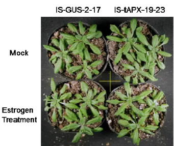
Surface-sterilized Arabidopsis thaliana wild-type (Col-0) and transgenic seeds
were sown on Murashige & Skoog medium containing 3% (w/v) sucrose. Plates were
stratified in darkness for 2 days at 4 °C and then transferred to a growth chamber. After
7 days, seedlings were potted in soil and grown in the same growth chamber. In this
study we use a continuous light (NL: 100 µmol of photons m-2 s-1, 25 °C) condition for
plant growth to abolish light/dark effect on the production of ROS in chloroplasts.
Seventeenday-old IS-tAPX-19 and IS-GUS-2 plants were treated with a 100 µM estrogen
solution containing 0.1% (v/v) Tween 20 under light.
Preparation of Total RNA and cDNA Synthesis
Total RNA was semi-automatically purified from leaves of Arabidopsis plants
using a QuickGene RNA cultured cell kit and Quick-Gene-Mini80 (FUJIFILM Corp.,
Tokyo, Japan). The first strand cDNA was synthesized using reverse transcriptase
(ReverTra Ace; Toyobo) with an oligo(dT) primer. These analyses were performed
according to the manufacturer’s instructions.
Quantitative Real-Time PCR Experiments
Quantitative Real-Time PCR (q-PCR) experiments were performed according to
Nishizawa et al. (2006). Primer sequences were described in Table II-3. The q-PCR
experiments were also repeated at least three times with cDNA prepared from three
Table II-3. Primer sequences used for q-PCR analysis.
AGI code Forward Primer Reverse Primer
At3g18780 5'-GGTGGTTCCATTCTTGCTTC CC-3' 5'-TCATACTCGGCCTTGGAGAT CC-3' At5g59845 5'-ACTCTCAACCGCGGATTCG-3' 5'-TCTTCCTGCCTTTGAACATC TCA-3' At1g60110 5'-TGGAGCACATTTCTGTCCGT TAC-3' 5'-CCCAAGAAGCTCCTCCATCA C-3'
At2g20825 5'-GCACCAGCCACCGTTACG-3' 5'-GGTGATTTGAAGTTCTCCAT CTGAA-3' At3g14680 5'-CAATGGTTCTGTTGAGCCA ACA-3' 5'-GCCAAAAACTTGCTTCACTT CCT-3' At4g13190 5'-AGCTGTAGCAATAGCGGCA ATG-3' 5'-TAAGAGCGGTTCCCGTTAG ACC-3' At4g25490 5'-TTTGGGATGCCGACTTTGTT -3' 5'-CATCTCCTTCGCCGTCATAA TTA-3' At4g25470 5'-GGCTATTTATACGCCGGAA CAG-3' 5'-CCTTCGGCCATGTTATCCAA -3' At5g15970 5'-GCAAAGCTGAGGAGAAGAG CAA-3' 5'-CGATATACTCTTTCCCGCCT GTT-3' At2g42530 5'-GCTTCTGATTTCGTGACGGA TAA-3' 5'-CGCAGTTTCATTGGCTTCAA -3' At1g29395 5'-GGGAGAGTATGGTGTATGG G-3' 5'-CCAAGAGTAATGCGATGAA TGG-3' At1g29390 5'-GGGTGAATATGGCATCTGG G-3' 5'-CCAAAAGCAAAGCAATGAA GGG-3' At1g18870 5'-CCATTTTCTGGGCACAAACC -3' 5'-GGCGAGAGACACGAGAGG AT-3' At4g01870 5'-GGTGGTTCCATTCTTGCTTC CC-3' 5'-CCGTGAACTTCAACGCTCA A-3' At4g11340 5'-CGTCTCGCTTGGGCTTAACA -3' 5'-TGAGGAAACGTTGTCAAGC ATT-3' At4g47890 5'-AAATGAGCTTCCTTATTCGT TCCA-3' 5'-TTGGTCGGAATGACACAAG TG-3' At2g32680 5'-GTTCGACGAAGGCGGCTAT-3' 5'-CAATGGCGGCGTAGGAAGT -3' At3g11010 5'-ACACGCCAGCATCACATCA A-3' 5'-ACCAGGTCCGAATCCTATT GC-3' At3g24900 5'-GGTCACTCGACGTTGGCTA CA-3' 5'-GATTCCGTTGTGGTCCACAC T-3'
CA-3' -3' At1g09415 5'-AGCAGAGGCCAACAACATT GA-3' 5'-CGTTAGTCTCAGATCCTTCT TGCTT-3' At2g04450 5'-GGTCCAGGAGATTGATGGT CAT-3' 5'-CCCTAAGTGCTCCTTCCCAA A-3' At2g02130 5'-ATCTCCGCTGCTCTTCTCTT GT-3' 5'-TTGAACCTATGGCTTTTTGA CTCA-3' At2g02120 5'-TCGCAGAGCCAAAGATTCA A-3' 5'-CGGAATCCACGGCAATCA-3' At2g26150 5'-GTGTTGAGGTTGGGCAATA CG-3' 5'-TTGCTGTTGCCTCAACCTAA CTAC-3' At3g09640 5'-AGGACCGTTTGGGACGATA AG-3' 5'-TAACGGCAATATCAAGACC ATTGTT-3' At5g59720 5'-GGTTACCGGAGAATGCAAA GATG-3' 5'-CGATGGACTTGACTTGAGG CTTC-3' At1g74710 5'-CTAATCTCCGCCGTCTCTGA ACT-3' 5'-TTGGAACCTGTAACCGAAC GA-3' At2g14160 5'-CGAAAGCTCAAGATAGCCC ACA-3' 5'-TTCTGCGTAGCTCCGAGCAT AG-3' At3g57260 5'-CACGGCCAACATCCATCTA GAC-3' 5'-AACCGAGTCGAGATTTGCG TC-3'
Measurements of Chlorophyll Fluorescence
Chlorophyll fluorescence was measured following Maruta et al. (2010). The
maximum quantum yield of photosystem II (Fv/Fm), and quantum yield of photosystem
II (PSII) in Arabidopsis leaves was determined after dark adaptation for 20 min.
Chlorophyll fluorescence in the Arabidopsis leaves was measured at 25 °C with a Closed
FluorCam 800MF (Photon Systems Instruments, Brno, Czech Republic).
Quantification of Free and Total SA
Free and total SA were extracted and measured according to Malamy et al. (1992)
methanol and centrifuged at 7,000 x g for 15 min. The pellet was resuspended with 3
mL of 90% (v/v) methanol and centrifuged. Methanol extracts were combined,
centrifuged at 7,000 x g for 15 min, and dried at 35 °C under vacuum. The dried
methanol extract was resuspended in 5 mL of water at 80 °C, and the solution was
divided into two equal portions. An equal volume of 0.2 M sodium acetate buffer (pH
4.5) was added to one portion for assaying free SA, and an equal volume of 0.2 M sodium
acetate buffer (pH 4.5) containing 0.24 mg/mL -glucosidase (16.9 unit/mg; Roche) was
added to the other for assaying total SA. Both samples were incubated at 37 °C
overnight. After digestion, samples were acidified to pH 1–1.5 with HCI. The fractions
were then extracted with two volumes of ethyl acetate:cyclopentane:isopropyl alcohol
(50:50:1 [v/v]), dried down, resuspended in 23% (v/v) methanol/sodium acetate (pH 5.0),
and filtered through a centrifugal filter (0.2 µm; Millipore). SA was quantified by
reverse-phase HPLC on a 5C18 column (COSMOSIL; AR-II, 4.6ID x 150 mm) and
detected using a Shimazu RF-10AXL fluorescence detector (excitation energy 313 nm,
emission energy 405 nm).
Data Analysis
The significance of differences between data sets was evaluated with a t test.
Calculations were carried out with Microsoft Excel software.
Suppression of Cold-responsive Genes by tAPX Silencing Causes Plant Sensitivity to Cold Stress
Previous microarray data using IS-tAPX indicated that the expression of
cold-responsive genes was suppressed by tAPX silencing under NL, which was consistent
with the results of the q-PCR analysis (Figure II-1A). These genes included
C-repeat/DRE binding factor/dehydration-responsive element binding factors
(CBF1/DREB1B and CBF2/DREB1C), and coldregulated genes (COR6.6, COR15B,
COR414-TM1, and COR414-TM2). To investigate the involvement of the H2O2
signaling derived from chloroplasts in the regulation of cold response, the effect of tAPX
silencing on plant sensitivity to cold was checked. Seventeen-day-old IS-GUS-2-17 and
IS-tAPX-19-23 plants were incubated under light for 48 h after the treatment with or
without estrogen and then subjected to cold stress (continuous light of 100 µmol of
photons m-2 s-1, 4 °C) for 2 weeks. Both transgenic lines showed visible symptoms
under cold stress, but only the estrogen-treated IS-tAPX-19-23 plants exhibited brown
leaves (Figure II-1B). In addition, Fv/Fm decreased significantly in the IS-tAPX-19-23
plants compared with the IS-GUS-2-17 plants under cold stress (Figure II-1C). The
cold sensitivity of IS-tAPX-19-23 plants depended on the treatment with estrogen
(Figure II-1B and 1C). On the other hand, the cold-sensitive phenotypes of the
tAPX-silenced plants were not observed under low light (LL; 10 µmol photons m-2 s-1)
(Figure II-2), because the production rate of chloroplastic H2O2 appears to be limited
under LL. These findings demonstrated that tAPX silencing negatively affects cold
Figure II-1. Effect of tAPX silencing on cold acclimation.
(A) Seventeen-day-old IS-GUS-2-17 and IS-tAPX-19-23 plants were sprayed with a 100 µM estrogen and kept under NL. At 48 h after the estrogen treatment, the transcript levels of RTS genes (CBF1/DREB1B, CBF2/DREB1C, COR6.6, COR15B, COR414-TM1, and COR414-TM2), known to be involved in cold acclimation, were measured by q-PCR. Error bars indicate SD (n
= 3). Significant differences: *P < 0.05 vs. the value for IS-GUS-2-17 plants. (B and C)
17-day-old IS-GUS-2-17 and IS-tAPX-19-23 plants were sprayed with a 100 µM estrogen solution or water (mock) and transferred to cold stress conditions (100 µmol photons m-2 s-1, 4 °C) for 2 weeks. The treatment with estrogen was performed every 3 days to maintain the tAPX silencing. (B) Fourteen days after cold stress, the IS-GUS-2-17 and IS-tAPX-19-23 plants were photographed. The same results were obtained in four independent experiments. (C) Fv/Fm values in the leaves of IS-GUS-2-17 and IS-tAPX-19-23 10 days after cold stress were measured using a Closed FluorCam 800MF. Error bars indicate SD (n = 3). Significant differences: *P < 0.05 vs. the value for IS-GUS-2-17 plants.
Figure II-2. Effect of lack of tAPX on cold acclimation under LL.
Seventeen-day-old IS-GUS-2-17 and IS-tAPX-19-23 plants were sprayed with a 100 µM estrogen solution or water (mock), and transferred to cold stress conditions (10 µmol photons m-2 s-1, 4°C) for 2 weeks. The treatment with estrogen was performed every 3 days in order to maintain the tAPX silencing. At 14 days after cold stress, the IS-GUS-2-17 and IS-tAPX-19-23 plants were photographed. The same results were obtained in three independent experiments.
The sensitivity of KO-tAPX plants to cold stress was also analyzed. Although
the transcript levels of CBF2/DREB1C and COR414-TM1 were significantly lower in the
KO-tAPX plants than wild-type plants, those of other cold-responsive genes were similar
in the two lines (Figure II-3). In addition, there was no visible difference in sensitivity
to cold stress between the wild-type and KO-tAPX plants. Furthermore, the Fv/Fm in
the leaves of wild-type plants was almost the same as that in the KO-tAPX plants under
cold stress (Figure II-3). The activity of APX in the membrane fraction was much
lower in the KO-tAPX plants than in the estrogen-treated (48 h) IS-tAPX-19-23 plants
signaling and not due to a decrease in ROS scavenging capacity. Thus, the H2O2
signaling derived from chloroplasts seems to be involved in the suppression of cold
acclimation.
Figure II-3. Effect of lack of tAPX on cold acclimation.
(A) The wild-type and KO-tAPX plants were grown under light for 17 days. The transcript levels of RTS genes (CBF1/DREB1B, CBF2/DREB1C, COR6.6, COR15B, COR414-TM1, and
COR414-TM2) were measured by q-PCR. Error bars indicate SD (n = 3). Significant
differences: *P < 0.05 vs. the value for wild-type plants. (B) Seventeen-day-old wild-type and KO-tAPX plants were transferred to cold stress conditions (100 µmol photons m-2 s-1, 4°C). At 2 weeks after cold stress, the wild-type and KO-tAPX plants were photographed. (C) Fv/Fm values in the leaves of wild-type and KO-tAPX at 2 weeks after cold stress were measured using a Closed FluorCam 800MF. Error bars indicate SD (n = 3).
tAPX Silencing Activates Expression of Disease-resistance Genes and SA Response Some RTS genes are known to be involved in pathogen response/resistance,
suggesting that H2O2 derived from chloroplasts plays a role in plant responses to biotic
stress. For example, low molecular weight cysteine-rich proteins (LCR68 and LCR70)
are defensin-type pathogenesis-related (PR) proteins (Sels et al., 2008). Receptor-like
proteins (RLP7, -23, -34, -39, and -41) as well as Toll-interleukin resistance (TIR)
domain protein (At4g11340) and a related protein (At4g01870, annotated as TolB-related
protein) are generally thought to be associated with plant responses to pathogen attack
(Kruijt et al., 2005; Burch-Smith and Dinesh-Kumar, 2007). Cytosolic Nudix hydrolase
(AtNUDX6) and NIM1-interacting 3 (NIMIN-3) were recently identified as regulators of
the SA response in Arabidopsis (Ogawa et al., 2005; Ishikawa et al., 2010a, 2010b;
Weigel et al., 2001). Furthermore, an isochorismate synthase (ICS), ICS2, is involved in
the biosynthesis of SA (Garcion et al., 2008). The q-PCR analysis confirmed that the
transcription of these genes was up-regulated by the tAPX silencing (Figure II-4). In
addition to the induction of ICS2, the levels of free and total SA were slightly but
significantly increased in the IS-tAPX-19-23 plants at 48 h after estrogen treatment
(Figure II-5A). Arabidopsis has another ICS gene, ICS1, that is highly responsive to a
pathogen infection which significantly enhances levels of SA (Wildermuth et al., 2001).
As shown in Figure II-6, tAPX silencing was not effected transcript level of ICS1.
Figure II-4. Effect of tAPX silencing on the transcript levels of disease-resistance genes.
Seventeen-day-old IS-GUS-2-17 and IS-tAPX-19-23 plants were sprayed with a 100 µM estrogen and kept under NL. At 48 h after the estrogen treatment, the transcript levels of RTS genes (ICS2, TolB, TIR, RLP7, RLP23, RLP34, RLP39, RLP41, NIMIN3, NUDX6, LCR68, LCR70),
PR1, and PR2, known to be involved in disease resistance, were measured by q-PCR. Error bars indicate SD (n = 3). Significant differences: *P < 0.05 vs. the value for IS-GUS-2-17
Figure II-5. Effect of tAPX silencing on the response to SA.
Seventeen-dayold IS-GUS-2-17 and IS-tAPX-19-23 plants were sprayed with a 100 µM estrogen and kept under NL for 48 h. (A) levels of free and total SA in the IS-GUS-2-17 and IS-tAPX-19-23 plants before and after estrogen treatment were measured as described in
Materials and Methods. (B) 48 h after treatment with estrogen, IS-GUS-2-17 and
IS-tAPX-19-23 plants were sprayed with a 100 µM SA. The transcript levels of PR1 and PR2, SA-responsive genes, were measured by q-PCR. Error bars indicate SD (n = 3). Significant differences: *P < 0.05 vs. the value for IS-GUS-2-17 plants.
Figure II-6. Effect of tAPX silencing on the transcript levels of ICS1 genes.
Seventeen-day-old IS-GUS-2-17 and IS-tAPX-19-23 plants were sprayed with a 100 µM estrogen and kept under NL. At 48 h after the estrogen treatment, the transcript levels of ICS1 was measured by q-PCR. Error bars indicate SD (n = 3). Significant differences: *P < 0.05 vs. the value for IS-GUS-2-17 plants.
As shown in Figure II-4, however, the transcript levels of pathogenesis-related
genes (PR1 and PR2), known to be SA-responsive genes (Reuber et al., 1998), were not
affected by the tAPX silencing in the IS-tAPX-19-23 plants. Therefore, the effect of
tAPX silencing on the SA response was analyzed. At 48 h after the treatment with
estrogen, the IS-GUS-2-17 and IS-tAPX-19-23 plants were sprayed with 50 µM SA. The
transcript levels of PR1 increased in both plants under the SA treatment but were higher
in the IS-tAPX-19-23 plants (Figure II-5B). Furthermore, the transcript levels of PR2
were also higher in the IS-tAPX-19-23 plants at 3 h after SA treatment. These findings
suggest that the H2O2 signaling derived from chloroplasts activates SA biosynthesis and
SA-inducible gene expression. In contrast, the levels of ICS2 transcripts and SA in the
not higher in the KO-tAPX plants but, rather, lower in the mutants than in the wild-type
plants (Figure II-7). Thus, as was the case for the cold acclimation response, the plant
responses to SA and pathogen were not enhanced in the KO-tAPX plants.
Figure II-7. Effect of lack of tAPX on the transcript levels of RTS genes involved in disease resistance.
The wild-type and KO-tAPX plants were grown under normal light for 17 days. The transcript levels of RTS genes (ICS2, TolB, TIR, RLP7, RLP23, RLP34, RLP39, RLP41, NIMIN3, NUDX6, LCR68, and LCR70) were measured by q-PCR. Error bars indicate SD (n = 3). Significant differences: *P < 0.05 vs. the value for wild-type plants.
Figure II-8. Effect of lack of tAPX on the levels of SA.
The wild-type and KO-tAPX plants were grown under normal light for 17 days. The levels of free and total SA in the wild-type and KO-tAPX plants were measured as described in Materials
and Methods.
To study the effect of chloroplastic H2O2 on the SA response, the sensitivity of
tAPX-silenced plants to SA treatment was analyzed. At 48 h after the treatment with
estrogen, the IS-GUS-2-17 and IS-tAPX-19-23 plants were further treated with a high
concentration (5 mM) of SA for 96 h. As shown in Figure II-9, the leaves of IS-GUS
plants and estrogen-untreated IS-tAPX plants were visibly damaged by SA treatment to
the same degree. Conversely, the leaves of estrogen-treated IS-tAPX plants were
insensitive to the treatment, suggesting that chloroplastic H2O2 causes SA insensitivity.
As shown in Figure II-5, tAPX silencing enhances the levels of SA and the SA response.
However, the SA-insensitive phenotype of the tAPX-silenced plants strongly supports the
possibility that chloroplastic H2O2 is involved in the regulation of the SA response. It is
possible that chloroplastic H2O2 induces the expression of gene(s) involved in the
Figure II-9. Effect of tAPX silencing on SA sensitivity.
Seventeen-day-old IS-GUS-2–17 and IS-tAPX-19–23 plants were sprayed with 100 μM estrogen. At 48 h after the treatment, plants were further sprayed with 5 mM SA. The plants 96 h after SA treatment were photographed. The same results were obtained in three independent experiments. A representative photograph is shown.
Interplay of Chloroplastic H2O2 and HL in Gene Regulation
HL is one of the most characterized stresses to enhance ROS production in
chloroplasts. Therefore, the interplay between chloroplastic H2O2 and HL in gene
regulation was investigated. First, to check the effect of tAPX silencing on HL
sensitivity, at 48 h after estrogen treatment, the IS-GUS-2-17 and IS-tAPX-19-23 plants
were exposed to HL at 1,000 µmol photons m-2 s-1. As shown in Figure II-10A, there
with our previous work using the KO-tAPX plants (Maruta et al., 2010). Fv/Fm was
similarly decreased by HL in both IS-GUS-2-17 and IS-tAPX-19-23 plants (Figure
II-10B). Furthermore, the sensitivity of the tAPX-silenced plants to various concentrations of methylviologen (MV), a ROS-producing agent in chloroplasts and
mitochondria, treatment was almost the same as that of the control plants (data not
shown). These findings suggest that the tAPX silencing in the IS-tAPX-19-23 plants
had no effect on tolerance to HL- and MV treatment-induced photooxidative stress.
Figure II-10. Effect of tAPX silencing on HL sensitivity.
Seventeen-day-old IS-GUS-2-17 and IS-tAPX-19-23 plants were sprayed with a 100 µM estrogen solution or water (mock), and kept under NL. At 48 h after the treatments, IS-GUS-2-17 and IS-tAPX-19-23 plants were exposed to HL (1000 µmol photons m-2 s-1). (A) At 72 h after HL, the IS-GUS-2-17 and IS-tAPX-19-23 plants were photographed. The same results were obtained in three independent experiments. (B) Fv/Fm values in the leaves of IS-GUS-2-17 and IS-tAPX-19-23 were measured using a Closed FluorCam 800MF. Error bars indicate SD (n = 5).
Next, to determine the effect of HL on the expression of five RTS genes (GRFP,
JLFP, ULT2, CYP72A14, and PK), 19-day-old wild-type plants were exposed to HL. The transcript levels of GRFP were significantly increased by HL (Figure II-11),
suggesting that chloroplastic H2O2 acts as a positive signal for HL response. However,
the transcript levels of the other RTS genes were decreased or not affected by HL
exposure (Figure II-11). The effect of tAPX silencing on expression of these genes
under moderate light (ML; 400 µmol photons m-2 s-1) was also analyzed. One-week-old
IS-GUS-2-17 and IS-tAPX-19-23 plants, grown under NL, were further grown for 10
days under ML and then treated with estrogen. There was no difference in phenotype
and Fv/Fm between IS-GUS-2-17 and IS-tAPX-19-23 plants (Figure II-12A and 12B).
At 48 h after estrogen treatment, the transcript levels of GRFP, ULT2, and PK were
significantly suppressed by tAPX silencing. Furthermore, although the transcript levels
of JLFP and CYP72A14 were increased by tAPX silencing, the induction levels of these
genes under ML were largely lower than those under NL (Figure II-12C). In the
IS-GUS-2-17 and IS-tAPX-19-23 plants, the expression of these genes was similar under
both ML and NL (data not shown). Thus, it seems likely that there are not only
Figure II-11. Effect of HL on the expression of RTS genes.
Nineteen-day-old wild-type plants were exposed to HL (1000 µmol of photons m-2 s-1). The transcript levels of RTS genes (GRFP, JLFP, ULT2, CYP72A14, and PK) were measured by q-PCR. Error bars indicate SD (n = 3). Values without a common letter are significantly different according to t tests (P < 0.05).
Figure II-12. Effect of tAPX silencing on the expression of RTS genes under ML.
One-week-old IS-GUS-2-17 and IS-tAPX-19-23 plants grown under NL were further grown for 10 days under ML (400 µmol of photons m-2 s-1). The plants were then sprayed with estrogen and kept under ML for 72 h. (A) 48 h after estrogen treatment the IS-GUS-2-17 and IS-tAPX-19-23 plants were photographed. (B) Fv/Fm values in the leaves of IS-GUS-2-17 and IS-tAPX-19-23 after estrogen treatment were measured using a Closed FluorCam 800MF. Error
bars indicate SD (n = 3). (C) 48 h after estrogen treatment, the transcript levels of RTS genes
(GRFP, JLFP, ULT2, CYP72A14, and PK) were measured by q-PCR. Error bars indicate SD (n
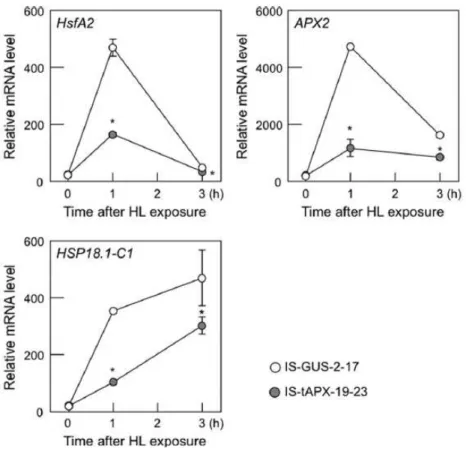
Finally, the effect of tAPX silencing on the expression of known ROS-responsive
genes, HsfA2, APX2, and heat shock protein 18.1-C1 (HSP18.1-C1) under HL was
analyzed (Maruta et al., 2010; Nishizawa et al., 2006). In both IS-GUS-2-17 and
IS-tAPX-19-23 plants, the transcript levels of ROS-responsive genes were drastically
increased by HL (Figure II-13). However, the induction of ROS-responsive genes by
HL was significantly inhibited by tAPX silencing (Figure II-13), supporting the
antagonistic effects of chloroplastic H2O2 on HL Response.
Figure II-13. Effect of tAPX silencing on expression of ROS-responsive genes under HL.
Seventeen-day-old IS-GUS-2-17 and IS-tAPX-19-23 plants were sprayed with a 100 µM estrogen and kept under NL. At 48 h after treatment with estrogen, IS-GUS-2-17 and IS-tAPX-19-23 plants were exposed to HL (1,000 µmol photons m-2 s-1). The transcript levels of ROS-responsive genes (HsfA2, APX2, and HSP18.1-C1) were measured by q-PCR. Error bars
Isolation of stress-sensitive and/or -insensitive mutants from RTS mutant lines
Because RTS genes might be chloroplastic H2O2-responsive genes, functional
analysis of these genes could help to clarify the molecular mechanism underlying the
chloroplastic H2O2-mediated signaling. Hence, the isolation of stress-sensitive and
insensitive mutants from knockout and/or dominant-negative mutants for RTS genes was
conducted. List of knockout and/or dominant-negative mutant lines used for the mutant
screening is shown in Table II-1 and 2. By comparing the sensitivity of these RTS
mutants and wild-type plants to MV-induced oxidative stress and elicitor peptide flg22,
some methylviologen-susceptible and -resistant (mvs and mvr), and flg22-sensitive (fs)
mutants were screened. The causative genes of these mutants were listed in Table II-4.
In a large number of these genes, the physiological function of responses to stress was
unclear. Thus, functional analyses of these genes will be interesting for understanding
the molecular mechanism and further physiological role of ROS-mediated oxidative
signaling in stress response.
Table II-4. List of stress-sensitive and -insensitive RTS mutant lines.
methylviologen susceptible (mvs)
Line No. AGI code Annottion State
mvs1 At5g13730 sigma factor 4 (SIG4) knockout
mvs2 At3g11430 glycerol-3-phosphate acyltransferase 5 knockout
mvs5 At4g28530 NAC domain containing protein 74 knockout
mvs7 At4g17640 Homeobox-leucine zipper protein 4 (HAT1) knockout
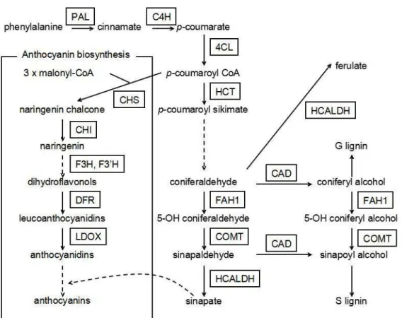
mvs8 At4g36220 ferulic acid 5-hydroxylase 1 (FAH1) knockout
mvs9 At5g04150 basic helix-loop-helix (bHLH) DNA binding super family protein: AtbHLH101
dominant negative
super family protein: AtbHLH11
mvs12 At4g29230 NAC domain containing protein 75 dominant negative
mvs13 At2g17040 NAC domain containing protein 36 dominant negative
mvs14 At1g15580 indole-3-acetic acid inducible 5 (IAA5) dominant negative
mvs15 At1g09540 myb domain protein 61 dominant negative
mvs16 At1g33760 Integrase-type DNA-binding superfamily protein
dominant negative
mvs17 At4g25490 C-repeat/DRF binding factor 1 (CBF1) dominant negative
mvs18 At4g14560 indole-3-acetic acid inducible 1 (IAA1) dominant negative
methylviologen resistance (mvr)
Line No. AGI code Annottion State
mvr1 At1g49230 RING/U-box superfamily protein (ATL78) knockout
mvr2 At5g17330 glutamate decarboxylase knockout
mvr3 At5g63160 BTB and TAZ domain protein 1 knockout
mvr4 At5g15660 F-box and associated interaction domains-containing protein
knockout
mvr5 At1g21750 PDI-like 1-1 knockout
mvr6 At5g59090 subtilase 4.12 knockout
mvr7 At4g25350 EXS (ERD1/XPR1/SYG1) family protein knockout
mvr9 At2g02450 NAC domain containing protein 34/35 dominant negative
flagellin sensitive (fs)
Line No. AGI code Annottion State
fs1 At5g06839 bZIP transcription factor family protein
(AtbZIP65: TGA10)
knockout
fs2 At3g11280 MYB family transcription factor dominant negative
fs3 At5g26170 WRKY DNA binding protein 50 dominant negative
fs4 At5g61430 NAC domain containing protein 100 dominant negative
fs5 At1g72210 basic helix-loop-helix (bHLH) DNA binding super family protein: AtbHLH096