INTRODUCTION
Cavernous hemangioma of the liver is the most common benign neoplasm (1). Cavernous heman-gioma usually presents as solitary well-delineated, subcapsular, and disclosed nodules, and these dis-tinctive structures show a characteristic hemody-namic pattern on enhanced computed tomography (CT). However, it was already reported that some cases contain parts of hyaline degeneration, secon-dary to thrombus, necrosis, or cicatrization, mim-icking the hepatic malignancies such as metastatic liver tumor or cholangiocarcinoma (2). A hepatic
sclerosed hemangioma is the ultimate form having all these degenerative changes (3). We report herein a case of such a rare hepatic tumor, sclerosed he-mangioma, which was misdiagnosed as a liver me-tastasis of gastric cancer.
CASE REPORT
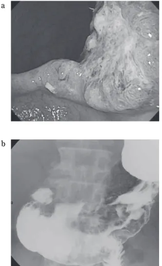
A 75 years old male was referred to our institute for gastric cancer and a hepatic space occupied le-sion (SOL). He had visited an affiliated hospital of our university for routine examination of prostatic hyperplasia, and was pointed out anemia. For fur-ther examinations, he took gastroscopy and fur-there was type 3 tumor at posterior wall of gastric angle (Fig 1a). Biopsied specimens were diagnosed as well differentiated adenocarcinoma (tub1). Gastric fluoroscopy showed tumor and induration of lesser
CASE REPORT
Hepatic screlosed hemangioma which was misdiagnosed
as metastasis of gastric cancer : report of a case
Shinichiro Yamada, Mitsuo Shimada, Tohru Utsunomiya, Yuji Morine, Satoru Imura,
Tetsuya Ikemoto, Hiroki Mori, Jun Hanaoka, Shuichi Iwahashi, Yu Saitoh, and
Michihito Asanoma
The Department of Surgery, the University of Tokushima, Tokushima, Japan
Abstract : A screlosed hemangioma of the liver is rare among hepatic tumors. A 75 years old male was referred to our hospital for gastric cancer and a hepatic tumor. The histo-logical finding of gastric cancer was revealed to be well differentiated adenocarcinoma. The liver tumor was 1.1 1.0 cm in size and located in segment 8 of the liver. Computed tomography (CT) showed it to be a tumor with ring enhancement. Magnetic resonance imaging (MRI) showed the tumor to have a low signal on T1-weighted and slightly high signal on T2-weighted images. Level of hemoglobin was 7.8 g/dl. It was thought to be per-sistent bleeding from gastric cancer. With diagnosis of liver metastasis from gastric can-cer, chemotherapy is recommended. However, to control the bleeding from gastric cancan-cer, we performed distal gastrectomy and wedge resection of liver (S8). The histological exami-nation of the liver tumor revealed to be a hepatic sclerosed hemangioma with hyalinized tissue and collagen fibers. We report herein a case of the rare tumor which was misdiag-nosed as a liver metastasis of gastric cancer. J. Med. Invest. 59 : 270-274, August, 2012
Keywords : cavernous hemangioma, liver, screlosed hemangioma, gastric cancer, metastatic tumor
Received for publication March 8, 2012 ; accepted April 11, 2012. Address correspondence and reprint requests to Mitsuo Shimada, MD, FACS, Department of Surgery, the University of Tokushima, 3 - 18 - 15 Kuramoto, Tokushima City, Tokushima, 770 - 8503, Japan and Fax : + 81 - 88 - 631 - 9698.
curvature (Fig 1b). Enhanced CT showed a small mass in segment 8 of the liver, and this hepatic SOL was suspected to be metastasis of gastric cancer.
The initial laboratory data were as follows : 5300 /μl white blood cell count, 7.8 g/dL hemoglo-bin, 430,000 /μl platelet count, 19 IU/L aspartate aminotransferase, 13 IU/L alanine aminotransferase, 11 mg/dL blood urea nitrogen, 0.87 mg/dL cre-atinin, 3.7 g/dL albumin. Tumor markers were as follows : 7 ng/mL alpha-fetoprotein, 1.7 ng/mL car-cinoembryonic antigen (CEA), and 38 U/mL carbo-hydrate antigen (CA) 19-9. He was positive for hepa-titis B (HB) surface antibody (HBsAb), HBeAb and HBcAb, and negative for HBsAg and HBeAg.
Plain CT showed a low density mass, 1.1
!
1.0 cm in size, in segment 8 of the liver. Enhanced CT showed that the mass lesion was gradually enhanced mainly in the marginal border between normal pa-renchyma and SOL, and the mass was not enhancedcompared to parenchyma (iso-density) in the de-layed phase (Fig 2). Magnetic resonance imaging (MRI) showed that the tumor had a low signal on T1-weighted and high signal on T2-weighted im-ages. Hepatobiliary phase of ethoxybenzyl (EOB)-enhanced MRI showed no uptake in the correspond-ing area, as a defect (Fig 3). Fluoro-deoxy-glucose positron emission tomography (FDG-PET) showed
a
b c
Figure 2 :Abdominal computed tomography. (a) Plain CT. (b) Contrast CT, early phase. (c) Contrast CT delayed phase. Arrows indicate the tumor.
a b
c
Figure 3 :Abdominal magnetic resonance imaging. (a) MRI, T1 -weighted image. (b) MRI, T2 - -weighted image. (c) EOB en-hanced MRI, hepatocyto phase. Arrows indicate the tumor. aa
b b
Figure 1 :Gastric examinations. (a) findings of gastroscopy. There is type 3 tumor at posterior wall in gastric angle. The his-tological examination revealed the tumor to be well differentiated adenocarcinoma (tub1). (b) findings of Gastric fluoroscopy. This shows tumor and induration of the lesser curvature.
that FDG accumulated only in gastric tumor (SUV-max 6.8). With these findings, we diagnosed the liver mass as metastasis of gastric cancer (Stage IV). In this case, chemotherapy is recommended accord-ing to gastric cancer treatment guideline. However, we performed distal gastrectomy in order to con-trol the bleeding from gastric cancer.
Operative fingngs and resected specimens
With fully informed consents for a possible he-patic malignant tumor, we performed distal gastrec-tomy. The edge of the liver was sharp, and liver sur-face was smooth. As intraoperative echography re-vealed that the liver mass of segment 8 was near the liver surface, wedge resection of segment 8 of the liver was also performed. The resected specimen of the liver showed a well demarcated homogenous white solid mass, 0.8
!
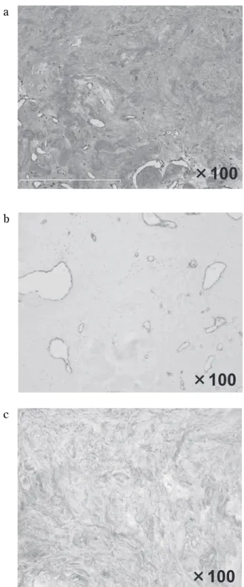
0.7 cm in size, elastic hard (Fig 4). Histrogical examinations showed that mostarea comprised hyalinized tissue and collagen fibers with small and thin-walled vascular space (Fig 5a). Immunohistochemical staining revealed some vas-cular components, as shown by positive CD34 in its peripheral region (Fig 5b). Masson’s trichrome stain highlighted the sclerotic condition of the mass
aa
b b
Figure 4 :Macroscopic findings of the resected tumor. The tu-mor was a well - demarcated homogenous white solid nodule, 0.8
!
0.7 cm in size. (a) resected specimen. (b) after formalin preservation.aa
b b
cc
Figure 5 :Microscopic findings of the resected tumor. The tu-mor was composed of fibrous connective tissue and various sizes of cavernous hemangioma tissue, HE staining (a), CD34 positive vascular components (b), Sclerosis is highlighted with collagen fiber, Masson’s trichrome (c).
(Fig 5c). From these characteristics, the tumor was finally diagnosed as a hepatic sclerosed heman-gioma. Final diagnosis of gastric cancer was T3(ss), N2, ly2, v1, M0 (Stage IIIb).
DISCUSSION
Cavernous hemangioma of the liver is the most frequent, the reported incidence being as high as 20% in liver tumor (4). The high incidence of he-mangioma increases the probability of encounter-ing atypical manifestations, includencounter-ing necrosis, scar-ring, and calcification. A sclerosed hemangioma is the ultimate form having all these degenerative changes, and such varaeties of pathological charac-teristics make precisely radiological diagnosis very difficult (5).
In our case, plain CT showed hepatic screlosed hemanangioma as low density, and enhanced CT showed a ring enhancement pattern, which is char-acteristic of adenocarcinoma. Although cavernous hemangioma shows typical features : low density on plain CT and delayed pooling enhancement, many previous reports have not shown this feature in scle-rosed hemangioma (5-12). MRI finding in our case was fit to typical feature of cavernous hemangioma, such as low on T1 and high on T2 weighted images, but this feature can be also observed in the cases of metastatic liver tumors and cholangiocellular car-cinoma. Others also reported that MRI findings of sclerosed hemangioma were different from the typi-cal cavernous hemangioma (5-11). It was reported that nodular areas of reduced signal intensity on T2-weighted images corresponded to the histological finding of fibrosis (13). A complicating factor is the contribution to signal heterogeneity of hemorrhage, thrombosis, hyalinization, calcification, and cystic cavities (13). On the otherhand, in our case, FDG accumulated only in gastric tumor, not in hepatic tumor in FDG-PET scan. However, the size of he-patic tumor was so small that this finding could not eliminate the malignancy. With the fact that there was advanced gastric cancer and these radiological findings, metastatic liver tumor from gastric cancer could not be excluded. It has reported that the find-ings such as geographical pattern, capsular-retrac-tion, decrease in size in follow-ups, and vanishing of previous enhanced lesion should raise the pos-sibility of the presence of screlosed hemangioma, and it can be diagnosed with biopsy before surgi-cal intervention (5). However, we do not agree the
universal application of perctaneous needle biopsy to make a definite diagnosis of hepatic tumor such as present case. Becouse a percutaneous biopsy has been reported to have a potential risk of abdomi-nal dissemination by needle tract implantation (14). As hepatic resection has been safe procedure in re-cent years (15, 16), hepatic resection should be cho-sen as a first strategy for tumors with unknown pathologically malignant potential.
Although sclerosed hemangioma is a rare disease among hepatic tumors, it needs to be taken into con-sideration among differential diagnosis of hepatic tu-mors. As it was extremely difficult to differentiate the lesion from liver metastasis or cholangiocellar-carcinoma, if the malignancy of the tumor cannot be denied, hepatic resection should be chosen for diagnostic therapy.
CONFLICT OF INTEREST STATEMENT
The authors declare no conflict of interest related to this case report.
REFERENCE
1. Aibe H, Hondo H, Kuroiwa T, Yoshimitsu K, Irie H, Tajima T, Shinozaki K, Asayama Y, Taguchi K, Masuda K : Sclerosed hemangioma of the liver. Abdom Imaging 26(5) : 496-9, 2001
2. Cheng HC, Tsai SH, Chiang JH, Chang CY : Hyalinized liver hemangioma mimicking malig-nant tumor at MR imaging. AJR Am J Roent-genol 165(4) : 1016-17, 1995
3. Makhlouf HR, Ishak KG : Sclerosed heman-gioma and sclerosing cavernous hemanheman-gioma of the liver : a comparative clinicopathologic and immunohistochemical study with empha-sis on the role of mast cells in their histogene-sis. Liver 22(1) : 70-8, 2002
4. Karhunen PJ : Benign hepatic tumours and tu-mour like conditions in men. J Clin Pathol 39 (2) : 183-188, 1986
5. Doyle DJ, Khalili K, Guindi M : Imaging fea-tures of sclerosed hemangioma. AJR Am J Roentgenol 189(1) : 67-72, 2007
6. Mathieu D, Rahmouni A, Vasile N, Jazaerli N, Duvoux C, Tran JV, Zafrani ES : Sclerosed liver hemangioma mimicking malignant tumor at MR imaging : pathologic correlation. J Magn
Reson Imaging 4(3) : 506-8, 1994
7. Haratake J, Horie A, Nagafuchi Y : Hyalinized hemangioma of the liver. Am J Gastroenterol 87(2) : 234-6, 1992
8. Yamashita Y, Shimada M, Taguchi K, Gion T, Hasegawa H, Utsunomiya T, Hamatsu T, Matsumata T, Sugimachi K : Hepatic sclerosing hemangioma mimicking a metastatic liver tu-mor : report of a case. Surg Today 30(9) : 849-52, 2000
9. Shim KS, Suh JM, Yang YS, Kim JG, Kang SJ, Jeon JS, Kim BS : Sclerosis of hepatic cavern-ous hemangioma : CT findings and pathologic correlation. J Korean Med Sci 10(4) : 294-7, 1995
10. Takayasu K, Moriyama N, Shima Y, Muramatsu Y, Yamada T, Makuuchi M, Yamasaki S, Hirohashi S : Atypical radiographic findings in hepatic cavernous hemangioma : correlation with histologic features. AJR Am J Roentgenol 146(6) : 1149-53, 1986
11. Vilgrain V, Boulos L, Vullierme MP, Denys A, Terris B, Menu Y : Imaging of atypical heman-giomas of the liver with pathologic correlation.
Radiographics 20(2) : 379-97, 2000
12. Mori H, Ikegami T, Imura S, Shimada M, Morine Y, Kanemura H, Arakawa Y, Kanamoto M, Hanaoka J, Sugimoto K, Tokunaga T : Scle-rosed hemangioma of the liver : Report of a case and review of the literature. Hepatol Res 38(5) : 529-533, 2008
13. Ros PR, Lubbers PR, Olmsted WW, Morillo G : Hemangioma of the liver : heterogeneous appearance on T2-weighted images. AJR Am J Roentgenol 149(6) : 1167-1170, 1987
14. Caturelli E, Ghittoni G, Roselli P, De Palo M, Anti M : Fine needle biopsy of focal liver le-sions : the hepatologist’s point of view. Liver Transpl 10(2 Suppl 1) : 26-9, 2004
15. Tsai MK, Lee PH, Tung BS, Yu SC, Lee CS, Wei TC : Experiences in surgical management of cavernous hemangioma of the liver. Hepa-togastroenterology 42(6) : 988-92, 1995 16. Fan ST, Lo CM, Liu CL, Lam CM, Yuen WK,
Yeung C, Wong J : Hepatectomy for hepatocel-lular carcinoma : toward zero hospital deaths. Ann Surg 229(3) : 322-30, 1999