Functional evolution of haloalkane
dehalogenases for the degradation of
persistent environmental pollutants
著者
Chen Nannan
学位授与機関
Tohoku University
学位授与番号
11301
博士論文
Functional evolution and engineering of
haloalkane dehalogenases for the
degradation of persistent organic
pollutants
(ハロアルカンデハロゲナーゼの難分解性環境汚染物質分解
能の機能進化に関する研究)
令和 2 年度
東北大学大学院生命科学研究科
分子化学生物学専攻
陳楠楠
Ph.D. Thesis
Functional evolution and engineering of
haloalkane dehalogenases for the
degradation of persistent organic
pollutants
Laboratory of Microbial Evolution and Function Research,
Graduate School of Life Sciences, Tohoku University
Contents
Background ... 5
0-1 γ-Hexachlorocyclohexane ... 5
0-2 Biodegradation of γ-HCH ... 6
0-3 The lin genes involved in the γ-HCH degradation ... 7
0-4 Haloalkane dehalogenases (HLDs) ... 7
0-4-1 Introduction of HLDs ... 7
0-4-2 Structure and reaction mechanism of HLDs ... 8
0-4-3 HLDs and its related proteins used in this study ... 9
0-4-4 Ancestral proteins ... 12
0-5 Mutagenesis of enzymes ... 13
0-5-1 Mutagenesis of HLDs ... 14
0-5-2 Random mutagenesis ... 15
0-6 Purposes of this study ... 16
Chapter 1 Construction and characterization of the linB-replacement strains ... 17
1-1 Background... 17
1-2 Materials and methods... 17
1-2-1 Strains, plasmids, medium composition and culture condition ... 17
1-2-2 DNA manipulations ... 17
1-2-3 Construction of the linB-deletion and replacement strains ... 18
1-2-4 GC analysis for the γ-HCH degradation ... 18
1-2-5 Assay for the -HCH utilization activity on solid medium (spot assay) ... 19
1-3 Results ... 26
1-3-1 Construction of the linB-replacement strains ... 26
1-3-2 γ-HCH degradation activity of the linB-replacement strains ... 29
1-3-3 γ-HCH utilization activity of the linB-replacement strains ... 31
1-4 Discussion... 34
Chapter 2 Construction of in vivo and in vitro evolution systems of HLDs toward the -HCH utilization ... 36
2-1 Background... 36
2-2 Materials and methods... 36
2-2-1 Strains, plasmids, medium composition and culture condition ... 36
2-2-2 DNA manipulations ... 36
2-2-3 Construction of plasmids ... 39
2-2-4 Construction of the hypermutator strains ... 39
2-2-5 Screening for clones having the improved γ-HCH utilization ability ... 39
2-2-6 Error-prone PCR ... 39
2-2-7 Construction of mutant libraries of HLD and its related genes ... 40
2-3 Results ... 42
2-3-1 In vivo evolution system ... 42
Chapter 3 Purification and characterization of the putative evolved HLDs ... 54
3-1 Background... 54
3-2 Materials and methods... 54
3-2-1 Strains, plasmids, medium composition and culture condition ... 54
3-2-2 DNA manipulations ... 54
3-2-3 Construction of plasmids ... 56
3-2-4 Expression of His-tagged proteins in E. coli ... 58
3-2-5 Purification of His-tagged proteins ... 58
3-2-6 SDS-PAGE ... 59
3-2-7 Concentration of purified protein ... 60
3-2-8 Assay for dehalogenase activity ... 60
3-2-9 Assay for the LinB-like activity ... 61
3-3 Results ... 62
3-3-1 Expression and purification of the putative evolved HLDs ... 62
3-3-2 HLD activity of the putative evolved HLDs ... 63
3-3-3 LinB-like activity of the putative evolved HLDs ... 65
3-3-4 Expression, purification and characterization of Rluc and Rluc-43... 69
3-3-5 Expression, purification, and characterization of the putative evolved proteins obtained by the 2nd round screening ... 70
3-4 Discussion... 74
Discussions ... 75
Acknowledgement ... 78
Background
Pesticides, which are effective in pest and disease control management, are important for agriculture and public health purposes, but their excessive use affects food security and concurrent health threats for humans (Macdonald et al., 2000). Most of traditional pesticides are recalcitrant organic compounds which are not easily degraded by natural means and they are referred to as persistence organic pollutants (POPs). POPs are of two types, the organophosphate pesticides and the organochloride pesticides, and are of great environmental and health concerns due to their toxic, persistent and bio-accumulative capacities (Barber et al., 2005). Many of them may form residual compounds which are more toxic in the soil and can be accumulated in living tissue through direct or indirect means, and thus they can get into the food chain of an ecosystem and affect wide range of organisms.
0-1 γ-Hexachlorocyclohexane
γ-Hexachlorocyclohexane (γ-HCH), which is a broad-spectrum organochloride insecticide, was one of the most popular organochloride pesticides that had been used extensively worldwide for the control of agricultural pests and mosquitoes in malaria health programs prior to the 1990‟s (Li et al., 2003). -HCH production by chlorination of benzene under suitable conditions leads to a mixture of isomers, and -HCH and its isomers were extensively applied since the 1940s and were added to the list of persistent organic pollutants (POPs) in 2009 (Vijgen et al., 2011). HCH is available in two formulations: technical-grade HCH (60-70 % α-HCH, 5-12 % β-HCH, 10-15 % γ-HCH, 6-10 % δ-HCH, and 3-4 % ε-HCH) and lindane (almost pure γ-HCH) (Abhilash et al., 2008). HCH isomers differ not only in the spatial orientation of the chlorine atoms bound to the aliphatic carbon ring (Fig. 0-1), but also in toxicity, water solubility (and thus mobility and bioavailability) and recalcitrance. Among the HCH isomers, α-HCH and γ-HCH dominate in the atmosphere due to their higher volatility and lower partition coefficient, while β-HCH is the most persistent in nature and less volatile isomer and tends to accumulate in soils (Vijgen et al., 2011). Only γ-isomer exhibited insecticidal activity, and it was widely used since 1953 as a cheap and effective insecticide especially in developing countries (Lal et al., 2010). Lindane is extremely toxic to humans and deleterious for environment. It is rapidly absorbed from the gastrointestinal tract of mice or rats and gets extensively distributed in fat, liver, ovarian tissues and brain. Although it has been banned in many countries because of its toxicity and recalcitrance (Lal et al., 2010), it is still being used in developing countries because of its efficacy and low cost. Thus it has caused seriously environmental problems.
Fig. 0-1 Axial versus equatorial arrangements of chlorine atoms in the five major isomers of HCH. α-HCH exists in two enantiomeric (+and -) forms (Lal et al., 2010).
0-2 Biodegradation of γ-HCH
γ-HCH is usually degraded under both aerobic and anaerobic environments, but it can be mineralized only in aerobic condition (Naqvi et al., 2014). Many bacteria has been reported for γ-HCH degradation (Böltner et al., 2005), and the γ-HCH degradation pathway and genes and enzymes involved in the degradation have been well studied in Sphingobium japonicum UT26 which was isolated from γ-HCH-polluted soil (Nagata et al., 2007). -HCH degradation and mineralization was also reported by other species of Sphigobium, such as S.
indicum strain B90 (Kumari et al., 2002) and B90A from India (Dogra et al., 2004) and S. francense strain
Sp+ from France (Cérémonie et al., 2006).
The microbial aerobic degradation pathway of γ-HCH was revealed in S. japonicum UT26 (Fig. 0-2) (Nagata et al., 2011). In this pathway, γ-HCH is converted to 2,5-dichlorohydroquinone (2,5-DCHQ) by sequential reactions catalyzed by LinA (γ-HCH dehydrochlorinase), LinB (1,3,4,6-tetrachloro-1,4-cyclohexadiene chlorohydrolase), and LinC (2,5-dichloro-2,5-cyclohexadiene-1,4-diol dehydrogenase). 2,5-DCHQ is then dechlorinated to chlorohydroquinone (CHQ) by LinD (2,5-dichlorohydroquinone dechlorinase), and CHQ is further transformed to β-ketoadipate by LinE (chlorohydroquinone 1,2-dioxygenase) and LinF (maleylacetate reductase). β-Ketoadipate is further degraded by the β-ketoadipate pathway that is generally found in environmental bacteria.
Fig. 0-2 Degradation pathway of γ-HCH in Sphingobium japonicum UT26. Compounds: 1, γ-HCH; 2, γ-pentachlorocyclohexene; 3, 1,3,4,6-tetrachloro-1,4-cyclohexadiene; 4, 1,2,4-trichlorobenzene; 5, 2,4,5-trichloro-2,5-cyclohexadiene-1-ol; 6, 2,5-dichlorophenol; 7, 2,5-dichloro-2,5-cyclohexadiene-1,4-diol; 8, 2,5-dichlorohydroquinone; 9, chlorohydroquinone; 10, acylchloride; 11, hydroquinone; 12, c-hydroxymuconic semialdehyde; 13, maleylacetate; 14, b-ketoadipate; 15, 3-oxoadipyl-CoA; 16, succinyl-CoA; 17, acetyl-CoA. TCA,
citrate/tricarboxylic acid cycle (Tabata et al., 2016)
0-3 The lin genes involved in the γ-HCH degradation
The linA to linF genes in S. japonicum UT26 are dispersed on the three large circular replicons: the linA, linB, and linC genes on the 3.6-Mb chromosome I; the linF gene on the 670-kb chromosome II; and the linDE operon with its regulatory gene (linR) on a 185-kb plasmid, pCHQ1 (Nagata et al., 2006).
Nearly identical lin genes have also been identified in other HCH-degrading bacterial strains, such as S.
indicum B90 (Kumari et al., 2002) and B90A (Dogra et al., 2004) from India and S. francense Sp+ from
France (Cérémonie et al., 2006); most of the lin genes in these strains are closely associated with an insertion sequence (IS), IS6100 (Lal et al., 2006). pCHQ1 is conjugally transferable from S. japonicum UT26 to another
Sphingomonas paucimobilis strain (Nagata et al., 2006), and another report showed that the linA and linB
genes in other strains are also located on plasmids (Cérémonie et al., 2006). These observations indicate that
lin genes must be spread by mobile genetic elements (MGEs).
0-4 Haloalkane dehalogenases (HLDs)
aliphatic hydrocarbons, which are produced in several industrial processes (Ang et al., 2018). Nowadays, various practical applications of HLDs are known, and the number is increasing with the growing knowledge of their properties and structure-function relationships: bioremediation of environmental pollutants, biosensors for toxic chemicals (Ang et al., 2018), industrial biocatalysis (Janssen, 2007), decontamination of warfare agents (Prokop et al., 2006), as well as cell imaging and protein analysis (Los et al., 2008). HLDs belong to the α/β-hydrolase fold superfamily, a very large and diverse group of structurally related hydrolytic enzymes with esterase, lipase or epoxide hydrolase activities (Koudelakova et al., 2011). Phylogenetic study of HLD sequences revealed that HLDs were subdivided into three subfamilies HLD-I, HLD-II, and HLD-III (Chovancová et al., 2007). The composition of amino acid residues that are important for the reaction (see below) is different among the subfamilies: Asp-His-Asp (catalytic triad) and Trp-Trp (halide-stabilizing residues) in HLD-I, Asp-His-Glu and Asn-Trp in HLD-II, and Asp-His-Asp and Asn-Trp in HLD-III. In contrast to HLDs belonging to HLD-I and HLD-II, those belonging HLD-III are poorly characterized experimentally (Chovancová et al., 2007).
0-4-2 Structure and reaction mechanism of HLDs
HLDs have a globular structure and are composed of two domains: a large central catalytic domain with an α/β-hydrolase fold structure and the second domain which lies like a cap on the main domain. The latter domain emerges as a large R-helical excursion between β-strands 6 and 7 of the catalytic core. The interface of the two domains forms the hydrophobic active site. The catalytic triad residues are a nucleophilic aspartate, a base catalyst histidine, and an aspartate or glutamate as the third member. These amino acids form the basis of the dehalogenation reaction and are located in the main domain. Whereas there is significant sequence similarity in the catalytic core, the sequence and structure of the cap domain diverge considerably between different HLDs. The cap domain was proposed to play a prominent role in determining substrate specificity (Koudelakova et al., 2013).
HLDs perform catalysis using an SN2 (nucleophilic substitution) reaction and subsequent hydrolysis by the addition of water, in which only water is required as a cofactor (Fig. 0-3). This catalytic mechanism involves the catalytic triad of Asp-His-Asp/Glu. The carboxylate oxygen of aspartate initially launches a nucleophilic attack on the partially positive carbon atom of the halogen-bound substrate to produce a halide ion and alkyl-enzyme intermediate with an ester bond. The nearby His-Asp/Glu (acid-base pair) subsequently hydrolyzes a water molecule to produce a nucleophilic hydroxide that will attack the carbon of the ester bond. This generates a tetrahedral intermediate that immediately decomposes to form RCH2O− and grabs a proton from the nucleophile to form RCH2OH (Jong et al., 2003). HLDs possess halide-binding residues, also known as halide-stabilizing residues, which is their unique feature (Chovancová et al., 2007). These residues are critical for the catalytic activity of HLDs as they help to stabilize the halide during formation of the enzyme-substrate complex.
Fig. 0-3 Simplified reaction mechanism of HLDs. a, The ester is formed by SN2 nucleophilic substitution, and the transition state formed in this step is stabilized by two halide-stabilizing residues. b, A water molecule activated by a histidine-acid pair attacks the ester intermediate to produce an alcohol and halide ion (Nagata et al., 2015)
0-4-3 HLDs and its related proteins used in this study
0-4-3-1 LinA
LinA was initially identified as an enzyme that catalyzes the first step of γ-HCH degradation in S. japonicum UT26. LinA catalyzes dehydrochlorination of γ-HCH and γ-PCCH to produce 1,2,4-TCB (Fig. 0-2), and is not a member of HLDs. LinA can also degrade α-HCH and δ- HCH in addition to γ-HCH, but has no activity for β-HCH because β-HCH lacks a 1,2-biaxial HCl pair (Trantírek et al., 2001). Degradation assays of various halogenated compounds by purified LinA showed that the substrate specificity of LinA is very narrow. Because no gene significantly homologous to the linA gene has been found, its origin is unknown. LinA is thought to be a unique dehydrochlorinase, and its reaction mechanism of dehydrochlorination is of great
rearrangements (Peisajovich et al., 2006). It has been shown that two copies of IS6100 located close to the
linA gene are involved in its loss in S. japonicum UT26 (Nagata et al., 2011).
0-4-3-2 LinB
LinB is one of archetypal HLDs that involved in the -HCH degradation pathway and has been well characterized (Marek et al., 2000) (Fig. 0-4a). Site-directed mutagenesis of LinB confirmed that Asp108, His272, and Glu132 comprise the catalytic triad in this enzyme (Oakley et al., 2004).
LinB has a broad substrate specificity, mainly due to a large active site volume, which includes monochloroalkanes (C3-C10), dichloroalkanes, bromoalkanes and chlorinated aliphatic alcohols (Koudelakova et al., 2011). Notably, LinBUT26 yields a significantly lower specificity constant for β-HCH (0.02 mM-1 s-1) as compared to another relatively well characterized LinB, namely, LinBB90A (identical to LinBMI1205, and LinBBHC-A and LinBpLB1) from S. indicum strain B90A (0.20 mM
-1
s-1) (Okai et al., 2013). LinBB90A hydrolytically dechlorinates the metabolite 2,3,4,5,6-pentachlorocyclohexanol (PCHL), whereas LinBUT26 does not (Ito et al., 2007). A molecular dynamics simulation study suggests that this is mainly due to a difference in the flexibility of the entrance of the substrate access tunnel mediated by six out of the seven amino acid differences between the two enzyme variants (Okai et al., 2013).
Fig. 0-4 Structures of LinBUT (a) (PDB code, 1CV2) and DbjA (b) (PDB code, 3A2M). Catalytic triads of LinB (Asp108, Glu132, and His272) and DbjA (Asp103, Glu127, and His280) are shown in red. The ERB fragment (138HHTEVAEEQDH150) of DbjA is shown in blue (Nagata et al., 2015)
0-4-3-3 DmmA
DmmA is a HLD with a known tertiary structure that was identified from a marine metagenomic consortium (Gehret et al., 2012). Inspection of its crystal structure revealed that its unusually large active site (Fig. 0-5)
DmmA exhibited an exceptionally broad substrate specificity and degraded several halogenated environmental pollutants that are resistant to other members of HLDs. In addition to having this unique substrate specificity, the enzyme was highly tolerant to organic cosolvents such as dimethyl sulfoxide, methanol, and acetone. Its broad substrate specificity, high overexpression yield, good tolerance to organic cosolvents, and a broad pH range make DmmA an attractive biocatalyst for various biotechnological applications (Buryska et al., 2018).
Fig. 0-5 Structures of DmmA. The stereo ribbon diagram is colored as a rainbow from blue at the N terminus to red at the C-terminus with catalytic pentad residues in stick form with magenta C (Gehret et al., 2012).
0-4-3-4 DbjA
DbjA, which was isolated from Bradyrhizobium japonicum USDA110 (Sato et al., 2005), possesses new substrate specificity with high catalytic activity towards β-methylated haloalkanes and sufficient enantioselectivity for industrial scale synthesis of optically pure compounds (Zbyněk Prokop et al., 2009). Comparison of the circular dichroism spectra of DbjA and other HLDs strongly suggested that DbjA contains more α-helices than the other HLDs (Sato et al., 2005) (Fig. 0-4b). A sequence comparison between DbjA and other HLDs has suggested that an 11-amino acid insertion between the main and cap domains of DbjA produces a unique active-site structure that results in the unique substrate specificity of DbjA (Sato et al., 2005). Compared with other characterized HLDs, DbjA possesses unique properties. Catalytic activity and structural stability in a broad range of pH conditions combined with high enantioselectivity with selected substrates make DbjA a very versatile biocatalyst (Chaloupkova et al., 2011). Interestingly, DbjA can kinetically discriminate between enantiomers of two distinct groups of substrates, α-bromoesters and β-bromoalkanes; it has enantioselectivity based on distinct molecular interactions, which can be modified
0-4-3-5 Rluc
Luciferase (Luc) from Renilla reniformis (Rluc) is not HLD, but is phylogenetically associated with HLD-II.
Renilla luciferase [Renilla-luciferin:oxygen 2-oxidoreductase (decarboxylating), EC 1.13.12.5] catalyzes the
oxidative decarboxylation of coelenterazine in the presence of dissolved oxygen to yield oxyluciferin, CO2, and blue light (λmax.=480 nm). The molecular weight of Rluc is 36 kDa (Lorenz et al., 1991). Luciferases have become important research tools over the last two decades, due to their ability to emit light and therefore be monitored easily. These bioluminescent proteins are utilized widely as reporter genes in cell culture experiments and more recently in the context of small animal imaging (Contag et al., 1997).
The Rluc mutant (Rluc8) was screened using a consensus sequence-driven strategy, and the results obtained showed that it was 200-fold more resistant to inactivation in murine serum and its light output was 4-fold higher than the wild type. Furthermore, the structure for Rluc8, a luciferase that utilizes coelenterazine as a substrate, was clarified for the first time, demonstrating a typical α/β-hydrolase folding at 1.4 Å resolution (Loening et al., 2006).
0-4-4 Ancestral proteins
The main goal of many protein engineering strategies is to improve enzyme properties for particular industrial or medical applications. One of these strategies is ancestral sequence reconstruction (ASR) (Wijma et al., 2013), in which a hypothetical ancestral sequence of a given set of related present-day sequences is predicted from a phylogenetic tree and reconstructed in a laboratory. This work has been covered in excellent reviews (Harms & Thornton, 2010). ASR has been used to enhance enzyme thermostability (Wijma et al., 2013), solubility (Gonzalez et al., 2014), and activity (Takenaka et al., 2013), and to modify substrate specificity (Smith et al., 2013).
In the last few decades, ASR has been widely used to study the evolution and structure-function relationships of many protein families, such as GFP-like proteins (Ugalde et al., 2004), opsins (Yokoyama, 2002), steroid receptors (Ortlund et al., 2007), G-protein receptors (Babkova et al., 2017), and others (Dean et al., 2007). Using ancestral protein resurrection, two permissive and five restrictive mutations played important roles in the loss of aldosterone sensitivity in the modern glucocorticoid receptors (Ortlund et al., 2007). By introducing five conserved amino acids that were different in red and green vertebrate opsins into the ancestral background, Yokoyama et al. (Yokoyama et al., 2008) successfully recapitulated the shift in the opsin absorbance spectrum from red to green, whereas previous mutagenesis studies using modern proteins had resulted in contradictory results concerning the functional importance of key mutations.
To the best of our knowledge, only a few researches reported ASR for HLDs. Sequences of dehalogenases DbjA (Sato et al., 2007), DbeA (Chaloupkova et al., 2014), DhaA (Newman et al., 1999), DmxA (Tratsiak et al., 2013), and DmmA (Gehret et al., 2012) were predicted by ASR. The present-day enzymes display considerable functional variations even though they are all closely evolutionary related and share similar structural topology, thus providing good models to investigate structural and functional divergence in the HLD-II subfamily. Characterization of the resurrected ancestral enzymes revealed unique functional properties, including enhanced thermostability, improved specific activity, or modified substrate specificity. This study highlights that the ASR method represents a powerful strategy for constructing highly active, stable,
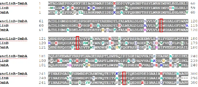
DmbA is 80% and 83%, respectively (Figure 0-6). The differences between the predicted ancestor and both present-day enzymes were mapped on LinB_dmbA_anc homology model (Figure 0-7).
Fig. 0-6 Comparison of LinB_dmbA_anc sequence with LinB and DmbA sequences (red square represents catalytic residue of LinB)
Fig. 0-7 Homology model of ancLinB-DmbA. Amino acid positions occupied by different residues in ancLinB-DmbA and LinB (A) and in ancLinB-DmbA and DmbA (B) are highlighted by red and cyan, respectively.
0-5 Mutagenesis of enzymes
protein engineering is to construct the enzymes with improved activity and modified specificity. The introduction of mutations into the genes, gene expression and protein purification take considerable effort and it is desirable to extensively characterize constructed mutants to detect even subtle changes in the specificity of the constructs (Marvanová et al., 2001).
0-5-1 Mutagenesis of HLDs
HLDs are attractive targets for protein-engineering studies aimed at improving catalytic efficiency and at broadening the range of substrate specificity for important environmental pollutants. It appears that libraries of structurally and mechanistically related enzymes will play an increasing role in biotransformation reactions, because each biocatalyst has its own characteristic substrate specificity, enantioselectivity, stability, and product inhibition data. Searching of sequenced genomes for putative HLD genes in conjunction with the overexpression and characterization of proteins encoded by these genes is one possible way for meeting the increasing demand for novel HLDs (Chan et al., 2010). Partial improvement in the catalytic properties and modification of the substrate specificities of HLDs by rational design (Chaloupková et al., 2003) and directed evolution approaches (Bosma et al., 2002) have been reported.
A variant of LinB, LinBMI from Sphingobium sp. MI1205, which is 98%identical (having a difference in only 7 of the 296 amino acid residues) to LinBUT (Fig. 0-8), can catalyze the two-step conversion of β-HCH to 2,3,5,6-tetrachlorocyclohexane-1,4-diol (TCDL) with the first conversion step being an order of magnitude more rapid than that by LinBUT (Ito et al., 2007), while LinBUT cannot convert the PCHL (Nagata et al., 2005). The substitution of the residues forming the catalytic pocket of LinBUT (I134 V/A247H) resulted only a weak effect on β-HCH conversion activity. Furthermore, the reciprocal double mutant of LinBMI (V134I/H247A) retained relatively high LinBMI-type activity (Ito et al., 2007). These results indicated that some of the five other residues are also important for the LinBMI-type activity. Site-directed mutagenesis and X-ray crystallographic studies (Okai et al., 2013) indicated that all seven residues are important for LinBMI-type catalytic activity.
Fig. 0-8 Structure of LinBMI (PDB code 4H77) and location of catalytic triad (D108, E132, and H272; shown in red) and the seven dissimilar amino acid residues between LinBMI and LinBUT: V134 and V112 (in magenta), L138, H247, and I253 (in cyan), T135 (in green), and T81 (in blue) (Moriuchi et al., 2014).
0-5-2 Random mutagenesis
“Random mutagenesis” is a technique that allows researchers to develop large libraries of variants of a particular DNA sequence. Once developed, these libraries can then be used for several purposes, including structure-function and directed evolution studies. Random mutagenesis is different from other mutagenesis techniques in that it does not require the researcher to have any prior knowledge about the structural properties of the DNA sequence being targeted, thus allowing for the unbiased discovery of novel or beneficial mutations. For this reason, random mutagenesis is especially useful for protein evolution studies (Forloni et al., 2018).
Error-prone PCR introduces random copying errors by imposing imperfect, and thus mutagenic, or „sloppy‟, reaction conditions (e.g. by adding Mn2+ or Mg2+ to the reaction mixture). This method has proven useful both for generation of random libraries of nucleotide sequences, and also for the introduction of mutations during the expression and screening process in a mutagenesis step (Pritchard et al., 2005). Many researches had obtained excellent mutants with higher activity, thermostability, specific activity by using error-prone PCR combine with site directed mutagenesis (Varriale et al., 2018). Fig. 0-9 showed a model of selecting good evolved protein by using error-prone PCR. The researcher begins with the gene for the parent protein. This parent gene is randomly mutagenized by using error-prone PCR or some similar technique. The library of mutant genes is then used to produce mutant proteins, which are screened or selected for the desired target property (e.g., improved enzymatic activity or increased stability). Mutants that fail to show improvements in the screening/selection are typically discarded, while the genes for the improved mutants are used as the parents for the next round of mutagenesis and screening. This procedure is repeated until the evolved protein exhibits the desired level of the target property.
Fig. 0-9 Schematic outline of a typical directed evolution experiment (Bloom et al., 2009).
0-6 Purposes of this study
Various natural HLDs are known, and their activities can be changed dramatically by only small number of mutations, and many fundamental knowledge related to the reaction mechanisms of HLDs has been accumulated. Thus, HLDs are good materials not only for demonstrating the process and mechanism of functional evolution of enzymes but also for engineering of enzymes with novel catalytic activity. It is also suggested that function of HLDs can be evolved rapidly in sphingomonads. LinB is one of prototypical HLDs and was originally identified as an enzyme necessary for utilization of γ-HCH. There are various γ-HCH degraders have been isolated from HCH-isomers-contaminated sites around the world, and they also have identical or almost identical LinBs. Until now, no γ-HCH degrader has been reported that uses other HLDs besides LinB for the γ-HCH utilization. To get some insights into the process and mechanisms of functional evolution of HLDs toward the γ-HCH utilization, the followings are conducted in this study:
1. Construction and characterization of the linB-replacement strains.
2. Construction of in vivo and in vitro evolution system of HLDs toward the -HCH utilization. 3. Purification and characterization of the putative evolved HLDs.
Chapter 1 Construction and characterization of the linB-replacement
strains
1-1 Background
As described in background section, sphingomonads seem to have ideal background for functional evolution of catabolic enzymes for various recalcitrant hydrophobic compounds. In this study, Sphingobium
japonicum UT26 was used as a host for in vivo evolution system, because functional evolution of HLDs is
expected to occur rapidly in sphingomonads represented by the case of LinB variants. The linB gene has variants whose protein products are different with a small number of amino-acid residues, and LinBUT and LinBMI are 98% identical but their -HCH degradation activity are remarkably different. The important point is that the sequence variations in such variants are non-synonymous substitutions, which strongly suggests that the linB gene are still evolving at high speed under strong selection pressures (Nagata et al., 2015). In addition, although LinB is the only HLD to date involved in the γ-HCH degradation, other HLDs seem to have a chance to evolve toward the γ-HCH degradation by a small number of mutations.
In this chapter, as the first step to get some insights into the evolution process of HLDs toward -HCH utilization, S. japonicum UT26-derivative strains, in which the linBUT gene was replaced by other HLD or its homologue genes including the putative ancestral genes, were constructed and characterized.
1-2 Materials and methods
1-2-1 Strains, plasmids, medium composition and culture condition
The strains and plasmids used in this chapter were shown in Table 1-1. E. coli cells were incubated by using LB medium and Sphingobium strains were incubated by using 1/3LB medium. Spot assay for estimating the
-HCH utilization ability was conducted by using W minimal salt medium containing 750 ppm of γ-HCH at final concentration as a sole carbon source. Compositions of these mediums were shown in Table 1-2. The solid medium was prepared by the addition of 1.5% (w/v) agar. Antibiotics were used at the final concentrations of 25 μg/mL for kanamycin (Km), 100 μg/mL for ampicillin (Ap), 50 μg/mL for streptomycin (Sm), and 10 μg/mL gentamycin (Gm). The incubation temperature of E. coli and Sphingobium cells was 37℃ and 30℃, respectively. Strains were stocked by addition of 15% glycerol at -80℃.
1-2-2 DNA manipulations
Established methods were employed for DNA manipulations. Plasmids were extracted by using LaboPassTM Plasmid Mini (COSMO Genetech) according to the attached instruction. Ligation of DNA was conducted by using Takara Ligation kit Mighty Mix (Takara). Gibson Assembly kit (New England BioLabs) was also used for assembling of DNA fragments. HIT Competent E.coli DH5 618 cells (RBC Bioscience) were used for transformation of E. coli. Hot Start Taq (NEB) and Q5 High-Fidelity DNA Polymerase (NEB) were used for polymerase chain reaction (PCR). When conducting colony PCR, a little cells were picked by toothpicks and
version 13 (SDC Inc., Tokyo). The bacterial cells were transformed by electroporation (EP). Cells grown on 1/3LB agar medium for two days were collected by an inoculation loop, washed three times with ice-cold EP buffer (1 mM MOPS and 10% glycerol), diluted appropriately, and mixed with DNA solution. The suspension was transferred to an EP cuvette with a 1 mm gap. EP was conducted under the conditions of 1.8 kV, 200 Ω and 25 µF. After the pulse, 1 mL of ice-cold 1/3LB medium was immediately added, then incubated for 2-10 h and spread onto a 1/3LB agar medium containing appropriate antibiotics.
1-2-3 Construction of the linB-deletion and replacement strains
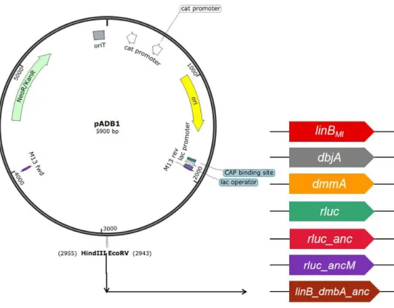
The linB-deletion mutant, in which just open reading frame of the linB gene has been deleted, was constructed by allelicexchange mutagenesis of S. japonicum UT26 using pK18mobsacB (Schäfer et al., 1994), which has the sacB gene for counter selection (Schweizer, 1992). The 1.5-kb upstream and downstream regions of the linB gene in S. japonicum UT26 were cloned into pK18mobsacB, and the resultant plasmid pK18mobsacB::linB_up_down was introduced into UT26 by EP, and the Kmr transformant into which the plasmid had been integrated via single crossover-mediated homologous recombination was selected. The Kmr SucS transformant was inoculated on a 1/3LB plate containing sucrose (10%), and the Kms Sucr clones were selected. Finally, the linB-deletion strain was selected by PCR, and named UTDB2. For introduction of other HLD genes into the linB site, allelicexchange mutagenesis of S. japonicum UTDB2 was carriedout by using pAK405, which has the streptomycin-sensitive rpsL allele (rpsL1) as a counterselection marker (Kaczmarczyk et al., 2012). Firstly, a plasmid pADB1 (Fig. 1-1) was constructed, which has the 1-kb upstream and downstream regions of the linB gene in pAK405. The linBMI, dbjA, dmmA, rluc, rluc_anc, rluc_ancM, and linB_dmbA_anc genes were introduced into pADB1, and the resultant plasmids were named pAMM1, pABJ1,
pAMM1, pARL1, pALA1, pALA2, and pABA1, respectively. These plasmids were introduced into UTDB2 by EP, and the Kmr Sms transformants into which these plasmids had been integrated via single crossover-mediated homologous recombination were selected. The Kmr Sms transformants was inoculated on a 1/3LB plate containing Sm, and the Kms Smr clones were selected (Fig. 1-2). Finally, the strains that have the linBMI, dbjA, dmmA, rluc, rluc_anc, rluc_ancM, and linB_dmbA_anc genes in the linBUT site were selected by PCR among the Kms Smr clones and designated as UTBM1, UTBJ1, UTMM1, UT2RL1, UTLA1, UTLA2, and UTBA1, respectively. Primers used for amplification and plasmids used as templates of the genes are shown in Table 1-3. The primer sets were designed by NEBuilder (http://nebuilder.neb.com) for assembly with EcoRV and HindIII-digested pADB1 by using a Gibson Assembly system (NEB). UTDB2DAX, in which both the linB and adhX genes were deleted, was constructed from UTDB2 by the same procedure using pAAXD1, which is a pAK405-based plasmid for deletion of the adhX gene (Inaba et al., 2020). DAX series strains, UTBM1DAX, UTBJ1DAX, UTMM1DAX, UTRL1DAX, UTLA1DAX, UTLA2DAX, and UTBA1DAX having the linBMI, dbjA, dmmA, rluc, rluc_anc, rluc_ancM and linB_dmbA_anc genes, respectively, in the linBUT site were constructed from UTDB2DAX by the same procedure using pABM1, pABJ1, pAMM1, pARL1, pALA1, pALA2, and pABA1, respectively.
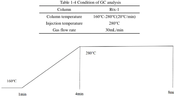
1-2-4 GC analysis for the γ-HCH degradation
electron capture detector (ECD) and Rtx-1 capillary column (30 m×0.25 mm×0.25 μm; Restek) was used, and condition for the analysis is shown in Table 1-4. The concentration of -HCH and intermediates were quantified from peak area by using standard chemicals.
1-2-5 Assay for the -HCH utilization activity on solid medium (spot assay)
Bacterial cells grown on 1/3LB agar medium were collected by inoculation loop and washed three times with PBS. The bacterial cell suspension was diluted by PBS and adjusted to 100 mg cells/mL. This suspension was diluted 10 (10 mg cells/mL) and 102 (1 mg cells/mL) fold, and each 10 L aliquots of each dilution were spotted on solid W minimal salt medium containing -HCH (750 ppm) or glucose (0.2%), or without adding any carbon sources, and incubated for 5 days at 30oC.
Table 1-1 Bacterial strains and plasmids used in this chapter
Strains or plasmid Relevant characteristics Source or reference
Sphingomonads
Sphingobium japonicum UT26S γ-HCH degrader (Nagata et al., 2011)
Sphingobium japonicum UTDB2 linB This study
Sphingobium japonicum UTBM1 linB -> linBMI (Ito et al., 2007)
Sphingobium japonicum UTBJ1 linB -> dbjA (Sato et al., 2005)
Sphingobium japonicum UTMM1 linB -> dmmA (Gehret et al., 2012)
Sphingobium japonicum UTLA1 linB -> rluc_ancM (Chaloupkova et al.,
2019)
Sphingobium japonicum UTLA2 linB -> rluc_anc (Chaloupkova et al.,
2019)
Sphingobium japonicum UTRL1 linB -> rluc (Loening et al., 2006)
Sphingobium japonicum UTBA1 linB -> linB_dmbA_anc (Jesenská et al., 2005)
Sphingobium japonicum UT26DAX γ-HCH degrader, adhX (Inaba et al., 2020)
Sphingobium japonicum UTDB2DAX linB, adhX This study
Sphingobium japonicum UTBM1DAX linB -> linBMI, adhX This study
Sphingobium japonicum UTBJ1DAX linB -> dbjA, adhX This study
Sphingobium japonicum UTMM1DAX linB -> dmmA, adhX This study
Sphingobium japonicum UTLA1DAX linB -> rluc_ancM, adhX This study
Sphingobium japonicum UTLA2DAX linB -> rluc_anc, adhX This study
Sphingobium japonicum UTRL1DAX linB -> rluc, adhX This study
Sphingobium japonicum UTBA1DAX linB -> linB_dmbA_anc, adhX This study
E.coli
DH5α recA1 endA1 gyrA96 thi-1 hsdR17 supE44
relA1 (lacZYA-argF) Φ80lacZM15 (Marietta et al., 1988)
Plasmid
pK18mobsacB Suicide plasmid for gene deletion, Kmr Schgfer et al., 1994
pK18mobsacB_linB_up_down pK18mobsacB::linB_up_down This study
pAK405 oripBR322, RP4 oriT, rpsL1, Kmr (Kahm et al., 2010)
pADB1 pAK405::linB_up_down_EPH This study
pBDQ1 pBBR MCS-1 (Cm) -UT26dnaQexo This study
pABM1 pAK405::linB_up_down_linBMI This study
pABJ1 pAK405::linB_up_down_dbjA This study
pAMM1 pAK405::linB_up_down_dmmA This study
pALA1 pAK405::linB_up_down_rluc_ancM This study
pALA2 pAK405::linB_up_down_rluc_anc This study
pARL1 pAK405::linB_up_down_rluc This study
Table 1-2 Compositions of medium
1/3LB broth Per liter.
Bacto tryptone 3.3g Bacto yeast extract 1.7g
NaCl 5g pH 7.0 W medium Per liter KH2PO4 1.7g Na2HPO4 9.8g (NH4)2SO4 1.0g MgSO4 48.7mg FeSO4 0.52mg MgO 10.75mg CaCO3 2.0mg ZnSO4 0.81mg CuSO4 0.16mg CoSO4 0.15mg H3BO3 0.06mg
1/10 W was made in which the concentrations of KH2PO4, Na2HPO4 and (NH4)2SO4 were diluted to 1/10 those in the W medium.
LB broth Per liter.
Bacto tryptone 10g
Bacto yeast extract 5g
NaCl 5g
Table 1-3 Primers used in this chapter
Primer Sequence (5‟→3‟) Purpose
linB_up_FW CTAGAGTCGACCTGCACCGGGTTTCCCC GCCGACCCCGTC synthesis of pK18mobsacB::linB_up_down and pK18mobsacB::linB_up_MI_down linB_up_RV GTTCCGGACGATATTCTCCTTGAGCGATT TTC synthesis of pK18mobsacB::linB_up_down and pK18mobsacB::linB_up_MI_down linB_down_FW GAATATCGTCCGGAACCGGCTCATTTTC TAAG synthesis of pK18mobsacB::linB_up_down and pK18mobsacB::linB_up_MI_down linB_down_RV GTGCCAAGCTTGCATGTGGCCTTCGGCA TTGCCGAGATGC synthesis of pK18mobsacB::linB_up_down and pK18mobsacB::linB_up_MI_down pADB1_rluc_F ctcaaggagaatatcgATGACTTCGAAAGTTTAT
GATC Amplification of rluc
pADB1_rluc_R tgagccggttccggaTTATTGTTCATTTTTGAGA
ACTCG Amplification of rluc
pADB1_rluc_anc_opt_F ctcaaggagaatatcgATGGTTAGCGCAAGCCAG
CG Amplification of rluc_anc
pADB1_rluc_anc_opt_R tgagccggttccggaTTATTTGGTCAGTTCGTTC
AGAAAATCG Amplification of rluc_anc
pADB1_linB_dmbA_anc_F cgctcaaggagaatatcgATGACCGCACTGGGTGC
AG Amplification of linB_dmbA_anc
pADB1_linB_dmbA_anc_R gaaaatgagccggttccggaTTAAACACCGGCTGC
TGCACG Amplification of linB_dmbA_anc
linB_up_1000_CF GGTATCATGTCAACTGGGGC Construction of pARL1, pALA2
and pABA1
linB_down_1000_CR TGGCATGGCACCGAGAAGGC Construction of pARL1, pALA2
and pABA1
linB_down_1000_CR2 GGCCACGTCGAGCACAAGCTC Construction of pARL1, pALA2
and pABA1
linB_down_1000_CR3 GATAATAGGCTTCCCGCCCGGAG Construction of pARL1, pALA2
and pABA1
M4out GCTGCAAGGCGATTAAG Construction of pARL1, pALA2
and pABA1
RVout GGCTCGTATGTTGTGTG Construction of pARL1, pALA2
Table 1-4 Condition of GC analysis
Column Rtx-1
Column temperature 160℃-280℃(20℃/min)
Injection temperature 280℃
1-3 Results
1-3-1 Construction of the linB-replacement strains
Firstly, the linB-deletion strain UTDB2 was constructed, in which just open reading frame of the linB gene was deleted. This strain can be used as a negative control for the cell having no LinB activity. Indeed, UTDB2 showed neither the LinB activity in the -HCH degradation pathway nor the -HCH-utilization activity on the
-HCH plate (see below). Then, pADB1 (Fig. 1-1), which has the 1-kb upstream and downstream regions of
linB, was constructed by using pAK405 as a base to make it easier to construct plasmids for introduce of
various genes into the linB site. The resultant plasmids were introduced into UTDB2 and the strains that have the linBMI, dbjA, dmmA, rluc, rluc_anc, rluc_ancM and linB_dmbA_anc genes (Table 1-5) in the linB site were constructed (Fig. 1-2), and named UTBM1, UTBJ1, UTMM1, UT2RL1, UTLA1, UTLA2, and UTBA1, respectively (Fig.1-3).
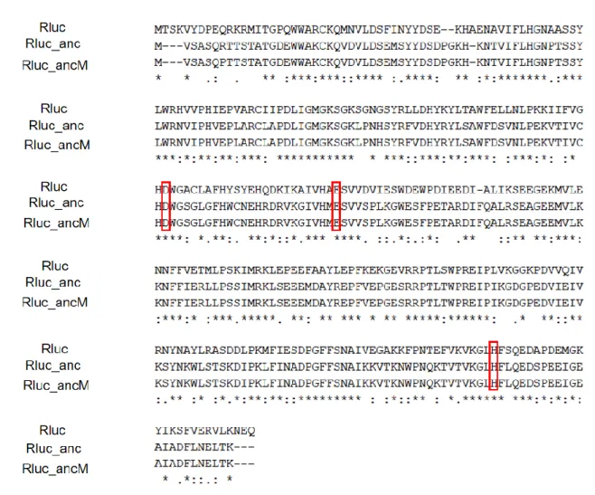
The phylogenetical relationships of HLDs or HLD homologues used in this study is shown in Fig. 1-4. LinBMI from Sphingobium sp. MI1205 is 98% identical (7 amino acid differences among total 296 amino acids) with LinBUT but shows higher activity toward -HCH than LinBUT (Ito et al., 2007). DbjA is a HLD from Bradyrhizobium japonicum USDA110, which prefers bulky substrates (Sato et al., 2005). DmmA is a HLD from a marine metagenome and has an unusually large active site, and thus shows the most versatile substrate specificity among known HLDs (Gehret et al., 2012). Rluc is Renilla-luciferin 2-monooxygenase from Renilla reniformis (Lorenz et al., 1991), which has luciferase activity toward coelenterazine by monooxygenation mechanism. Rluc is monooxygenase, whose reaction mechanism is completely different from that of HLD, but its amino acid sequence is phylogenetically close to the HLD-II subfamily of HLDs (Fig. 1-6). To date, HLD activity of Rluc toward any HLD substrates has not been detected, but Rluc is considered to be an excellent candidate for investigating the functional evolution of HLDs (Nagata et al., 2015). Rluc_anc is putative ancestral protein of LinB and Rluc (Fig. 1-4) that have been designed in silico (Chaloupkova et al., 2019). Rluc_ancM, which was unexpectedly produced on the cloning process, has just one amino acid difference R7P with Rluc_anc (Fig. 1-5). LinB_dmbA_anc is a putative ancestral protein of LinB and DmbA (Fig. 1-4). DmbA is a HLD from Mycobacterium bovis 5033/66 and only single amino acid is different with DmtA (K120 is N in DmbA) from Mycobacterium tuberculosis (Jesenská et al., 2005).
DAX-series strains, UTBM1DAX, UTBJ1DAX, UTMM1DAX, UTRL1DAX, UTLA1DAX, UTLA2DAX, and UTBA1DAX, were also constructed from the strain UTDB2DAX, in which the adhX gene is also deleted in addition to the linB gene. If the adhX gene is expressed by spontaneous mutation, the strain become to be able to grow on the solid minimal salt medium without adding any carbon sources (Inaba et al., 2020), and thus DAX series strains have a merit to avoid the selection of false positive mutants that grow well on the
Fig. 1-3 The linB-replacement strains construed in this chapter
Fig. 1-4 Phylogenetic tree of HLDs and Rluc. HLDs, Rluc, and putative ancestral proteins used in this study are shown in red.
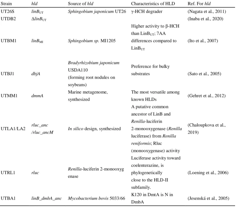
Table 1-5 Specific information of linB replacement strains and their corresponding hlds
Strain hld Source of hld Characteristics of HLD Ref. For hld
UT26S linBUT Sphingobium japonicum UT26 γ-HCH degrader (Nagata et al., 2011)
UTDB2 ΔlinBUT (Inaba et al., 2020)
UTBM1 linBMI Sphingobium sp. MI1205
Higher activity to β-HCH than LinBUT; 7AA differences compared to LinBUT (Ito et al., 2007) UTBJ1 dbjA Bradyrhizobium japonicum USDA110
(forming root nodules on soybeans)
Preference for bulky
substrates (Sato et al., 2005)
UTMM1 dmmA Marine metagenome,
synthesized
The most versatile among
known HLDs (Gehret et al., 2012)
UTLA1/LA2 rluc_anc
/rluc_ancM In silico design, synthesized
A putative common ancestor of LinB and
Renilla-luciferin
2-monooxygenase (Renilla luciferase) from Renilla
reniformis; Rluc
(monooxygenase) activity
(Chaloupkova et al., 2019)
UTRL1 rluc Renilla-luciferin 2-monooxyg
enase
Luciferase activity toward coelenterazine, is
phylogenetically close to the HLD-II subfamily.
(Loening et al., 2006)
UTBA1 linB_dmbA_anc Mycobacterium bovis 5033/66 K120 in DmtA is N in
Fig. 1-5 Amino acid sequence alignment of Rluc, Rluc_anc and Rluc_ancM (red squares represent catalytic residues)
1-3-2 γ-HCH degradation activity of the linB-replacement strains
S. japonicum UT26 degrades γ-HCH through the pathway shown in Fig. 1-6. γ-HCH is converted by two
steps of LinA-catalyzed dehydrochlorination via γ-pentachlorocyclohexene (γ-PCCH) to 1,3,4,6-tetrachloro-1,4-cyclohexadiene (1,4-TCDN), and this compound is productively metabolized by two steps of LinB-catalyzed hydrolytic dehalogenation via 2,4,5-trichloro-2,5-cyclohexadiene-1-ol (2,4,5-DNOL) to 2,5-dichloro-2,5-cyclohexadiene-1,4-diol (2,5-DDOL). 2,5-DDOL is converted to 2,5-dichlorohydroquinone (2,5-DCHQ) by dehydrogenase LinC, and 2,5-DCHQ is further metabolized. In this pathway, two substrates of LinB, 1,4-TCDN and 2,4,5-DNOL, are unstable and have not been directly detected, and their production is predicted by the production of two dead end products, 1,2,4-trichlorobenzene (1,2,4-TCB) and 2,5-dichlorophenol (2,5-DCP), respectively (Nagata et al., 1993). By the GC assay used in this study, we can detect -HCH, -PCCH, 1,2,4-TCB, 2,5-DCP, 2,5-DDOL, and 2,5-DCHQ, and the important point is that the production of 2,5-DCP, 2,5-DDOL, and 2,5-DCHQ means that the cells have the LinB activity.
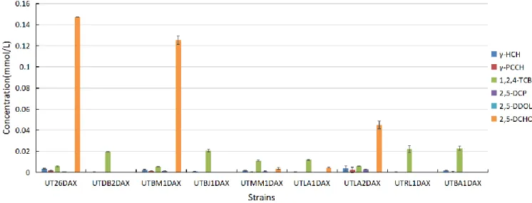
The -HCH degradation activity of the constructed strains was examined by GC analysis and the concentration of remaining -HCH and metabolites produced after the incubation for 60 min are shown in Fig.
UTMM1, UTLA1, and UTLA2, indicating that DmmA, Rluc_anc, and Rluc_ancM have the LinB-like activity. The same tendency was obtained in the experiment using the DAX-series strains (Fig. 1-8), supporting the conclusion that DmmA, Rluc_anc, and Rluc_ancM have the LinB-like activity
Fig. 1-6 Upstream degradation pathway of γ-HCH in S. japonicum UT26
Fig. 1-8 GC analysis of Sphingobium japonicum UT-DAX-series strains
1-3-3 γ-HCH utilization activity of the linB-replacement strains
γ-HCH utilization activity of the constructed strains was examined to see the growth of cells on the solid W minimal salt medium containing -HCH as a sole carbon source (-HCH plate). As positive and negative control, cells were also spotted on the solid W minimal salt medium containing glucose (Glucose plate) and adding no carbon source (w/o C plate), respectively. Strains that utilize -HCH increase the cell number to the visible state accompanying with clear zone around the spotted area on the -HCH plate. Three different concentrations of cells (100, 10 and 1 mg cells/mL) were spotted to distinguish the small difference. As predicted, all the strains grew well on Glucose plate (Fig. 1-10A), but not on w/o C plate (Fig. 1-9A). UT26 and UTBM1 showed obvious -HCH utilization activity at the cell concentration of 10 and 1 mg cells/mL, while UTDB2, UTBJ1, and UTRL1 showed no -HCH utilization activity (Fig. 1-11A). UTLA2 showed
-HCH utilization activity at the cell concentration of 10 mg cells/mL, and UTLA1 and UTMM1 formed larger clear zone around the spotted area than UTDB2 at the cell concentration of 100 mg cells/mL (Fig. 1-11A). The same tendency was observed in the experiment using the DAX-series strains (Fig. 1-11B). These results indicate that Rluc_anc, Rluc_ancM, and DmmA have weak LinB-like activity for the -HCH utilization.
Fig.1-9 Spot assay of Sphingobium japonicum linB replacement strains on W plate without carbon source (2 days incubation). A. Sphingobium japonicum UT series strains. B. Sphingobium japonicum UT-DAX series strains.
Fig.1-10 Spot assay of Sphingobium japonicum linB replacement strains on W plate with glucose (1 day incubation). A.
Fig.1-11 Spot assay of Sphingobium japonicum linB replacement strains on W-γ-HCH plate (14 days incubation). A.
Sphingobium japonicum UT series strains. B. Sphingobium japonicum UT-DAX series strains.
1-4 Discussion
In this chapter, the linB-replacement strains of S. japonicum UT26 were constructed by using homologous recombination, in which the linBUT gene was replaced with linBMI, dbjA, dmmA, rluc, rluc_anc, rluc_ancM
(Koudelakova et al., 2011), but it was experimentally confirmed for the first time in this study. Especially, it is important that 'natural' HLD DmmA showed the LinB activity.
DmmA is a HLD from marine metagenome and belongs to HLD-II subfamily, but its biological source is unknown. DmmA possesses an unusually large active-site cavity comparing with other structurally characterized HLDs (Gehret et al., 2012), and shows unusual broad substrate specificity. DmmA showed activity toward all 29 substrates constituting a set of representative HLD substrates (Koudelakova et al., 2011). Additionally, DmmA is active toward all poorly degradable chlorinated environmental pollutants, e.g., 1,2-dichloroethane, 1,2-dichloropropane, 1,2,3-trichloropropane, and chlorocyclohexane as well as toward newly identified substrates of this enzyme family (Daniel et al., 2015). The broad substrate specificity of DmmA may be linked to its large active site and readily accessible active site. Analysis of access tunnels using CAVER identified the widely open mouth without any sign of bottleneck, which is unique to DmmA and has never been observed with other family members (Gehret et al., 2012). This wide opening provides easy access of a large spectrum of diverse molecules to the enzyme active site. While complementary analysis of LinB revealed clear bottlenecks which separate the active site from the surrounding water solvent. All of these results suggested that DmmA possesses a combination of several unique properties attractive for practical applications.
On the other hand, it should be also noted that DbjA did not show the LinB activity, indicating that not all HLDs with broad substrate specificities show the LinB activity. DbjA from Bradyrhizobium japonicum USDA110 has been intensively analyzed because it shows unique substrate specificity such as a high catalytic activity for β-methylated haloalkanes and high enantioselectivity with β-brominated alkanes (Sato et al., 2005). Since this enzyme possessed unique catalytic activity, structural stability in a broad pH range, combined with high enantioselectivity with particular substrates, it still be used in the protein engineering analysis and further mutations on this enzyme will make it a very versatile biocatalyst. Determinants for the LinB activity will be revealed by comparing HLDs that show the LinB activity and those not.
Rluc_ancM seems to have higher LinB-like activity than Rluc_anc and DmmA, since (i) UTLA2 (UTLA2DAX) produced larger amount of 2,5-DCHQ and 2,5-DCP than UTLA1 (UTLA1DAX) and UTMM1 (UTMM1DAX) (Fig. 1-12 and 1-13), and (ii) UTLA2 (UTLA2DAX) grew well at the cell concentration of 10 mg cells/mL than UTLA1 (UTLA1DAX) and UTMM1 (UTMM1DAX) (Fig. 1-11). It is interesting because only one amino acid residue is different between Rluc_anc and Rluc_ancM. This result strongly suggest that (i) HLDs can change their LinB-like activity only by small number of amino acid residue substitution, and (ii) the assay system used in this study is sensitive enough to detect the difference.
Taken together, strains constructed in this study can be used as starting materials in the functional evolution and engineering studies. Especially, DAX-series strains are usefully for avoiding false positive clones that grow well on the solid minimal salt medium without adding any carbon sources in the screening process.
Chapter 2 Construction of in vivo and in vitro evolution systems of
HLDs toward the -HCH utilization
2-1 Background
In Chapter 1, it was revealed that some HLDs besides LinB can potentially be involved in the -HCH utilization. Furthermore, it was suggested that (i) HLDs can change their LinB-like activity only by small number of amino acid residue substitution, and (ii) the assay system used in this study is sensitive enough to detect the difference.
In this chapter, to get some insights into HLDs evolution toward the optimized -HCH utilization, experimental evolution systems of HLDs were constructed. As in vivo evolution system, the engineered strains constructed in Chapter 1 were directly used for the screening. Considering the possibility that mutation rate is too low to obtain the evolved genes in the in vivo evolution system, (i) hypermutator strains were constructed for the in vivo evolution system by the introduction of the mutated dnaQ gene into the linB replacement strains, and (ii) in vitro evolution system was constructed, in which error-prone PCR was used for random mutagenesis. Strategies used in this study are summarized in Fig. 2-2.
2-2 Materials and methods
2-2-1 Strains, plasmids, medium composition and culture condition
The strains and plasmid used in this chapter were shown in Table 2-1. The medium and culture conditions were in accordance with Chapter 1. In addition, chloramphenicol (Cm) was used at the final concentration of 25 μg/mL.
2-2-2 DNA manipulations
The basic DNA manipulations were in accordance with Chapter 1. Primers used in this chapter were shown in Table 2-2. HIT Competent E.coli DH5 619 cells (RBC Bioscience) showing higher efficient transformation rate than 618 cells were used for construction of mutant libraries of HLD or its related genes in
Table 2-1 Bacterial strains and plasmids used in this chapter
Strains or plasmid Relevant characteristics Source or reference
Sphingomonads
Sphingobium japonicum UT26DAX γ-HCH degrader, adhX (Inaba et al., 2020)
Sphingobium japonicum UTDB2DAX linB, adhX (Inaba et al., 2020)
Sphingobium japonicum UTBM1DAX linB -> linBMI, adhX This study
Sphingobium japonicum UTBJ1DAX linB -> dbjA, adhX This study
Sphingobium japonicum UTMM1DAX linB -> dmmA, adhX This study
Sphingobium japonicum UTLA1DAX linB -> rluc_ancM, adhX This study
Sphingobium japonicum UTLA2DAX linB -> rluc_anc, adhX This study
Sphingobium japonicum UTRL1DAX linB -> rluc, adhX This study
Sphingobium japonicum UTBA1DAX linB ->linB_dmbA_anc, adhX This study
Sphingobium japonicum UT26DAX/pBDQ1 γ-HCH degrader, adhX, pBDQ1 This study
Sphingobium japonicum UTDB2DAX/pBDQ1 linB, adhX, pBDQ1 This study
Sphingobium japonicum UTBM1DAX/pBDQ1 linB -> linBMI, adhX, pBDQ1 This study
Sphingobium japonicum UTBJ1DAX/pBDQ1 linB -> dbjA, adhX, pBDQ1 This study
Sphingobium japonicum UTMM1DAX/pBDQ1 linB -> dmmA, adhX, pBDQ1 This study
Sphingobium japonicum UTLA1DAX/pBDQ1 linB -> rluc_ancM, adhX, pBDQ1 This study
Sphingobium japonicum UTLA2DAX/pBDQ1 linB -> rluc_anc, adhX, pBDQ1 This study
Sphingobium japonicum UTRL1DAX/pBDQ1 linB -> rluc, adhX, pBDQ1 This study
Sphingobium japonicum UTBA1DAX/pBDQ1 linB ->linB_dmbA_anc, adhX, pBDQ1 This study
Sphingobium japonicum UTDB2DAX/pBLB1 linB, adhX, pBBR5TP::linBUT This study
Sphingobium japonicum UTDB2DAX/pBLB2 linB, adhX, pBBR5TP::linBMI This study
Sphingobium japonicum UTDB2DAX/pBBJ1 linB, adhX, pBBR5TP::dbjA This study
Sphingobium japonicum UTDB2DAX/pBMM1 linB, adhX, pBBR5TP::dmmA This study
Sphingobium japonicum UTDB2DAX/pBLA1 linB, adhX, pBBR5TP::rluc_ancM This study
Sphingobium japonicum UTDB2DAX/pBLA2 linB, adhX, pBBR5TP::rluc_anc This study
Sphingobium japonicum UTDB2DAX/pBRL1 linB, adhX, pBBR5TP::rluc This study
Sphingobium japonicum UTDB2DAX/pBBA1 linB, adhX,
pBBR5TP::linB_dmbA_anc This study
E.coli
DH5α
recA1 endA1 gyrA96 thi-1 hsdR17 supE44 relA1 (lacZYA-argF) Φ80lacZM15
(Marietta et al., 1988) Plasmid
pBDQ1 pBBR MCS-1 (Cm) -UT26dnaQexo (Inaba et al., 2020)
pBBR5T pBBR1MCS-5_terminator This study
pBBR5TP pBBR1MCS-5 carrying T1 This study
pBLB1 pBBR5TP::linBUT This study
pBLB2 pBBR5TP::linBMI This study
pBRL1 pBBR5TP::rluc This study
pBBA1 pBBR5TP::linB_dmbA_anc This study
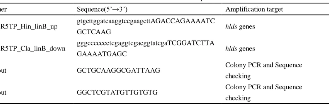
Table 2-2 Primers used in this chapter
Primer Sequence(5‟→3‟) Amplification target
pBBR5TP_Hin_linB_up gtgcttggatcaaggtccgaagcttAGACCAGAAAATC
GCTCAAG hlds genes
pBBR5TP_Cla_linB_down gggccccccctcgaggtcgacggtatcgaTCGGATCTTA
GAAAATGAGC hlds genes
M4out GCTGCAAGGCGATTAAG Colony PCR and Sequence
checking
RVout GGCTCGTATGTTGTGTG Colony PCR and Sequence
2-2-3 Construction of plasmids
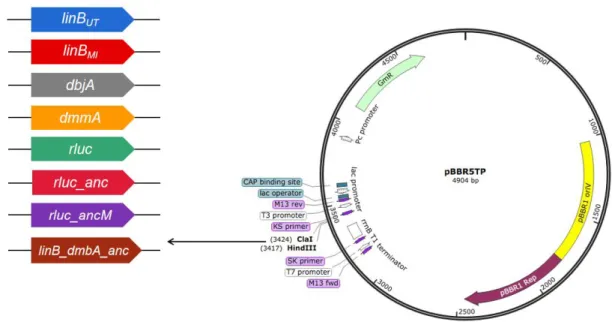
The terminator sequence was introduced into the broad-host-range vector pBBR1-MCS-5, and the resultant plasmid was named pBBR5T. The promoter sequence Pu necessary for constitutive expression of linA gene in
S. japonicum UT26 was introduced into pBBR5T, and the resultant plasmid was named pBBR5TP. The linBUT, linBMI, dbjA, dmmA, rluc, rluc_anc, rluc_ancM, and linB_dmbA_anc genes were introduced into pBBR5TP, and the resultant plasmids were named pBLB1, pBLB2, pBBJ1, pBMM1, pBRL1, pBLA1, pBLA2, and pBBA1, respectively (Fig. 2-1).
Fig. 2-1 Construction of plasmids for expression of HLD and its homologue genes in Sphingobium strains.
2-2-4 Construction of the hypermutator strains
Hypermutator strains were constructed by introduction of pBDQ1, which carries the mutated dnaQ gene of UT26 (dnaQexo) (Inaba et al., 2020), into the linB-replacement strains by using electroporation.
2-2-5 Screening for clones having the improved γ-HCH utilization ability
Cells (Table 2-3) cultured by appropriate medium were collected, washed, and suspended in PBS at the concentration of 10 mg/mL. 100 μL of cell suspension was spread on W-γ-HCH plate and incubated at 30℃ for two weeks. Colonies grew well with larger clear zone than others were selected for further analysis.
2-2-6 Error-prone PCR
Random mutagenesis of the HLD and its related genes was conducted by error-prone PCR. The composition of the reaction solution is shown in Table 2-4. The reaction condition consisted of a denaturation step for 1 min at 94℃, followed by 30 cycles of denaturation at 94℃ for 30 sec, annealing at 56℃ for 30 sec, and extension at 72℃ for 30 sec, with a final extension step at 72℃ for 10 min. Mutation rate was adjusted to
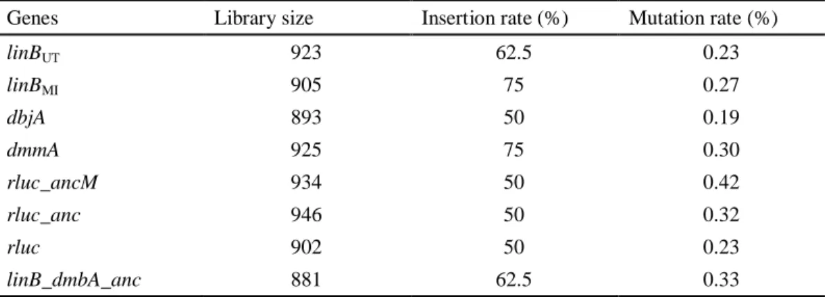
2-2-7 Construction of mutant libraries of HLD and its related genes
Mutated genes generated by error-prone PCR were cloned into pBBR5TP by using Gibson Assembly kit and transformed into E. coli DH5, and the resultant transformants were used as mutant libraries in E. coli. Insertion rate of the PCR-amplified fragments and their mutation rate were estimated by using plasmids extracted from clones of the libraries that were randomly selected. The mixture of plasmids were extracted from the libraries in E. coli, and introduced by EP into S. japonicum UTDB2DAX (linB, adhX) to obtain
the libraries in Sphigobium.
Table 2-3 Strains used in the in vivo evolution system
Strain Relevant characteristics Growth on HCH
(low cell density)
Growth on HCH (high cell density)
Sphingobium japonicum UT26DAX γ-HCH degrader Yes Yes (death)
Sphingobium japonicum UTBM1DAX linB -> linBMI, adhX Yes Yes (death)
Sphingobium japonicum UTBJ1DAX linB -> dbjA, adhX No No
Sphingobium japonicum UTMM1DAX linB -> dmmA, adhX No Yes (weak)
Sphingobium japonicum UTLA1DAX linB -> rluc_ancM, adhX No Yes (weak)
Sphingobium japonicum UTLA2DAX linB -> rluc_anc, adhX No Yes(weak)
Sphingobium japonicum UTRL1DAX linB -> rluc, adhX No No
Sphingobium japonicum UTBA1DAX linB->linB_dmbA_anc
adhX No Yes
Sphingobium japonicum
UT26DAX/pBDQ1 γ-HCH degrader Yes Yes (death)
Sphingobium japonicum
UTBM1DAX/pBDQ1 linB -> linBMI, adhX Yes Yes (death)
Sphingobium japonicum
UTBJ1DAX/pBDQ1 linB -> dbjA, adhX No No
Sphingobium japonicum
UTMM1DAX/pBDQ1 linB -> dmmA, adhX No Yes (weak)
Sphingobium japonicum
UTLA1DAX/pBDQ1
linB -> rluc_ancM,
adhX No Yes (weak) Sphingobium japonicum
UTLA2DAX/pBDQ1 linB -> rluc_anc, adhX No Yes(weak)
Sphingobium japonicum
UTRL1DAX/pBDQ1 linB -> rluc, adhX No No
Sphingobium japonicum
UTBA1DAX/pBDQ1
linB->linB_dmbA_anc,
adhX No Yes
Sphingobium japonicum
UTDB2DAX/pBLB1 pBBR5TP::linBUT Yes Yes (death)
Sphingobium japonicum
UTDB2DAX/pBLB2 pBBR5TP::linBMI Yes Yes (death)
Sphingobium japonicum
UTDB2DAX/pBBJ1 pBBR5TP::dbjA No No
Sphingobium japonicum
UTDB2DAX/pBMM1 pBBR5TP::dmmA No Yes (weak)
Sphingobium japonicum
UTDB2DAX/pBLA1 pBBR5TP::rluc_ancM No Yes (weak)
Sphingobium japonicum
UTDB2DAX/pBLA2 pBBR5TP::rluc_anc No Yes(weak)
Sphingobium japonicum
Table 2-4 Compositions of solution for error-prone PCR
Reagents Volume
Template 0.5 μL (plasmid was diluted by TE
buffer* to 100 fold) pBBR5TP_Hin_linB_up (50pmol/μl) 0.5 μL pBBR5TP_Cla_linB_down (50pmol/μl) 0.5 μL rTaq (5U/μl) 0.5 μL 10×buffer (Mg2+ free) 5 μL dNTP mixture 4 μL DMSO 2.5 μL MgCl2 (25mM) 3 μL MnCl2 (10mM) 0.5 μL Sterilized water Up to 50 μL *TE buffer: 1M Tris (pH 8.0) 2 mL 0.5M EDTA (pH 8.0) 400 μL Sterilized water up to 200 mL
2-3 Results
2-3-1 In vivo evolution system
The linB-replacement UT26 (wild type)- and UT-DAX-series strains constructed in Chapter 1 were incubated on the W--HCH plate, and clones that grew well with larger clear zone than others were selected. Considering the possibility that spontaneous mutation rate is too low to obtain the evolved genes in this system, hypermutator strains were also constructed by the introduction of the mutated dnaQ gene into the UT-DAX-series strains. The -HCH utilization ability of the resultant strains was assayed on the W--HCH plate (Fig. 2-3). These strains showed the same tendency with UT-DAX-series strains (Fig. 1-11).
Some candidate clones were obtained by the screening for further analysis. HLD or its related genes of such candidates were amplified by PCR and sequenced. However, they carried the same gene as original or the linB gene. The former indicates that mutation(s) in the genome other than HLD or its related genes improved the
Fig. 2-3 Spot assay of UT-DAX(pBDQ1)-series strains (9 days incubation). Concentration of cells from outer to inner is 100 mg/mL, 10 mg/mL and 1 mg/mL.
2-3-2 Introduction of HLD or its related genes into UTDB2DAX by using a broad-host-range vector
The linBUT, linBMI, dbjA, dmmA, rluc, rluc_anc, rluc_ancM, and linB_dmbA_anc genes were cloned into a broad-host-range vector pBBR5TP, and the resultant plasmids were named pBLB1, pBLB2, pBBJ1, pBMM1, pBRL1, pBLA1, pBLA2, and pBBA1 (Fig. 2-1).These plasmids were introduced into UTDB2DAX (linB,
adhX), and the -HCH utilization ability of the resultant strains was assayed on the W--HCH plate ( Fig. 2-4). The positive control strains, UTDB2DAX (pBLB1) and UTDB2DAX (pBLB2), showed obvious -HCH utilization activity, while the negative control strain UTDB2DAX showed no -HCH utilization activity (Fig. 2-4A). As expected from the results of Chapter 1, UTDB2DAX (pBBJ1) and UTDB2DAX (pBRL1) showed no -HCH utilization activity, and UTDB2DAX (pBMM1), UTDB2DAX (pBLA1), and UTDB2DAX (pBLA2) showed weak -HCH utilization activity (Fig. 2-4). Unexpectedly, UTDB2DAX (pBBA1) showed weak -HCH utilization activity (Fig. 2-4B), suggesting that LinB_dmbA_anc has faint LinB-like activity. The activity was detected only in UTDB2DAX (pBBA1), probably because LinB_dmbA_anc was expressed at higher level in UTDB2DAX (pBBA1) than in UTBA1.