N-(3-oxododecanoyl)-L-homoserine-lactone, a quorum sensing molecule, causes a bell-shaped change in nonprotein thiol content in rat thymic lymphocytes: Its relation with oxidative stress and intracellular Zn2+
Yumiko Nishimura-Danjobara#, Keisuke Oyama##, Kaori Kanemaru, Keiko Takahashi###, Kumio Yokoigawa, Yasuo Oyama
Department of Food Science, Faculty of Bioscience and Bioindustry, Tokushima University, Tokushima 770-8513, Japan
Present address:
# Graduate School of Biomedical and Health Sciences, Hiroshima University, Hiroshima 734-8553, Japan
## Intensive Care Unit, Osaka University Hospital, Osaka 565-0871, Japan
### Faculty of Human Life Science, Shikoku University, Tokushima 770-1192, Japan
Corresponding author Yasuo Oyama, Ph.D. [email protected]
Highlights
· N-(3-oxododecanoyl)-L-homoserine-lactone (ODHL) is a quorum sensing molecule. · ODHL induced a ‘bell-shaped’ change in the glutathione content of rat thymocytes. · ODHL concentration-dependently elevated intracellular Zn2+ levels.
· ODHL increased cellular O2- content in a concentration-dependent manner. · ODHL may exhibit diverse actions on host cells.
Abstract
Cellular actions of N-(3-oxododecanoyl)-L-homoserine-lactone (ODHL), a quorum sensing molecule of bacteria, were studied on rat thymocytes using a flow cytometer with appropriate fluorescent dyes to elucidate the effects of ODHL on host cells. A bell-shaped concentration-response relation was observed in the ODHL-induced changes in cellular glutathione content ([GSH]i). ODHL concentration-dependently increased intracellular Zn2+ levels ([Zn2+]i) and cellular O2- content ([O2-]i). The bell-shaped relation induced by ODHL can be explained as follows: a low concentration of ODHL is expected to induce moderate oxidative stress that intracellularly releases Zn2+ by converting thiols to disulfides. A slight elevation of [Zn2+]i may increase the [GSH]i. On the other hand, it is likely that a high concentration of ODHL causes severe oxidative stress that further causes both the decrease in [GSH]i and the increase in [Zn2+]i. Excessive increase in [Zn2+]i may augment oxidative stress that further decreases the [GSH]i. Other notable actions induced by ODHL included the elevation of [Zn2+]i by Zn2+ influx and the increase in [GSH]i under Zn2+-free conditions. Therefore, it is suggested that ODHL elicits diverse actions on host cells.
Keywords:
Quorum sensing molecule
N-(3-oxododecanoyl)-L-homoserine-lactone Lymphocytes
1. Introduction
Quorum sensing (QS) molecules are signal mediators for cell-to-cell communication in bacteria to monitor cell population density (Bassler, 1999; Williams, 2007). QS allows bacteria to synchronize biological events in a group. Since QS molecules are secreted in considerable amounts during bacterial growth, several types of host cells respond to these molecules (Holm and Vikström, 2014). N-(3-oxododecanoyl)-L-homoserine-lactone (ODHL), a QS molecule, disrupts the barrier of human epithelial colorectal adenocarcinoma Caco-2 cells (Vikström et al., 2006; Eum et al., 2014) and enters membranes of primary murine T cells (Ritchie et al., 2007); it also causes direct cytotoxicity in human pancreatic carcinoma cells (Kumar et al., 2014). Furthermore, ODHL treatment increases intracellular Ca2+ release in both P815 mastocytoma and HMC-1 human mast cells (Li et al., 2009). In case of lymphocytes, ODHL inhibits cell differentiation (Ritchie et al., 2005) and induces apoptosis (Tateda et al., 2003). Thus, ODHL is expected to exert diverse actions on mammalian cells. It is important to examine the cellular actions of ODHL on host cells. In this study, using a flow cytometer and rat thymic lymphocytes as a cell model, we first confirmed the threshold concentration of ODHL to induce cell death. Thereafter, the cellular actions of ODHL at sublethal concentrations were examined. We found that ODHL induced a bell-shaped change in cellular GSH content ([GSH]i) that was associated with changes in cellular O2- content ([O2-]i) and intracellular Zn2+ concentration ([Zn2+ ]i), and also discussed the underlying mechanism.
2. Materials and methods
ODHL was purchased from Sigma-Aldrich Corporation (St. Louis, Missouri, USA). Fluorescent probes used to measure cellular parameters are listed in Table 1. Other chemical reagents were obtained from Wako Pure Chemicals (Osaka, Japan).
(Table 1 near here)
2.2. Cell preparation
The Animal Experiment Committee of Tokushima University approved this study using experimental animals (T29-54). Cell suspension was prepared as follows: thymus glands were dissected from 14 ether-anesthetized male Wistar rats (8–12-week-old). Razor-sliced glands were triturated in Tyrode's solution buffered with 4-(2-hydroxyethyl)-1-piperazineethanesulfonic acid to dissociate single cells (Chikahisa et al., 1996). The solution containing single cells was passed through a 50 µm diameter mesh, following which the cell suspension was used for the experiments.
2.3. Experimental procedures and cytometric measurements
All experiments using the cell suspension were carried out at a temperature of 36–37ºC. ODHL was dissolved in dimethyl sulfoxide (DMSO) and the ODHL solution (3–300 mM) was then added to the suspension to achieve final concentrations (3–300 µM) of ODHL.
Fluorescence analysis was done using flow cytometry (CytoACE-150; JASCO, Tokyo, Japan) and JASCO software (Version 3.06). Cell lethality was assessed by adding 5 µM propidium iodide (PI). Exposed phosphatidylserine (PS) on the outer surface of membrane was detected using FITC fluorescence after treating the cells with 10 µL/mL of annexin V- FITC and 5 µM PI for 30 min (Koopman et al., 1994). 5-CMF-DA was employed to estimate changes in [GSH]i (Chikahisa et al., 1996). The cells were treated with 500 nM 5-CMF-DA for 30 min before the measurement of 5-CMF fluorescence. FluoZin-3-AM was used to monitor changes in [Zn2+]i (Gee et al., 2002). The cells were treated with 1 µM FluoZin-3-AM for 1 h prior to the
FluoZin-3 fluorescence measurement. The [O2-]i was estimated by BES-So-AM (Maeda et al.,
2005). The cells were treated with 5 µM BES-SO-AM for 1 h before measuring BES-SO fluorescence. Excitation and emission wavelengths for the fluorescent probes are also listed in Table 1.
2.4. Statistical analysis
Numerical results were statistically analyzed using Tukey's multivariate analysis. P-values of 0.05 or less were considered significant. Experimental P-values were described as mean ± standard deviation of 4–6 samples. Each experiment was performed twice to validate the results.
3. Results
3.1. Threshold concentration of ODHL to induce cell death
The cells were treated with 30–300 µM ODHL for 3 h. ODHL at 30 µM did not change the population of cells with PI fluorescence, indicating that 30 µM ODHL did not alter cell viability. The population of cells exhibiting PI fluorescence (areas P and AP, dead cells) was increased by 300 µM ODHL (Fig. 1A). ODHL at 300 µM also increased the population of cells with FITC fluorescence and without PI fluorescence (area A), indicating that ODHL increased the population of living cells with PS-exposed membranes. In case of 100 µM ODHL the dead cell population was slightly increased. Therefore, the threshold ODHL concentration to induce cell death was > 30 µM and < 100 µM under present experimental conditions. Results are summarized in Fig. 1B.
(Figure 1 near here)
Cell treatment with 30 µM ODHL for 3 h shifted the fluorescence histogram of 5-CMF to a region of higher intensity while it was not the case for 300 µM ODHL (Fig. 2A). Treatment with 300 µM ODHL for 3 h did not cause an increase in fluorescence intensity. A bell-shaped concentration-response relation was observed in the ODHL-induced changes in 5-CMF fluorescence (Fig. 2B). In a previous study (Kinazaki et al., 2011), the elevation of [Zn2+]i increased [GSH]i. Therefore, to determine whether Zn2+ contributed to the ODHL-induced increase in [GSH]i, the changes in 5-CMF fluorescence by 10–30 µM ODHL treatment were examined in the presence of N,N,N',N'-tetrakis(2-pyridylmethyl)ethylenediamine (TPEN), a chelator of intracellular Zn2+. TPEN at 10 µM did not affect the control fluorescence level. However, TPEN attenuated the ODHL-induced increase in intensity, suggesting the contribution of Zn2+ (Fig. 2C).
(Figure 2 near here)
3.3. ODHL-induced elevation of [Zn2+ ]i
The treatment of cells with 30–300 µM ODHL for 3 h shifted the fluorescence histogram of FluoZin-3 to a higher intensity region (Fig. 3A), suggesting the ODHL-induced elevation of [Zn2+]i . Treatment with ODHL at 10–300 µM for 3 h concentration-dependently increased the mean intensity of FluoZin-3 fluorescence (Fig. 3B), suggesting that ODHL at concentrations of 10 µM or higher increased the of [Zn2+]i. To determine if extracellular Zn2+ contributed to the ODHL-induced increase in FluoZin-3 fluorescence, the action of ODHL was examined in the presence of diethylenetriamine-N,N,N',N",N"-pentaacetic acid (DTPA), a chelator of extracellular Zn2+. DTPA application reduced the control fluorescence of FluoZin-3, suggesting the contribution of extracellular Zn2+ to the [Zn2+]i. The FluoZin-3 fluorescence of the control, as well as ODHL-treated cells, was lower in DTPA, although 30–100 µM ODHL increased the fluorescence even in the presence of DTPA (Fig. 3C), suggesting that the ODHL action is partly independent from extracellular Zn2+.
(Figure 3 near here)
3.4. ODHL-induced oxidative stress
The results presented in Fig. 3C suggest that ODHL increased oxidative stress, resulting in the release of intracellular Zn2+ from nonprotein thiols, leading to an increase in [Zn2+]i. Therefore, the [O2-]i level was estimated using BES-SO in the ODHL-treated cells. Treatment with 30–100 µM ODHL for 3 h shifted the histogram of BES-SO fluorescence to a region of higher intensity (Fig. 4A). Thus, ODHL is expected to increase [O2-]i , leading to an increase in oxidative stress. Results of the ODHL-induced increase in the mean intensity of BES-SO fluorescence are summarized in Fig. 4B.
(Figure 4 near here)
4. Discussion
4.1. Mechanism of the ODHL-induced bell-shaped change in [GSH]i
The bell-shaped response of ODHL-induced change in [GSH]i can be explained by the oxidative stress induced by ODHL. At concentrations of 10–100 µM, ODHL at 10–100 µM significantly increased the [O2-]i in a concentration-dependent manner (Fig. 4). A low concentration of ODHL is expected to induce moderate oxidative stress that induces an intracellular release of Zn2+ by converting thiols to disulfides (Maret, 1994). In rat thymic lymphocytes, the external application of micromolar concentrations of ZnCl2 concentration-dependently increases both [Zn2+]i and [GSH]i , with a positive correlation (correlation coefficient, 0.99) (Kinazaki et al., 2011). Zinc increases the [GSH]i either by activating the de
novo synthesis pathway of GSH or by increasing transcription of the catalytic subunit of
glutamate-cysteine ligase, a GSH synthetase (Ha et al., 2006; Cortese et al., 2008). Therefore, the increase in [GSH]i by zinc may exceed the decrease in [GSH]i by oxidative stress. On the
other hand, a high concentration of ODHL elicits severe oxidative stress that causes a further decrease in [GSH]i. Excessive increase in [Zn2+]i augments oxidative stress (Kim et al., 1999;
Matsui et al., 2010). Thus, the decrease in [GSH]i by oxidative stress may exceed the increase in [GSH]i by zinc. Consequently, ODHL induced a bell-shaped change in [GSH]i .
4.2. Experimental implication
Numerous bacteria use QS molecules as signals for cell-to-cell communication to sense a cell mass (Waters and Bassler, 2005). In addition, QS molecules affect the host cells to minimize host defense mechanisms and establish infection (Costerton et al., 1999; Yi-Hu et al., 2001). Therefore, it is not surprising that QS molecules exert adverse actions on host cells. The present study showed that ODHL at micromolar concentrations altered some cellular parameters in rat thymic lymphocytes. The change in [GSH]i is considered to be a secondary action of ODHL, as discussed above. The effects of ODHL on [Zn2+]i and [O
2-]i are important. ODHL increased the [Zn2+]i by intracellular Zn2+ release and Zn2+ influx (Fig. 3), in addition to [O2-]i (Fig. 4). An excessive increase in [Zn2+]i and [O
2-]i causes cell injury and death (Kim et al.,
1999; Weiss et al, 2000; Ryter et al., 2007; Ott et al., 2007). Oxidative stress elevates [Zn2+]i , which in turn induces oxidative stress. Therefore, it is likely that Zn2+ and oxidative stress synergistically damage the cells. The minimum concentration of ODHL necessary to simultaneously increase the [Zn2+]i and [O
2-]i was 10 µM (Figs. 3 and 4). Although the information on the local levels of ODHL around bacterial biofilms is unavailable, micromolar concentrations of ODHL are expected to be cytotoxic in mammalian cells.
This study was supported by the Tokushima University within the Research Cluster No. 1703021 (Tokushima, Japan) and the Grant-in-Aid for Scientific Research (C26340039) from the Japan Society for the Promotion of Science (Tokyo, Japan).
References
Bassler, B.L., 1999. How bacteria talk to each other: regulation of gene expression by quorum sensing. Curr. Opin. Microbiol., 2(6), 582–587.
Chikahisa, L., Oyama, Y., Okazaki, E., Noda, K., 1996. Fluorescent estimation of H2O2-induced changes in cell viability and cellular nonprotein thiol level of dissociated rat thymocytes. Jpn. J. Pharmacol., 71(4), 299–305.
Costerton, J.W., Stewart, P.S., Greenberg, E.P., 1999. Bacterial biofilms: a common cause of persistent infections. Science, 284(5418), 1318–1322.
Cortese, M.M., Suschek, C.V., Wetzel, W., Kröncke, K.D., Kolb-Bachofen, V., 2008. Zinc protects endothelial cells from hydrogen peroxide via Nrf2-dependent stimulation of glutathione biosynthesis. Rad. Biol. Med., 44(12), 2002–2012.
Eum, S.Y., Jaraki, D., Bertrand, L., András, I.E., Toborek, M., 2014. Disruption of epithelial barrier by quorum-sensing N-3-(oxododecanoyl)-homoserine lactone is mediated by matrix metalloproteinases. Amer. J, Physiol.-Gastroint. Liver Physiol., 306(11), G992–G1001. Gee, K.R., Zhou, Z.L., Qian, W.J., Kennedy, R., 2002. Detection and imaging of zinc secretion
from pancreatic β-cells using a new fluorescent zinc indicator. J. Amer. Chem. Soc., 124(5), 776–778.
Ha, K.N., Chen, Y., Cai, J., Sternberg, P., 2006. Increased glutathione synthesis through an ARE-Nrf2–dependent pathway by zinc in the RPE: implication for protection against oxidative stress. Invest. Ophthalmol. Vis. Sci., 47(6), 2709–2715.
Holm, A., Vikström, E., 2014. Quorum sensing communication between bacteria and human cells: signals, targets, and functions. Front. Plant Sci., 5, 309.
Kim, E.Y., Koh, J.Y., Kim, Y.H., Sohn, S., Joe, E., Gwag, B.J., 1999. Zn2+ entry produces oxidative neuronal necrosis in cortical cell cultures. Eur. J. Neurosci., 11(1), 327–334.
Kinazaki, A., Chen, H., Koizumi, K., Kawanai, T., Oyama, T. M., Satoh, M., Ishida, S., Okano, Y., Oyama, Y., 2011. Putative role of intracellular Zn2+ release during oxidative stress: a trigger to restore cellular thiol content that is decreased by oxidative stress. J. Physiol. Sci., 61(5), 403–409.
Koopman, G., Reutelingsperger, C.P., Kuijten, G.A., Keehnen, R.M., Pals, S.T., Van Oers, M.H., 1994. Annexin V for flow cytometric detection of phosphatidylserine expression on B cells undergoing apoptosis. Blood, 84(5), 1415–1420.
Kumar, A.S., Bryan, J.N., Kumar, S.R., 2014. Bacterial quorum sensing molecule N-3-oxo-dodecanoyl-L-homoserine lactone causes direct cytotoxicity and reduced cell motility in human pancreatic carcinoma cells. PLoS One. 9(9), e106480.
Li, H., Wang, L., Ye, L., Mao, Y., Xie, X., Xia, C., Chen, J., Lu, Z., Song, J., 2009. Influence of
Pseudomonas aeruginosa quorum sensing signal molecule N-(3-oxododecanoyl)
homoserine lactone on mast cells. Med. Microbiol. Immunol., 198(2), 113–121.
Maeda, H., Yamamoto, K., Nomura, Y., Kohno, I., Hafsi, L., Ueda, N., Yoshida, S., Fukuda, M., Fukuyasu, Y., Yamauchi, Y., Itoh, N., 2005. A design of fluorescent probes for superoxide based on a nonredox mechanism. J. Amer. Chem. Soc., 127(1), 68–69.
Maret, W., 1994. Oxidative metal release from metallothionein via zinc-thiol/disulfide interchange. Proc. Nat. Acad. Sci., 91(1), 237–241.
Matsui, H., Oyama, T. M., Okano, Y., Hashimoto, E., Kawanai, T., Oyama, Y., 2010. Low micromolar zinc exerts cytotoxic action under H2O2-induced oxidative stress: excessive increase in intracellular Zn+ concentration. Toxicol., 276(1), 27–32.
Ott, M., Gogvadze, V., Orrenius, S., Zhivotovsky, B., 2007. Mitochondria, oxidative stress and cell death. Apoptosis, 12(5), 913–922.
Ritchie, A.J., Jansson, A., Stallberg, J., Nilsson, P., Lysaght, P., Cooley, M.A., 2005. The
lactone inhibits T-cell differentiation and cytokine production by a mechanism involving an early step in T-cell activation. Infect. Immun., 73(3), 1648–1655.
Ritchie, A.J., Whittall, C., Lazenby, J.J., Chhabra, S.R., Pritchard, D.I., Cooley, M.A., 2007. The immunomodulatory Pseudomonas aeruginosa signalling molecule N-(3-oxododecanoyl)-L-homoserine lactone enters mammalian cells in an unregulated fashion. Immunol. Cell Biol., 85(8), 596–602.
Ryter, S.W., Kim, H.P., Hoetzel, A., Park, J.W., Nakahira, K., Wang, X., Choi, A.M., 2007. Mechanisms of cell death in oxidative stress. Antioxid. Redox Signal., 9(1), 49–89.
Tateda, K., Ishii, Y., Horikawa, M., Matsumoto, T., Miyairi, S., Pechere, J.C., Standiford, T.J., Ishiguro, M., Yamaguchi, K., 2003. The Pseudomonas aeruginosa autoinducer N-3-oxododecanoyl homoserine lactone accelerates apoptosis in macrophages and neutrophils. Infect. Immun., 71(10), 5785–5793.
Waters, C.M., Bassler, B.L., 2005. Quorum sensing: cell-to-cell communication in bacteria. Annu. Rev. Cell Dev. Biol., 21, 319–346.
Weiss, J.H., Sensi, S.L., Koh, J.Y., 2000. Zn2+: a novel ionic mediator of neural injury in brain disease. Trend Pharmacol. Sci., 21(10), 395–401.
Williams, P., 2007. Quorum sensing, communication and cross-kingdom signalling in the bacterial world. Microbiology., 153(12), 3923–3938.
Vikström, E., Tafazoli, F., Magnusson, K.E., 2006. Pseudomonas aeruginosa quorum sensing molecule N-(3-oxododecanoyl)-l-homoserine lactone disrupts epithelial barrier integrity of Caco-2 cells. FEBS Lett., 580(30), 6921–6928.
Yi-Hu, D.O.N.G., Lian-Hui, W.A.N.G., Jin-Ling, X.U., Hai-Bao, Z., 2001. Quenching quorum-sensing-dependent bacterial infection by an N-acyl homoserine lactonase. Nature, 411(6839), 813–817.
Figure legends
Fig. 1. ODHL-induced changes in cell populations classified with annexin V-FITC and PI. (A) Fluorescence (PI fluorescence versus FITC fluorescence) of control cells (CONTROL) and ODHL-treated cells (ODHL 300 µM). Each panel consisted of 2500 cells. Area N: intact living cells, area A: annexin V-positive living cells exhibiting strong FITC fluorescence, and areas P + AP: dead cells exhibiting strong PI fluorescence. (B) ODHL-induced changes in the percentage of populations of respective areas (N, A, and P + PA). Column and bar indicate the mean and standard deviation of 4 samples, respectively. Asterisks (**) indicate significant difference (P < 0.01) between the control cells and ODHL-treated cells.
Figure 2. ODHL-induced changes in 5-CMF fluorescence as a parameter of [GSH]i. (A) 5-CMF fluorescence of ODHL-treated cells. Each histogram was constructed using 2500 cells. (B) Concentration-response of ODHL-induced changes as mean intensity of 5-CMF fluorescence. Column and bar indicate the mean and standard deviation of four samples. Asterisks (*, **) indicate significant difference (P < 0.05, 0.01) between control cells and ODHL-treated cells. (C) ODHL-induced increase in mean intensity of 5-CMF fluoresce in absence (open column) and presence (filled column) of TPEN. Column and bar indicate the mean and standard deviation of four samples. Symbol (##) indicates significant difference (P < 0.01) between the groups of cells in absence and presence of TPEN. Asterisks (*, **) indicate significant difference (P < 0.05, 0.01) between the control cells and cells treated with ODHL in presence of TPEN.
Figure 3. ODHL-induced changes in FluoZin-3 fluorescence as a parameter of [Zn2+ ]i. (A) (A) Changes in the histogram of FluoZin-3 fluorescence by ODHL. Each histogram was constructed using 2500 cells. (B) Concentration-response relation of ODHL-induced changes in mean
intensity of FluoZin-3 fluorescence. Column and bar indicate the mean and standard deviation of four samples. Asterisks (*, **) indicate significant difference (P < 0.05, 0.01) between control cells and ODHL-treated cells. (C) ODHL-induced increase in mean intensity of FluoZin-3 fluorescence in the absence (open column) and presence (filled column) of DTPA. Column and bar indicate the mean and standard deviation of four samples. Symbol (##) indicates significant difference (P < 0.01) between the groups of cells in absence and presence of DTPA. Asterisks (*, **) indicate significant difference (P < 0.05, 0.01) between the control cells and cells treated with ODHL in presence of DTPA.
Figure 4. ODHL-induced changes in BES-So fluorescence as a parameter of [O2- ]i. (A) Changes in the histogram of BES-SO fluorescence by ODHL. Each histogram was constructed using 2500 cells. (B) Concentration-response relation of ODHL-induced changes in mean intensity of BES-SO fluorescence. Column and bar indicate the mean and standard deviation of four samples. Asterisks (*, **) indicate significant difference (P < 0.05, 0.01) between control cells and ODHL-treated cells.
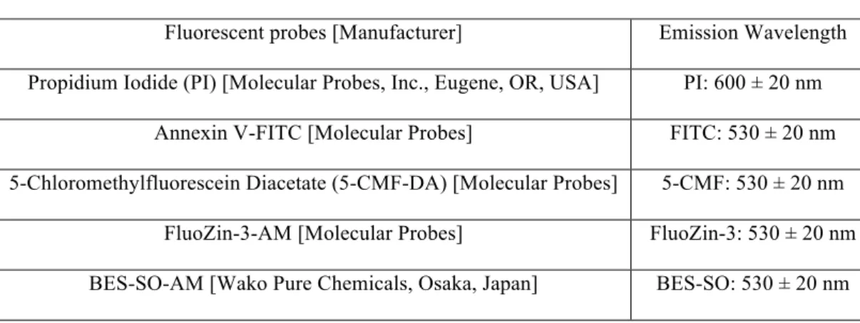
Table 1. Fluorescent probes used in this study
Excitation wavelength was 488 nm for all fluorescent probes.
Fluorescent probes [Manufacturer] Emission Wavelength Propidium Iodide (PI) [Molecular Probes, Inc., Eugene, OR, USA] PI: 600 ± 20 nm
Annexin V-FITC [Molecular Probes] FITC: 530 ± 20 nm 5-Chloromethylfluorescein Diacetate (5-CMF-DA) [Molecular Probes] 5-CMF: 530 ± 20 nm
FluoZin-3-AM [Molecular Probes] FluoZin-3: 530 ± 20 nm BES-SO-AM [Wako Pure Chemicals, Osaka, Japan] BES-SO: 530 ± 20 nm
Figure 1 (A)
Figure 2 (A)
(B)
Figure 3 (A)
(B)
Figure 4 (A)