1
Change in perception of the mandible caused by wearing dentures
Nobuhiko Furuse
(Director: Professor Yasuhiko Kawai)
2
Abstract
Purpose: Although many denture wearers often complain more about pain or irritation in their mandibular ridge mucosa, some patients do not complain pain or dullness even though they have prominet ulcers. However, no investigation has been done yet with the mandibular perception. Thus, this study aimed to investigate the influence of denture wearing on the mandibular nerve by measuring current perception threshold (CPT) and pain threshold (PT), and objectively understand the pain experienced by the denture wearers.
Methods: The CPT and PT values were obtained from the left mental foramen of 40 volunteers (20 edentulous and 20 dentulous) by using the Neurometer CPT/C® device to deliver electrical stimulation. The PT values were directly proportional to the CPT values.
Results and Discussions: The results showed that edentulous subjects exhibit a change in sensory nerve response to stimulation compared with dentulous subjects. The CPT value of the edentulous subjects was significantly higher. This indicated that the sensation is blunted in these subjects. If the CPT increases, there is a possibility that the subjects will not complain of symptoms even if there is excessive contact of the denture. Those with high CPT as well as PT values are presumed to be less likely to complain of the initial symptoms, but they may later complain of symptoms such as pain.
Conclusion: It suggests that perception is blunted by wearing a denture.
3
1. Introduction
Japan has the highest proportion of older population in the world, and is known as a
“super-aging” society (1). This increases in the elderly population of denture wearers (2, 3). Yoshikawa et al. showed that 90% of Japanese had experienced prosthodontic treatment and 88% of Japanese elders have been identified as needing dentures due to few remaining teeth (4).
Denture wearers experience several types of denture-related problems, such as inadequate mastication, cleaning difficulty, soreness or pain, poor retention, and problems with appearance. However, for new dentures, the most frequent problems are related to sore spots, painful gums, and discomfort, which are differently perceived by each patient (5). The difference in perception of pain might make denture management more difficult. Thus, precise prediction based on the diagnosis becomes essential (6). Therefore, diagnosing individual pain perception and predicting the required control measures and treatment planning is crucial.
Although many denture wearers often complain more about pain or irritation in their mandibular ridge mucosa (7), some patients do not complain pain or dullness even though they have prominet ulcers. Thus, it is essential to investigate individual perception and pain in the mandibular alveolar ridge. We have previously reported that the current perception threshold (CPT) and pain threshold (PT) are useful to evaluate individual perception in the maxillary alveolar ridge (8). It is known that denture wearing contributes to changes in responses to stimulations of the sensory nerve underlying the maxillary alveolar ridge (8). Furthermore, individuals wearing partial and complete dentures, and those not wearing dentures showed different responses to stimulation of the intraoral sensory nerve (9), which indicates that
4
denture wearing changes oral perception. Thus, the nerve of the mandibular mucosa may be influenced by wearing dentures. Pain in the supporting mucosal tissue after denture wearing is one of the most common and critical problems directly affecting function and treatment outcomes (6). Therefore, clarification of the influence of wearing mandibular dentures on the sensory nerve of the mandibular mucosa may contribute to an accurate prognosis of denture wearing.
However, no investigation has been done yet with the mandibular perception.
Therefore, the purpose of this study is to investigate the influence of denture wearing on the mandibular nerve by measuring CPT and PT.
2. Materials and Methods 2.1. Participants
Twenty edentulous patients who are willing to fabricate new dentures and having no problem with the new fabricated dentures and twenty dentulous students and employees of our institution were recruited for the study, and provided written informed consent prior to enrollment. Table 1 shows the participant characteristics.
Individuals with the following conditions were excluded: (a) general health problems that could affect the measurement of nerve activity, (b) signs and symptoms of orofacial pain disorders (e.g. trigeminal neuralgia or postherpetic neuralgia), (c) pacemaker wearers, (d) obvious cognitive impairment, (e) lack of understanding of written or spoken Japanese, (f) history of operation of the mandible, and (g) pregnancy. This study was approved by the institutional Human Ethics Committee of Nihon University School of Dentistry at Matsudo ( EC16-020 ).
2.2. Measurement environment
5
The participants were seated comfortably on dental chairs in a quiet room. The target region for measurement was the left mental foramen. To ensure contact between the mucosa and the stimulation electrodes, a measurement apparatus with Ø 1-mm thermoforming discs (Erkodur, Erkodent®, Pfalzgrafenweiler, Germany) was used for dentulous subjects and splint-resin (splint-resin LC®,GC Corp., Tokyo, Japan) was fabricated for edentulous participant. The plate (18×6×3 mm) with stimulation electrodes (Ø 2 mm) was mounted, using the part made of resilient denture liner (Sofliliner MS, Tokuyama, Japan), on an intraoral removable appliance (Fig. 1). The plate was firmly secured on the same measurement region: the left mental foramen.
The measurement region was blocked from saliva by using cotton rolls.
2.3. CPT and PT measurement
Neurometer CPT® (Neurotron, Inc., Towson, MD, USA) was used to measure peripheral nerve activity (10). This device can selectively stimulate different types of nerve fibers, A beta, A delta, and C fibers, at 2000-Hz, 250-Hz, and 5-Hz frequencies, respectively. A beta fibers transmit pressure and tactile signals, while A delta and C fibers transmit pain signals. These three types of nerve fibers comprise the majority of sensory nerve fibers. Therefore, it is possible to know the state of the sensory nerve by stimulating it at a frequency of 2000 Hz, 250 Hz, or 5 Hz. The time zone of measuring was set from 10AM to 3PM.
2.4. CPT
As per the manufacturer's instructions, the electrical current was slowly increased from 0.01 mA until the subjects reported a sensation for a given frequency. A
6
preliminary perception threshold was then determined. Subsequently, a microprocessor‐controlled forced‐choice method, which used 6 to 20 cycles of randomly selected true and false stimuli above and below the preliminary perception threshold, was implemented. This was double‐blinded until the exact CPT was determined.
2.5. PT
Similar to the CPT measurement procedure, the operator verbally explained the measurement process to each participant and exposed the participant to each stimulation frequency used to assess PT prior to the actual PT measurement.
Thereafter, the electrical current was gradually increased until the subjects reported pain for a given frequency.
2.6. Statistical analysis
After verifying the normality of the data using the Kolmogorov–Smirnov test, parametric statistical analysis was applied. The differences in the age, mucosal thickness, CPT, and PT between the edentulous and dentulous subjects were analyzed by using a t‐test. The relationship between CPT and PT was analyzed by Pearson's correlation coefficient. All statistical analyses were performed using SPSS® Statistics 21 (SPSS-IBM, MD, USA), with statistical significance set as P < .05.
3. Result 3.1. CPT
Fig. 2 shows CPT values for the respective frequencies. The CPT values were
7
significantly higher for the edentulous subjects than for the dentulous subjects at 2000 Hz (dentulous, 27.9 ± 17.3; edentulous, 49.0 ± 36.5; p=0.02), 250 Hz (dentulous, 17.1
± 13.1; edentulous, 27.3 ± 19.2; p=0.05), and 5 Hz (dentulous, 14.8 ± 11.3; edentulous, 28.7 ± 26.0; p=0.03).
3.2. PT
Fig. 3 shows the PT values for the respective frequencies. The PT values showed no significant difference between the edentulous and dentulous subjects at 2000 Hz (dentulous 96.4 ± 51.7 ; edentulous, 97.8 ± 41.3), 250 Hz (dentulous, 61.4 ± 32.0 ; edentulous, 63.2 ± 29.2), and 5 Hz (dentulous, 63.0 ± 30.9 ; edentulous, 72.5 ± 33.3).
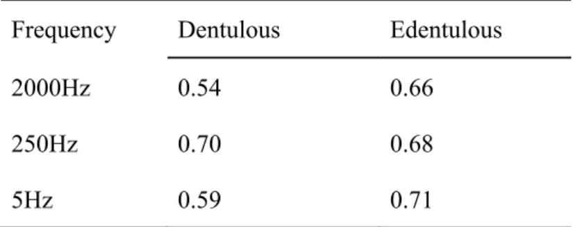
3.3. The Relationship between CPT and PT
Table 2 shows the correlation between CPT and PT. A statistically significant correlation was observed at all frequencies.
4. Discussion
This study shows that edentulous subjects exhibit a different sensory nerve response to stimulation compared with dentulous subjects. The CPT value of the edentulous subjects was significantly higher than those of the dentulous subjects. This suggests that the sensation is blunted in these subjects. Entrapment neuropathy due to carpal, cubital, and radial tunnel syndromes are caused by dynamic mechanical compression of a short segment of a single nerve at a specific site, as it passes through a fibro-osseous tunnel or an opening in fibrous or muscular tissue (11). Entrapment neuropathy and the condition of denture wearers are similar in that the alveolar
8
mucosa of the denture wearer is subjected to dynamic mechanical compression caused by the chewing force transmitted through the denture, and the mental nerve passes through the mental foramina and the alveolar mucosa under the denture. Furthermore, one of the characteristics of entrapment neuropathy is hypoesthesia, which is paralleled by increased CPT values (12, 13).
Kimoto et al. have reported that only the CPT value for 2000 Hz stimulation frequency is affected by wearing dentures (9). However, the results of this study showed a significant difference at all the tested frequencies (2000 Hz, 250 Hz, and 5 Hz). The mean denture-bearing area of the edentulous maxillae has more extensive area than that of the edentulous mandibles (14). Additionally, the nerves underlying the mandibular dentures might be subject to more severe compression because of occlusal force during mastication than those underlying the maxillary dentures owing to the smaller supporting area (15). Nishimura et al. discussed that due to dysfunctional sensory nerve fibers, caused by compression, changes in CPT began with larger fiber and extended to smaller ones in a stepwise manner as the dysfunction progresses (13). Therefore, it is suggested that the mandible dose greater nerve damage than the maxilla.
On the other hand, in the present study, PT values showed no significant difference between the edentulous and dentulous subjects at all frequencies. Reports are showing that pain is affected by many factors, such as psychological state and emotion (16, 17).
As a psychological factor influencing pain perception, it is well known that relaxation relieves pain; moreover, pain is associated with fear, and the pathological psychological state of anxiety and depression becomes stronger to increase pain perception (18, 19). However, a statistically significant correlation was observed
9
between CPT and PT at all frequencies. It was suggested that the person with a high CPT has a high PT. Therefore, there is a possibility that an ulcer may occur without feeling pain.
One of the limitations of this study was that the ages of the edentulous and dentulous subjects were not matched. In the future study, increased number of the subjects and enroll the edentulous and dentulous subjects by matching the age range should be planned. The analysis of relationship between age and CPT/PT would be feasible. Furthermore, in order to investigate the effect of denture wearing on the function of sensory nerves.
Within these limitations, however, the correlation coefficient of the edentulous tended to be higher than dentulous, which suggested that wearing of dentures may increase edentulous patient's mandibular CPT and PT.
5. Conclusion
1. The CPT of the edentulous subjects was significantly higher than dentulous subjects.
2. The PT values showed no significant difference between the edentulous and dentulous subjects at all frequencies.
3. A statistically significant correlation was observed between CPT and PT at all frequencies.
10
References
1.Chen BK, Jalal H, Hashimoto H, Suen S-C, Eggleston K, Hurley M, Schoemaker L, Bhattacharya, J: Forecasting trends in disability in a super-aging society: Adapting the Future Elderly Model to Japan. The Journal of the Economics of Ageing, 8:
42-51, 2016.
2. Douglass CW, Watson AJ: Future needs for fixed and removable partial dentures in the United States. The Journal of prosthetic dentistry, 87: 9-14, 2002.
3. Carlsson GE, Omar R: The future of complete dentures in oral rehabilitation. A critical review. J Oral Rehabil, 37: 143-156, 2010.
4. Yoshikawa M, Yoshida M, Nagasaki T, Tanimoto K, Tsuga K, Akagawa Y:
Influence of aging and denture use on liquid swallowing in healthy dentulous and edentulous older people. Journal of the American Geriatrics Society, 54: 444-449, 2006.
5. Szentpetery AG, John MT, Slade GD, Setz JM: Problems reported by patients before and after prosthodontic treatment. The International journal of prosthodontics, 18: 124-131, 2005.
6. Brunello DL, Mandikos MN: Construction faults, age, gender, and relative medical health: factors associated with complaints in complete denture patients. The Journal of prosthetic dentistry, 79: 545-554, 1998.
7. Beck CB, Bates JF, Basker RM, Gutteridge DL, Harrison A: A survey of the dissatisfied denture patient. The European journal of prosthodontics and restorative dentistry, 2: 73-78, 1993.
8. Ito N, Kimoto S, Kawai Y: Does wearing dentures change sensory nerve responses under the denture base? Gerodontology, 31: 63-67, 2014.
11
9. Kimoto S, Ito N, Nakashima Y, Ikeguchi N, Yamaguchi H, Kawai Y: Maxillary sensory nerve responses induced by different types of dentures. J Prosthodont Res, 57: 42-45, 2013.
10. Masson E, Boulton A: The Neurometer: validation and comparison with conventional tests for diabetic neuropathy. Diabetic Medicine, 8: S63-S66, 1991.
11. Dawson DM: Entrapment neuropathies of the upper extremities. New England Journal of Medicine, 329: 2013-2018, 1993.
12. Nishimura A, Ogura T, Hase H, Makinodan A, Hojo T, Katsumi Y, Yagi, K, Mikami, Y, Kubo, T: Objective evaluation of sensory function in patients with carpal tunnel syndrome using the current perception threshold. Journal of orthopaedic science, 8: 625-628, 2003.
13. Nishimura A, Ogura T, Hase H, Makinodan A, Hojo T, Katsumi Y, Yagi, K, Mikami, Y, Kubo, T: A correlative electrophysiologic study of nerve fiber involvement in carpal tunnel syndrome using current perception thresholds.
Clinical Neurophysiology, 115: 1921-1924, 2004.
14. Yanikoglu N, Ceylan G, Aladag Lİ: A comparison of the basal seat areas of the maxillary and mandibular dentures according to arch shapes. Atatürk Üniversitesi Diş Hekimliği Fakültesi Dergisi, 2005: 29-33, 2005.
15. Tanaka M, Ogimoto T, Koyano K, Ogawa T: Denture wearing and strong bite force reduce pressure pain threshold of edentulous oral mucosa. J Oral Rehabil, 31:
873-878, 2004.
16. Sjörs A, Larsson B, Persson AL, Gerdle B: An increased response to experimental muscle pain is related to psychological status in women with chronic non-traumatic neck-shoulder pain. BMC musculoskeletal disorders, 12: 230, 2011.
12
17. Meagher MW, Arnau RC, Rhudy JL: Pain and emotion: effects of affective picture modulation. Psychosomatic medicine, 63: 79-90, 2001.
18. Roykulcharoen V, Good M: Systematic relaxation to relieve postoperative pain. Journal of advanced nursing, 48: 140-148, 2004.
19. Keogh E, Ellery D, Hunt C, Hannent I: Selective attentional bias for pain-related stimuli amongst pain fearful individuals. Pain, 91: 91-100, 2001.
13
14
Legends to Figures
Fig. 1: Intraoral removable device with stimulation electrodes
Participants wore the measurement apparatus shown, with contact occurring between the mucosa and the stimulation electrodes
Fig. 2: Difference between the CPT of edentulous and dentulous subjects
The bar for each group (left and right sides) represents the mean CPT values at the left mental foramen and buccal shelf, respectively. CPT=current perception threshold
Fig. 3: Difference between the PT of edentulous and dentulous subjects
The bar in each group (left and right sides) represents the mean PT values at the left mental foramen and buccal shelf, respectively. PT=pain threshold
15
Fig. 1
16
Fig. 2
17
Fig. 3
18
Table 1. Participants’ characteristics
Variable Dentulous
Mean±SD
Edentulous Mean±SD
Sex(male/female) 10/10 10/10
Age(years) 26.4 ± 2.6 77.9 ± 6.1
Mucosal thickness(mm) 1.8±0.7 1.8±0.6
Duration of wearing denture (year) 18.4±9.5 SD=standard deviation
Table 2. Relationship between CPT and PT
Correlation coefficient
Frequency Dentulous Edentulous
2000Hz 0.54 0.66
250Hz 0.70 0.68
5Hz 0.59 0.71
CPT=current perception threshold, PT=pain threshold All P<0.05