The United Graduate School of Veterinary Science
Yamaguchi University
NZE NKOGUE Chimene
Contents Preface Dedication Acknowledgements Summary in Japanese List of abbreviations List of figures List of tables
Chapter I: General introduction
Chapter II: Molecular epidemiological study of adenovirus infecting western lowland
gorillas and humans in and around Moukalaba-Doudou National Park (Gabon)
Abstract
II.1. Introduction
II.2. Materials and Methods
II.2.2. DNA extraction and PCR
II.2.3. BLAST search
II.2.4. Sequencing and phylogenetic analysis
II.3 Results
II.3.1. Detection of AdV genes in western lowland gorillas in MDNP
II.3.2. Detection of AdV genes in local people living around the national park
II.3. 3. Phylogenetic analysis
II. 4. Discussion
Chapter III: Identification and molecular characterization of novel primate
bocaparvoviruses from wild western lowland gorillas of Moukalaba-Doudou National
Park, Gabon
Abstract
III.1. Introduction
III.2. Material and methods
III.2.2. Individual genetic identification
III.2.3. PCR detection of bocaparvovirus
III.2.4. Sequencing of the gorilla bocaparvoviruses
III.2.5. Phylogenetic analysis
III.2.6. Recombination analysis
III.3. Results
III.3.1. Detection of bocaparvoviruses in gorillas
III.3.2. Sequence analysis of the gorilla bocaparvoviruses
III.3.3. Phylogenetic analysis
III.3.4. Recombination analysis
III.4. Discussion
III. 5. Conclusions
Chapter IV. General discussion
IV.1. Genetic diversity and evolution of viruses
The current doctoral thesis in based on the following papers:
1- C. Nze Nkogue, M. Horie, S. Fujita, M. Ogino, Y. Kobayashi, K. Mizukami, T.
Masatani, S. Ezzikouri, A. Matsuu,T. Mizutani, M. Ozawa, O. Yamato, A. Ngomanda,
J. Yamagiwa, K. Tsukiyama-Kohara, 2016. Molecular epidemiological study of
adenovirus infecting western lowland gorillas and humans in and around
Moukalaba-Doudou National Park (Gabon). Virus Genes 52, 671-678.
2- C. Nze-Nkogue, M. Horie, S. Fujita, E. Inoue, E-F. Akomo-Okoue, M. Ozawa, A.
Ngomanda, J. Yamagiwa, K. Tsukiyama-Kohara, 2017. Identification and molecular
characterization of novel primate bocaparvoviruses from wild western lowland gorillas
of Moukalaba-Doudou National Park, Gabon. Infection, Genetics and Evolution 53,
It has been a long way to the achievement of this PhD. Many people have been
involved in so many ways from the field work in Gabon to the lab work in Japan. I would
like to express m
you find here the expression of my deep recognition.
First and foremost, I want to thank Dr. Juichi Yamagiwa who leaded the project
PROCOBHA (Project for Conservation of Biodiversity through Sustainable Coexistence
between Humans and Animals) in which I have made my first steps in the field, learning
about wild gorillas and from where I have developed my interest for the present study.
Dr. Shiho Fujita supported the present research work from the idea to the
accomplishment. I am grateful for her huge contribution.
My thesis committee: Dr. Kyoko Tsukiyama-Kohara, Dr. Masayuki Horie, Dr.
Makoto Ozawa, and Dr. Toshihiro Ito for being my main advisers. It was a great chance
that Dr. Tsukiyama-Kohara accepted me in her laboratory and supported my doctoral
research. The work in collaboration with Dr. Horie was the key to success of my doctoral
research. He polished my ideas and assisted technically throughout my research time. I
thinking about the next experiment plan in details. Dr. Ito guided and advised me about
paper-writing. Dr. Tatsunari Masatani and Dr. Aya Matsuu taught me advanced
techniques of molecular biology and cell culture. Their support is highly acknowledged.
To the lab staff: students, technicians and post-docs. All of you contributed to the
achievement of the present study. Khadija, although short, I miss the time you were
around. Kayesh- -san, Bouchra, Kawabata-san,
Yamaguchi-san, Nakagawa-san, Ueno-kun, Nori-chan, Okuya-kun, Kanda-kun,
Handa-kun, Haraguchi-san; I wish to all of you the best of luck.
: I thank Gabonese researchers (Dr. Etienne-Francois
Akomo-Okoue, Mr. Guy-Max Moussavou, Mr. Philippe Mbehang Nguema, Dr. Patrice
Makouloutou) and administrative (Dr. Alfred Ngomanda, Ms. Pamela Nang, Mr. Cedrick
Nguima, Mr. Didier Bineni), Japanese researchers (Dr. Kazunari Ushida, Dr. Yuji
Takenoshita, Dr. Shigeru Suzuki, Ms. Keiko Tsubokawa, Dr. Yuji Iwata, Dr. Yoshihiro
Nakashima, and Dr. Shun Hongo) and administrative (Ms. Chieko Ando, Ms. Naoko
To the people from Doussala, Konzi, and Mboungou: field assistants, trackers,
local people, you made my field trips always enjoyable and fruitful.
I would like to thank my family for their love and encouragements. The teachings
of my late grand father (Paul Mve Minko) and late grand-mother (Madeleine Avomo
Ovono), were an appetizer for an endless knowledge. My mother Mekui Mve rachel has
made me the woman I am. My father Nkogue Mba Raphael has been always behind the
scenes, and his presence is inestimable. Ntsame Ovono Dora rachel my sister and Ntsame
Mve Reine Livanne my little mum and friend, their
me feel always at home despite the distance. My two little princesses Sarah and
Bernancia, their love gave me wings to fly farer than I could expect. I thank Benjamin
Majanga Dotto for his blessing. I thank my friends Munde Vestergaard, Satomi
Hashimoto and Amina Moss for being such amazing active mothers.
I am grateful to the Research Institute in Tropical Ecology (IRET-CENAREST)
for allowing my study in Japan. The Ministry of Education, Culture, Sports, Science and
Technology, Japan (MEXT), for providing a scholarship for my PhD studies. The United
Graduate School of Veterinary Science, Yamaguchi University for allowing me to
Summary
The screening of infectious agents (viruses, parasites or bacteria) in wildlife
provides critical data regarding not only the presence of pathogens but also the diversity
and the natural history of the target microbes. The emergence and re-emergence of
diseases which originated from wildlife has emphasized the necessity for such pathogen
assessment for the conservation of endangered animal populations, controlling the risk on
the trade of wildlife or wildlife products, and preventing pathogen from spilling over into
livestock or human population. In addition, the studies on infectious pathogens in the
natural living great ape (gorillas, chimpanzees, etc.) populations has clarified the origins
of some human viruses (HIV, HTLV, etc) as well as raising the concern about pathogen
cross-species transmission between both hosts owing to the genetic relatedness between
great apes and humans.
I conducted the surveillance of adenovirus and bocaparvovirus infection in wild
western lowland gorillas in Moukalaba-doudou National Park (Gabon) in order to
investigate the presence, genetic diversity, and evolutionary history of these viruses, and
adenoviruses are widespread in humans and great apes, the data about the naturally
occurring infections remain rare. On the other hand, the evolutionary study of
adenoviruses infecting great apes has recently revealed that the Human mastadenovirus
B ( originated from ancient gorillas and had experienced several cross-species
(ape-ape and ape-human) transmission events. Bocaparvoviruses have been extensively
studied because of their frequent association with respiratory illness and/or and
gastroenteritis in humans. Although some bocaparvoviruses have been detected in
non-human primates (gorillas and chimpanzees), the presence, diversity, and evolution of
these viruses are not fully understood.
On the other
chimpanzee strains, which support the hypothesis of being a zoonotic virus. The
HAdV-E was clustered with the chimpanzee strains. This result indicates the possibility of an
ape-to-ape transmission of HAdV-E species because chimpanzees have been reported to
be the most probable ancestor hosts of these viruses.
Regarding the bocaparvorirus infection, I detected
. The
named Gorilla bocaparvovirus 2 (GBOV2) of this study is the first non-human primate
bocaparovirus within that species. The molecular evolutionary analyses of primate
bocaparvoviruses revealed the presence of inter and intra-species recombination events
which might lead to the emergence of new bocaparvovirus variants in human as well as
in non-human primate population.
The description of several adenoviruses and the identification of novel
bocaparvoviruses in wild western gorillas contribute to a better understanding of the
genetic diversity of these viruses as well as clarifies their evolutionary processes.
Although there is no evidence of gorilla-to-human interspecies transmission of the
as well as in humans (tourists, guides, local peoples, etc.) which potentially contact with
T
-24.1 35.0
HAdV-B HAdV-C
HAdV-E HAdV-C HAdV-D
HAdV-C
HAdV-B
HAdV-B
2 2 2 2 86.0 2 2 GBOV2
-°C. Degrees Celcius
µl. microliter
AdVs. Adenoviruses
bp. base pair
DPOL. DNA polymerase
G8. Group 8
GBOV. Gorilla bocavirus
GG. Group Gentil
HAdV. Human adenovirus
HBOV. Human bocavirus
HF. High Fidelity
HIV. Human Immunodeficiency Virus
HVR. Highly Variable Region
ICTV. International Committee of Taxonomy for Viruses
MDNP. Moukalaba-Doudou National Park
min. Minutes
mM. millimolar
ng. nanograms
ORF. Open Reading Frame
PCR. Polymerase Chain Reaction
SIV. Simian Immunodefiency Virus
U. Unit
WHO. World Health Organization
Figure 1.1. Location features of the sampling area
(A) Map of Gabon, showing MDNP. (B) The sampling area in the MDNP (blue line:
rivers; black line: roads; red line: hunting area limitation; green line with black strips:
national park limitation; dark green: primary forest; olive green: secondary forest; brown:
savanna; spotted green: swamp; black circle: sampling points of G8 pointed by an arrow;
gray circle pointed by an arrowhead: sampling points of GG; white circle: base camp;
black rectangle with a black flag: village; white squares: habitations).
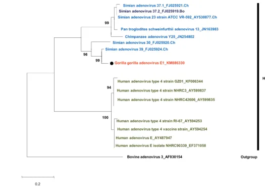
Figure 1.2. Phylogenetic tree of adenovirus (AdV) DPOL
The tree was constructed based on the alignment of AdV DPOL (539 bp) by using the
neighbor-joining bootstrap-confirmed method in MEGA 5.05 software with 100
replicates. The names of simian isolates include the serotype nomenclature and the animal
species of isolation (Ch: chimpanzee, Go: gorilla, Bo: bonobo). Names of novel
sequences obtained in this study are indicated with black dots. Bootstrap values less than
90% are omitted. Scale bar, nucleotide substitutions per site.
The tree was constructed based on the alignment of a 792-bp sequence of the hexon gene
by using the neighbor-joining bootstrap-confirmed method in MEGA 5.05 software with
100 replicates. The names of simian isolates include the serotype nomenclature and the
animal species of isolation (Ch: chimpanzee, Go: gorilla Bo: bonobo). Names of novel
sequences obtained in this study are indicated with black dots.
Figure 1.4. Phylogenetic tree of the partial sequence of DPOL
The tree was constructed based on the alignment of AdV DPOL (539 bp) by using the
neighbor-joining bootstrap-confirmed method in MEGA 5.05 software with 100
replicates. The names of simian isolates include the serotype nomenclature and the animal
species of isolation (Ch: chimpanzee, Go: gorilla, Bo: bonobo). Names of novel
sequences obtained in this study are indicated with black dots. Bootstrap values less than
90% are omitted. Scale bar, nucleotide substitutions per site.
Figure 1.5. Phylogenetic tree of the partial sequence of the hexon gene
The tree was constructed based on the alignment of a 792-bp sequence of the hexon gene
by using the neighbor-joining bootstrap-confirmed method in MEGA 5.05 software with
animal species of isolation (Ch: chimpanzee, Go: gorilla Bo: bonobo). Names of novel
sequences obtained in this study are indicated with black dots.
Figure 1.6. Phylogenetic tree of partial hexon of HAdV-D
The tree was constructed based on the alignment of a 792-bp sequence of hexon gene by
using the neighbor-joining bootstrap-confirmed method in MEGA 5.05 software with 100
replicates. The names of simian isolates include the serotype nomenclature and the animal
species of isolation (Ch: chimpanzee, Go: gorilla Bo: bonobo). Names of novel sequences
obtained in this study are indicated with black dots.
Figure 2.1. PCR strategy. (A) Schematic diagram of the PCR strategy. Scale, viral
genome, and primers are shown. Gray arrows indicate primers used for detection and
sequencing of bocaparvoviruses. The amplified regions are indicated by dashed gray
lines. (B) Gel electrophoresis results showing the amplicon sizes of both positive samples.
Figure 2.2. Genome organization of the gorilla bocaparvovirus 2. (A) The genome
organization of GBOV2 is shown. The predicted ORFs are shown in boxes. The gray
boxes are the regions shown in (B), (C), or (D). (B) A partial nucleotide alignment of the
partial alignment of deduced amino acid sequences of NS1 proteins. The conserved
ATP-binding Walker-Loop motif is indicated by the gray box. (D) An amino acid sequence
alignment of partial VP1 proteins. The phospholipase A2 motif, which consists of the
calcium binding region and catalytic residues, is indicated.
Figure 2.3. Phylogenetic tree based on the complete coding sequences of primate
bocaparvoviruses. The tree was reconstructed based on a nucleotide alignment of the
complete coding sequence of the indicated viruses using the maximum likelihood method
with 1000 bootstrap replicates. The GBOV2 sequences determined in this study are
indicated with black dots. Bootstrap values of >70% are indicated at nodes. Scale bar,
nucleotide substitutions per site.
Figure 2.4. Phylogenetic tree based on the partial NS and VP2 genes of primate
bocaparvoviruses. The tree was constructed based on the partial NS gene (486 b) (A) and
VP gene (486 b) (B) using the maximum likelihood method with 1000 bootstrap
replicates. The GBOV2 sequences obtained in this study are indicated with black dots.
Bootstrap values of >70% are indicated at nodes. Scale bar, nucleotide substitutions per
Figure 2.5. Recombination analysis. (A) Breakpoint detection using DualBrothers
applied to the complete coding sequences of representative primate bocaparvoviruses.
(B K) Phylogenetic trees were constructed based on partial alignments using the
maximum likelihood method with 1000 bootstrap replicates. The nucleotide positions
used for the phylogenetic inferences are indicated by blue letters. Bootstrap values of
>70% are indicated at nodes
Figure 2.6. A representation of individual gorillas constituting the target group based on
body size and age categories. The red circles show the infected infants.
Figure 2.7. Phylogenetic tree based on the complete NS1 sequence. The tree was
constructed based on the nucleotide sequences of complete NS1 using the maximum
likelihood method with 1000 bootstrap replicates. The GBOV2 sequences obtained in this
study are indicated with black dots. Bootstrap values of >70% are indicated at nodes.
Scale bar, nucleotide substitutions per site.
Figure 2.8. Phylogenetic tree based on the complete VP1 sequence. The tree was
reconstructed based on the nucleotide sequences of complete NP1 using the maximum
this study are indicated with black dots. Bootstrap values of >70% are indicated at nodes.
Scale bar, nucleotide substitutions per site.
Figure 2.9. Phylogenetic tree based on the complete NP1 sequence. The tree was
constructed based on the nucleotide sequences of the complete NP1 gene using the
maximum likelihood method with 1000 bootstrap replicates. The GBOV2 sequences
obtained in this study are indicated with black dots. Bootstrap values of >70% are
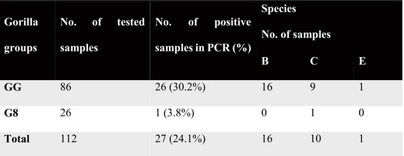
Table 1.1. Detection of adenovirus DPOL and hexon genes in samples from gorilla
groups in MDNP
Table 1.2. Adenovirus infection in humans
Table 1.3. Primers and probe sequences for amplification of DPOL and hexon genes
Table 1.4. Adenoviruses, accession number and hosts
Table 2.1. Bocavirus infection prevalence in gorillas in MDNP
Table 2.2. Primers used for sequencing the gorilla bocaparvovirus 2
Table 2.3. Bocavirus sequences used for the phylogenetic inference
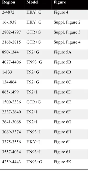
Table 2.4. List of models used for the Maximum Likelihood test
Table 2.5. Pairwise amino acid percentage identities of NS1 of GBOV2 and other primate
bocaparvovirus reference strains.
Epidemiological investigations of microorganisms (bacteria, parasites, viruses)
infecting wild animals contribute to the understanding of the diversity and the
evolutionary history of the target microbes. For virus screening, two different approaches
can be used: the antibody detection and the virus or viral nucleic acid detection
(Lehmkuhl and Hobbs, 2008; Luebcke et al., 2006; Mouinga-Ondémé et al., 2010;
Oberste et al., 2013; Wevers et al., 2011). Sero-epidemiological surveys inform about
which virus the animal has been exposed to (past or ongoing infections) (Makuwa et al.,
2006; Nidom et al., 2012; Rudicell et al., 2011; Starkman et al., 2003) and the virus
isolation or viral nucleic acid detection informs about the ongoing infections (Gál et al.,
2013; Wevers et al., 2010). The knowledge of the viral genetic sequence coupled with the
phylogenetic characterization will contribute to the classification and the understanding
of the diversity and the evolutionary history of the detected viruses.
Several molecular studies have revealed the close genetic relatedness between
non-human primates and human viruses (Ahuka-Mundeke et al., 2010; Duncan et al.,
adenoviruses infecting human and apes cluster in the species Human adenvovirus B, C,
E and F (Roy et al., 2009; Wevers et al., 2011). Similarly, primate bocaparvoviruses
include human and non-human primate strains
2010b; Sharp et al., 2010).
Additionally, approximately 75% of the diseases that have emerged or re-emerged over the past two decades have wildlife sources (Woolhouse et al., 2012) and great apes play a critical role regarding these zoonosis (Calvignac-Spencer et al., 2012). Ape-to-human zoonotic transmission is suggested for several viruses including the Human mastadenovirus-B (Hoppe et al., 2015), Ebolavirus (Leroy et al., 2011), the Human Immuno-Defiency virus (HIV)-I (Sharp and Hahn, 2010), the Human T-cell Leukemia
virus (HTLV)-I (Junglen et al., 2010) or the spumavirus (Betsem et al., 2011).
Despite the extensive research on the viruses infecting wild apes, their diversity and evolutionary history remain poorly understood.
The object of this study is to investigate adenovirus and bocaparvovirus naturally occuring infections in wild gorillas using PCR in order to understand their diversity and evolution.
In the first part of this thesis, I targeted the masdenoviruses infecting wild gorillas
of Moukalaba-Doudou NationalPark and human (local people leaving nearby the park
Mastadenoviruses infect human and non-human primates (Duncan et al., 2013;
Roy et al., 2009) and the human and non-human primate strains belong to the Human
adenovirus species A to G (Pantó et al., 2015; Roy et al., 2009). Additionally, a recent
report has documented about the Human mastadenovirus B originating from gorillas
(Hoppe et al., 2015), and switched to human population and chimpanzees (Hoppe et al.,
2015) which highlights the ape-human and ape-ape cross-species transmission.
On the other
hand, HAdV-B was clustered with other gorilla adenoviruses together with human and
chimpanzee strains, which support the hypothesis of being a zoonotic virus. The
ape-ape transmission of HAdV-E species because chimpanzees have been reported to be
the most probable ancestor hosts of these viruses.
In the second part, I targeted bocaparvoviruses in wild gorillas. The viruses infecting
human and non-human primates are grouped into 2 species named primate
bocaparvoviruses. To date, 2 species of primate bocaparvoviruses have been described.
Primate bocaparvovirus 1 and 2 (Allander et al., 2001; Arthur et al., 2009; Kapoor et al.,
2010b, 2009). A captive gorilla and a captive chimpanzee have been reported to be
infected by the members of the species Primate bocaparvovirus 1
Kapoor et al., 2010a); therefore the diversity of bocaparvoviruses infecting wild apes and
their evolutionary history remain poorly understood.
I detected
. The named Gorilla bocaparvovirus 2 (GBOV2) of this
study is the first non-human primate bocaparovirus within that species. The molecular
intra-species recombination events which might lead to the emergence of new
bocaparvovirus variants in human as well as in non-human primate population.
The findings of this study contribute to a better understanding of the genetic diversity of mastadenoviruses and bocaparvoviruses infecting gorillas and provide insights to the evolutionary history of primate bocaparvoviruses.
II.1. Introduction
Adenoviruses (AdVs) are non-enveloped icosahedral double-stranded DNA viruses.
They belong to the family of Adenoviridae, which is divided into 5 genera:
Mastadenovirus, Atadenovirus, Aviadenovirus, Siadenovirus, and Ichtadenovirus.
Members of species belonging to genera Mastadenovirus and Atadenovirus are known to
infect mammalian hosts (Lehmkuhl and Hobbs, 2008; Pantó et al., 2015).
Mastadenoviruses infecting primates encompass 7 Human mastadenovirus species
(HAdV-A to G), the accepted species Simian mastadenovirus A and candidate species
SAdV-B to G (Chen et al., 2011), and further not yet classified mastadenoviruses (Chen
et al., 2011; Gál et al., 2013; Pantó et al., 2015). That classification into species or
subgroups is based on hemagglutination features, DNA (Deoxyribonucleic acid
homology, and genomic organization (Robinson et al., 2013).There are currently over 60
HAdV types with HAdV-D containing the most members (Robinson et al., 2013).
Adenoviruses were first isolated from humans and identified as the causative agent of
epidemic febrile respiratory disease among military recruits in the 1950s (Hilleman and
is seropositive for one or more serotypes of adenoviruses
Wadell et al., 1987). The molecular biology of human-derived adenoviruses has been
characterized extensively for the species C group, for which HAdV 2 (HAdV-2) and
HAdV-5 serve as prototypes (Fields, et al., 1996). Adenoviruses cause a variety of
nonlethal infectious diseases in humans, and lethal disseminated adenovirus infection
occurs in immunosuppressed patients (Fields, et al., 1996).
The first description of a simian adenovirus in the literature was of a chimpanzee AdV
(Rowe et al., 1956), today known as SAdV-21 within the species Human mastadenovirus
B. Later, when investigating chimpanzees suffering from kuru, four novel apes AdVs
were discovered (Rogers, et al., 1967). Ape AdVs have been detected or isolated from
African apes including chimpanzees, bonobos and gorillas (Duncan et al., 2013; Hoppe
et al., 2015; Roy et al., 2009; Seimon et al., 2015; Wevers et al., 2011, 2010). Gorilla
adenoviruses have been proposed to be members of HAdV-B, C, E, and F (Duncan et al.,
2013; Hoppe et al., 2015; Roy et al., 2009; Seimon et al., 2015; Wevers et al., 2011, 2010).
A recent report confirmed that the species HAdV-B which includes viruses from mixed
and to chimpanzees during 2 different host switch events (Hoppe et al., 2015). Serological
surveys have found that anti-AdV antibodies were prevalent in 96% of mountain gorillas,
suggesting that AdVs are circulating among these animals (Whittier, 2009). In addition,
Hoppe et al. recently reported a high prevalence of AdV in wild apes including gorillas
(45 to 100%) (Hoppe et al., 2015). Because AdVs are shed in the feces and saliva of
infected animals (Roy et al., 2009), these viruses could possibly be transmitted among
host animals via the fecal-oral route and inhalation of aerosols (Fong et al., 2010).
Comprehensive studies are still needed to clarify the origin and the diversity of
adenoviruses spread in human and non-human primate populations. Thus, to fill the gap,
understanding the evolution pattern of AdVs spread in non-human primates and in people
frequently coming in contact with these animals is critical. In this study, I investigated
AdV infection in 2 habituated western lowland gorilla groups in MDNP. In addition, I
assessed AdV infection in the local people living around the national park to evaluate
II.2. Materials and Methods
II.2.1. Sample collection and preparation
The study site (MDNP) is located in the south-western part of Gabon (Fig. 1.2).
MDNP has been reported to have a high gorilla density (more than 3 gorillas per square
kilometer) (Ando et al., 2008), and the absence of hunting pressure from local villagers
makes it a major habitat for western lowland gorillas in central Africa. From December
2010 to November 2011, during tracking, we collected 112 fresh fecal samples from 2
wild gorilla groups, which were named as Group Gentil (GG) and Group 8 (G8). GG and
G8 had been habituated to human observers since 2003 (Ando et al., 2008) and 2011,
respectively. During the study period, GG consisted of 20 21 individuals, including 1
6
years old) males, and 3 young females, and all members were individually identified. In
contrast, G8 was estimated to consist of 8 12 individuals, including 1 adult male, 2 adult
females, 5 8 young males and females. GG was mainly sampled near the village
Doussala, in the ancient plantations, where the forest has been formerly used in various
addition to the gorilla samples, 20 fecal samples were collected from villagers, including
trackers working for the habituation of gorillas. Upon collection, each fecal specimen was
immediately placed into a tube containing 2 ml of RNAlater (Ambion, Austin, TX, USA).
The tubes were kept at room temperature for at most 20 days at the field camp until the
samples were transported to the laboratory in Libreville, the capital city of Gabon. At the
laboratory, the tubes were stored at 20°C until DNA extraction.
II.2.2. DNA extraction and PCR
Total DNA was extracted from the sample by using the QIAamp DNA Stool Mini Kit
ructions. I used the
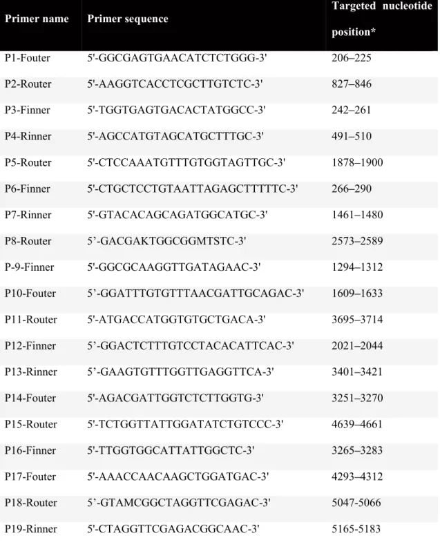
following primer sets for nested PCR: (1) 4431-s/4428-as and 4428-s/4429-as (Table 1.3),
targeting the HAdV DPOL gene (Wevers et al., 2010) and (2) AdhexF1/AdhexR1 and
AdhexF2/AdhexR2, targeting loop 1 encompassing the hypervariable region (HVR1
6) of the hexon gene of mastadenoviruses (Lu and Erdman, 2006). PCR for the DPOL
gene was performed in a total volume of 20 µl containing 10 µl of 2×GoTaq Green Master
Mix (Promega, Madison, WI, USA), 20 pmol of each primer, and 50 ng of DNA template.
2010) were used: 95°C for 2 min; 35 cycles of 95°C for 30 s, 55°C for 1 min, and 72°C
for 1 min; and a 7-min final extension step at 72°C. PCR amplification of the hexon gene
(HVR1 6) was performed in a total volume of 50 µl containing 200 µM of each dNTP,
20 pmol of each primer, 1.25 U of PrimeSTAR GXL polymerase (TaKaRa, Tokyo,
Japan), and 50 ng of DNA template. The cycling conditions were as follows: 98°C for 3
min; 35 cycles of 98°C for 10 s, 45°C for 1 min, and 72°C for 2 min; and a final extension
of 72°C for 7 min. For the nested reaction, 2 µl of the first PCR was amplified as above.
Amplified products were separated on 1.5% agarose gel and purified using the QIAquick
were then directly sequenced with the primers for the second PCR.
II.2.3. BLAST search
BLAST searches were carried out in the NCBI database
(http://blast.ncbi.nlm.nih.gov/Blast.cgi) using the determined nucleotide sequence as a
query in the BLASTN program. The queries with at least 90% identity with the deposited
II.2.4. Sequencing and phylogenetic analysis
Twenty-four of the 27 positive samples (DNA /µl), were subjected to
direct sequencing of DPOL gene fragments. Six samples were selected randomly for
cloning and sequencing of DPOL and hexon HVR1 6 gene fragments. The PCR products
were cloned into plasmid vector pCR-Blunt II-TOPO using the Zero Blunt TOPO PCR
Plasmid extraction was carried out using the Wizard Miniprep Kit (Promega), and the
extracted plasmids were sequenced by Big Dye terminator cycle sequencing (Applied
Biosystems, Foster City, CA, USA).
The hexon HVR1 6 and DPOL gene sequences were edited and aligned using
GENETYX software version 12.0 (Genetyx Co., Tokyo, Japan) and MEGA software
version 5.05 (Tamura et al., 2011). The nucleotide sequences of DPOL (528-bp,
corresponding to the position 29,200-29,727 in the reference simian adenovirus 21) and
782-bp fragments of the hexon gene (corresponding to the position 18, 867-19,635 in the
reference simian adenovirus 21) were aligned using MUSCLE, with the default
phylogenetic analyses. Phylogenetic trees were constructed using the maximum
likelihood method in MEGA 5.05 (Tamura et al., 2011). A statistical test for the
phylogeny was computed by means of bootstrapping. Percentages of 100 bootstrap
replicates at the node were calculated to ensure the reliability of the trees.
II.2.5. Nucleotide sequence accession numbers
Preliminary names were given to candidate novel HAdVs following the method used
by Wevers et al.(Wevers et al., 2010). The gorilla adenoviruses detected in this study were
named as follows: Gorilla gorilla AdV B11-B23 (KM886307- KM886309, KM886311,
KM886325- KM886328, KM886331- KM886335), Gorilla gorilla AdV C10-C18
(KM886310, KM886320- KM886324, KM886329), and Gorilla gorilla AdV E1
(KM886330). The sequences used as references for phylogenetic analysis are listed in
II.3 Results
II.3.1. Detection of AdV genes in western lowland gorillas in MDNP
To survey AdV infection in gorillas in MDNP, i collected fecal samples from 2 gorilla
groups (GG: well-habituated group, G8: newly habituated group), and analyzed them by
nested PCR targeting the DPOL and hexon genes.The DPOL and hexon genes were
detected in both groups (Table 1.1). The overall prevalence of AdV in the gorilla
population was 24.1% (27/112): of the 86 samples from GG, 21 were positive for both
genes, 4 were positive only for the DPOL gene, and 1 sample was positive only for the
hexon gene. In contrast, only 1 of the 26 samples was positive for both tested genes in G8
(Table 1.1). These data suggest that AdVs are naturally circulating among gorillas in
MDNP. To confirm the detected AdV species, I further determined the nucleotide
sequences of the amplicons and determined the species of the detected AdVs by BLAST
searches. Of the tested samples, 16 belonged to B; 10 to C; and 1 to
II.3.2. Detection of AdV genes in local people living around the national park
The prevalence of AdVs in well-habituated gorillas (30.2% in GG group) was higher
than that of newly habituated ones (3.8% in G8 group), raising 2 possibilities either the
AdVs in gorillas are derived from humans during the habituation process or AdVs are
ubiquitous in the environment in and around areas of human habitation. Therefore, I
screened the local people (village Doussala in Fig. 1.2) for AdV infection. The prevalence
in the local people was 35.0 % (7/20): 2 samples were positive for both DPOL and hexon
genes, and 5 were positive only for the hexon gene (Table 1.2). These results revealed
that the local people including trackers were also infected with AdVs.
I sequenced the detected virus genes and identified the species of AdVs: 1 sample
was infected with HAdV-C, and the others harbored HAdV-D.
II.3. 3. Phylogenetic analysis
HAdV-C genes were detected in both gorillas and humans in MDNP, suggesting
zoonotic transmission of AdV between the human and gorilla populations. To investigate
this possibility, as well as to gain insights into the genetic diversity of adenoviruses in
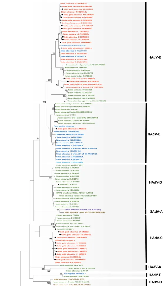
In gorillas, in the tree based on the DPOL gene, the 14 AdV genes identified in this
study were divided into 2 groups; they clustered with SAdV-28.2, SAdV-46, SAdV-47,
and gorilla AdV strains 6589 and 6575, which are representative strains of HAdV-B in
gorillas, and unidentified simian adenoviruses recently described (Hoppe et al., 2015)
(Fig. 1.3 and 1.5). Nine AdV genes were clustered with simian 45 and simian
AdV-43, which are representative strains of HAdV-C in gorilla and new unidentified simian
adenoviruses (Hoppe et al., 2015) (Fig.1.3 and 1.5). In contrast, one AdV gene clustered
with SAdV-26 and chimpanzee AdV strain Y25, which are chimpanzee-specific strains
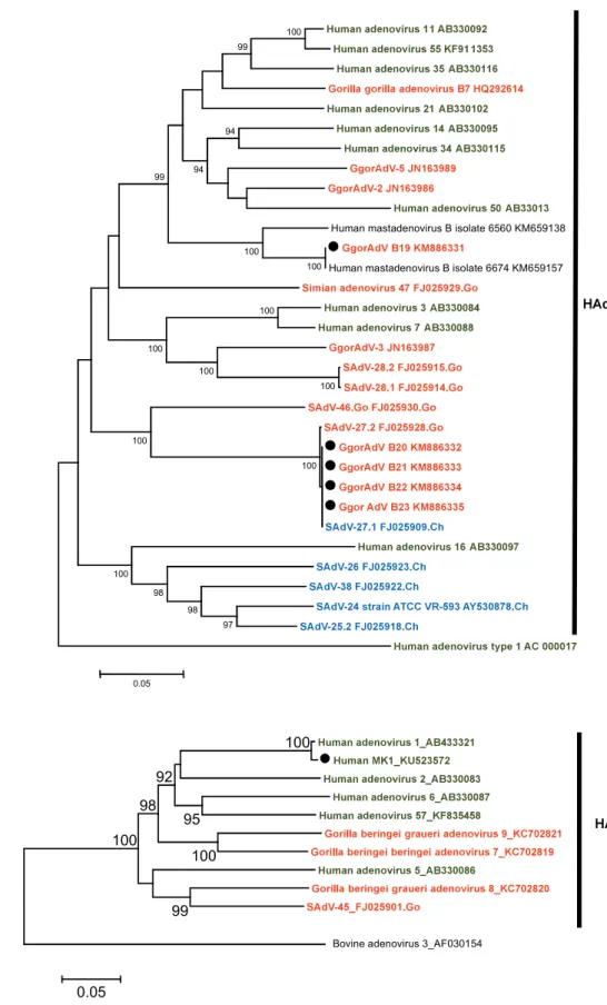
belonging to HAdV-E (Fig. 1.3 and 1.5). In the hexon gene-based trees, five HAdV-B
(Figure 1.6.A), 1 HAdV-C (Figure 1.6.B), and 1 HAdV-E (Fig. 1.4) strains were
identified among those isolated from gorillas. HADV-E is divided into 4 groups (Fig.
1.4): 2 groups of human origin and 2 of simian origin. The HAdV-E detected in gorillas
in this study belonged to the Human mastadenovirus E of simian origin (Fig. 1.4).
In the case of humans, the tree based on the DPOL gene showed 1 AdV gene clustered
with HAdV-1 (HAdV-C), which is genetically different from the strains detected in
AdV type 8, which belong to the HAdV-D (Fig. 1.3 and 1.7). The hexon gene-based tree
showed that the detected viruses belonged to 4 different serotypes in HAdV-D group
(Fig.1.7). The HAdV-D seems to be exclusively limited to the human population as
reported earlier (Hoppe et al., 2015).
II. 4. Discussion
In this study, I detected several species of AdVs in western lowland gorillas in MDNP
as well as in local people residing nearby. Interestingly, the positive rate in the
well-habituated group (30.2%) was higher than that of the newly well-habituated group (3.8%). In
addition, members of HAdV-C were detected in both gorillas and humans. However, the
phylogenetic analyses revealed that the AdVs detected from gorillas are genetically
distinct from those from local people living around the national park. Therefore, gorilla
viruses and human viruses may have been separately circulating in each population in this
region, and transmission between human and animals does not seem to happen easily in
both directions, although I cannot exclude the possibility that I just missed zoonotically
transmitted AdVs in this study. The difference in the prevalence between groups GG and
been fresher than the ones from G8; GG was sampled while following animals, but G8
was sampled on trails, sometimes without observing the animals. In contrast, AdVs were
reported to be transmitted between humans and non-human primates, indicating that
AdVs have zoonotic potential (Hoppe et al., 2015; Wevers et al., 2011) despite the belief
that AdVs have co-evolved with their hosts and are usually not transmitted to other
species.
populations as well as in other great apes (Hoppe et al., 2015; Seimon et al., 2015; Wevers
et al., 2011). In this study, the overall prevalence of AdV infection in gorillas was 24.1%,
which is lower than the previously reported figure of 44.9% in free-ranging gorillas in
Congo Republic (Seimon et al., 2015) or of 48 % in free-ranging gorillas in Loango
National Park (Gabon) (Hoppe et al., 2015). These differences might be due to the quality
of the samples and/or sensitivity of the PCR. In addition, the PCR systems used in this
study targeted the conserved DPOL gene of mastadenovirus or the hypervariable region
of the hexon gene, but in some samples, only 1 of the 2 genes was amplified. This shows
have been partially degraded, or simply natural differences (Hoppe et al., 2015).
Alternatively, AdV prevalence in the gorillas included in this study was low. Further
systematic studies are needed to assess these possibilities.
I detected members of 3 species: HAdV-B, HAdV-C, and HAdV-E in western
lowland gorillas in MDNP; these AdV species have been reported earlier (Duncan et al.,
2013; Hoppe et al., 2015; Seimon et al., 2015; Wevers et al., 2011) in western lowland
gorillas as well as in other gorilla sub-species in sub-Saharan Africa. The gorilla
adenoviruses of this study mainly belong to the HAdV-B (59%). This confirms the gorilla
as the major host of HAdV-B in sub-Saharan Africa. Based the hexon tree ( Fig.1.6 A),
the new virus named Gorilla gorilla adenovirus B19, together with the human
mastadenovirus B isolates 6560 and 6674 constitute a single clade probably originating
from gorillas. The pattern observed within the species Human mastadenovirus C (Fig. 1.6
B) is compatible with the host-pathogen divergence as previously reported (Hoppe et al.,
2015; Roy et al., 2009; Wevers et al., 2011). All the lineages in HAdV-C are host specific
(Hoppe et al., 2015). The only member of HAdV-E detected in this study clusters with
non-human primate AdVs members of the HAdV-E to originate from chimpanzees (Hoppe et
al., 2015). I can suspect the Gorilla gorilla adenovirus E1 of this study to be the result of
chimpanzee-to-gorilla transmission, as chimpanzees and gorillas are living sympatrically
in MDNP. Broader screening would clarify the evolution of viruses belonging to HAdV-
E.
On the other hand, the adenovirus-like genes detected in the human population around
MDNP are mainly members of the HAdV- D (85.71%) which confirms that the species
HAdV-D originated in humans (Hoppe et al., 2015) and so far has been exclusively
human specific. Therefore, 4 different serotypes were detected in this study; highlighting
the diversity of adenoviruses circulating in the target human population. Further
systematic studies should clarify the the circulation of AdVs in human population.
Taken together, my results show that AdVs are naturally present among gorillas and
humans in MDNP in Gabon. Although there is no evidence of zoonotic transmission of
AdVs in this region, my data shows de feasibility of monitoring viral agents in wild
habituated gorillas (Gilardi et al., 2015) and in local people living nearby for the safe
the evolution of virus. Since the zoonotic transmission of adenovirus already occurred
during hominin evolution, assessing the zoonotic transmission of that virus in the context
Table 1.1. Detection of adenovirus DPOL and hexon genes in samples from gorilla groups in Gorilla groups No. of tested samples No. of positive samples in PCR (%) Species No. of samples B C E GG 86 26 (30.2%) 16 9 1 G8 26 1 (3.8%) 0 1 0 Total 112 27 (24.1%) 16 10 1
Table 1.3. Primers and probe sequences for amplification of DPOL and hexon genes Primer set abbreviation Targeted gene Name of primer - PCR length DPOL-cons DPOL 1st round 4431-s 4428-as GTnTwyGAyAThTGyGGhATGTAyGC GAGGCTGTCCGTrTC(n/I)CCGTA# 956 2nd round 4428-s 4429-as CGGACGCCTCTGyTGGAC(n/I)AA GGCCAGCACrAA(n/I)GArGC 650 HVR(1-6) Hexon 1st round AdhexF1 AdhexR1 TICTTTGACATICGIGGIGTICTIGA CTGTCIACIGCCTGRTTCCACA 850 2nd round AdhexF2 AdhexR2 GGYCCYAGYTTYAARCCCTAYTC GGTTCTGTCICCCAGAGARTCIAGCA 774
Table 1.4.: Adenoviruses, accession number and hosts
Adenovirus Abbreviation GenBank
accession
number/reference
Host Wild Captivea
HAdV-B of this study
Gor. gorilla adenovirus B11 Ggor AdV
B11
KM886307 Gorilla +
Gor. gorilla adenovirus B12 Ggor AdV
B12
KM886308 Gorilla +
Gor. gorilla adenovirus B13 Ggor AdV
B13
KM886309 Gorilla +
Gor. gorilla adenovirus B14 Ggor AdV
B14
KM886311 Gorilla +
Gor. gorilla adenovirus B15 Ggor AdV
B15
KM886314 Gorilla +
Gor. gorilla adenovirus B16 Ggor AdV
B16
KM886315 Gorilla +
Gor. gorilla adenovirus B17 Ggor AdV
B17
KM886317 Gorilla +
Gor. gorilla adenovirus B18 Ggor AdV
B18
Gor. gorilla adenovirus B19 Ggor AdV
B19
KM886325 Gorilla +
Gor. gorilla adenovirus B20 Ggor AdV
B20
KM886326 Gorilla +
Gor. gorilla adenovirus B21 Ggor AdV
B21
KM886327 Gorilla +
Gor. gorilla adenovirus B22 Ggor AdV
B22
KM886328 Gorilla +
HAdV-C of this study
Gor. gorilla adenovirus C10 Ggor AdV
C10
KM886310 Gorilla +
Gor. gorilla adenovirus C11 Ggor AdV
C11
KM886312 Gorilla +
Gor. gorilla adenovirus C12 Ggor AdV
C12
KM886313 Gorilla +
Gor. gorilla adenovirus C13 Ggor AdV
C13
KM886320 Gorilla +
Gor. gorilla adenovirus C14 Ggor AdV
C14
KM886321 Gorilla +
Gor. gorilla adenovirus C15 Ggor AdV
C15
Gor. gorilla adenovirus C16 Ggor AdV
C16
KM886323 Gorilla +
Gor. gorilla adenovirus C17 Ggor AdV
C17
KM886324 Gorilla +
Gor. gorilla adenovirus C18 Ggor AdV
C18
KM886329 Gorilla +
HAdV E of this study
Gor. gorilla adenovirus E1 Ggor AdV E1 KM886330 gorilla + Reference sequences used for phylogeny
Gorilla gorilla adenovirus 1 Ggor AdV1 Wevers et
al.,2011
Gorilla +
6588 Gor. gor. adenovirus 6588 Ggor
AdV
Wevers et al.,2011
Gorilla +
6575 Gor. gor. adenovirus
Human adenovirus type 18
6575 Ggor AdV HAdV-A-18 Wevers et al.,2011 GU191010 Gorilla +
Human adenovirus type 31 HAdV-A-31 AM749299
Human adenovirus type 1 HAdV-C AF534906
Human adenovirus D-8 HAdV D-8 AB448767
Human adenovirus D-53 HAdV D-53 FJ169625
Human_adenovirus_D_isolate_hu4555_UG_ KF976533 Human_adenovirus_26_:_BP-2_ AB330107 Human_adenovirus_62 JN162671 Human_adenovirus_69 JN226748 Human_adenovirus_29 JN226754 Human_adenovirus_D KF976527 Human_adenovirus_54 AC000006 Human_adenovirus_9 NC010956 Human_adenovirus_10 NC012959 Human_adenovirus_15 AJ854486 Human_adenovirus_13 AB330091 Human_adenovirus_17 AB562586 Human_adenovirus_19_ JN226747 Human_adenovirus_20_ HQ910407 Human_adenovirus_22_ JQ326209 Human_adenovirus_22_ JN226749 Human_adenovirus_23_ FJ619037 Human_adenovirus_24_ KF279629 Human_adenovirus_25_ JN226751
Human_adenovirus_27_ JN226752 Human_adenovirus_28_ JN226753 Human_adenovirus_30_ FJ824826 Human_adenovirus_32_ JN226755 Human_adenovirus_33_ JN226756 Human_adenovirus_36_ JN226758 Human_adenovirus_37_ GQ384080 Human_adenovirus_38_ AB448778 Human_adenovirus_39_ JN226759 Human_adenovirus_42 JN226760 Human_adenovirus_43 JN226761 Human_adenovirus_44 JN226762 Human_adenovirus_45 JN226763
Human adenovirus type 4c HAdV-4 AY594253
Human adenovirus type 4d HAdV-4 AY594254
Human adenovirus type 4e HAdV-4 AY599835
Human adenovirus -E HAdV-E X74508
Human adenovirus-E HAdV-E AY487947
Human adenovirus type 4f HAdV-4 AY599837
Human adenovirus type 4g HAdV-4 KF006344
Human adenovirus F-40 HAdV F-40 NC_001454
Human adenovirus F-41 HAdV-41 DQ 315364
Human adenovirus G- 52 HAdV G-52 DQ 923122
Simian adenovirus 1 SAdV-1 AY771780 OWMb
Simian adenovirus 3 SAdV-3 AY598782.1 OWMb
Simian adenovirus 28.2 SAdV -28.2 FJ025915 Gorilla +
Simian adenovirus 46 SAdV-46 FJ025930 Gorilla +
Simian adenovirus 45 SAdV-45 FJ025901 Gorilla +
Simian adenovirus 48 SAdV-48 HQ241818.1 OWMb +
Simian adenovirus 24 SAdV-24 AY530878.1 Chimpanzee +
Simian adenovirus 37.2 SAdV-37.2 FJ025919 Bonobo +
Simian adenovirus 38 SAdV-38 FJ025922 Chimpanzee +
Simian adenovirus 30 SAdV-30 FJ025920 Chimpanzee +
Bovine adenovirus B BAdV-B-3 AC000002
Unidentified simian adenovirus strain u5753 LN829111 Chimpanzee +
Unidentified simian adenovirus strain u7312 LN829046 Gorilla +
Unidentified simian adenovirus strain u7289 LN829041 Gorilla +
Unidentified simian adenovirus strain u7283 LN829040 Gorilla +
Unidentified simian adenovirus strain u7280 LN829039 Gorilla +
Unidentified simian adenovirus strain u7264 LN829038 Gorilla +
Unidentified simian adenovirus strain u7261 LN829037 Gorilla +
Unidentified simian adenovirus strain u6208 LN829036 Chimpanzee +
Unidentified simian adenovirus strain u7259 LN829034 Bonobo +
Unidentified simian adenovirus strain u7258 LN829033 Bonobo + Unidentified simian adenovirus strain u7257 LN829032 Bonobo +
Unidentified simian adenovirus strain u7256 LN829031 Bonobo +
Unidentified simian adenovirus strain u7255 LN829030 Bonobo +
Unidentified simian adenovirus strain u7254 LN829029 Bonobo + Unidentified simian adenovirus strain u7253 LN829028 Bonobo +
Unidentified simian adenovirus strain u7252 LN829027 Bonobo +
Unidentified simian adenovirus strain u7251 LN829026 Bonobo +
Unidentified simian adenovirus strain u7250 LN829025 Bonobo + Unidentified simian adenovirus strain u7248 LN829024 Bonobo +
Unidentified simian adenovirus strain u7246 LN829023 Bonobo +
Unidentified simian adenovirus strain u7243 LN829021 Bonobo + Unidentified simian adenovirus strain u7242 LN829020 Bonobo +
Unidentified simian adenovirus strain u7241 LN829019 Bonobo +
Unidentified simian adenovirus strain u7239 LN829018 Bonobo +
Unidentified simian adenovirus strain u7237 LN829017 Bonobo + Unidentified simian adenovirus strain u7236 LN829016 Bonobo +
Unidentified simian adenovirus strain u7231 LN829015 Bonobo +
Unidentified simian adenovirus strain u7230 LN829014 Bonobo +
Unidentified simian adenovirus strain u7229 LN829013 Bonobo + Unidentified simian adenovirus strain u7228 LN829012 Bonobo +
Unidentified simian adenovirus strain u7227 LN829011 Bonobo +
Unidentified simian adenovirus strain u7226 LN829010 Bonobo +
Unidentified simian adenovirus strain u7225 LN829009 Bonobo + Unidentified simian adenovirus strain u7224 LN829008 Bonobo + Unidentified simian adenovirus strain u7315 LN829004 Gorilla +
Unidentified simian adenovirus strain u7287 LN828995 Gorilla +
Unidentified simian adenovirus strain u7273 LN828990 Gorilla + Unidentified simian adenovirus strain u7268 LN828988 Gorilla + Unidentified simian adenovirus strain u7265 LN828987 Gorilla +
Unidentified simian adenovirus strain u6776 LN828984 Gorilla +
Unidentified simian adenovirus strain u6588 LN828983 Gorilla + Unidentified simian adenovirus strain u6575 LN828982 Gorilla + Unidentified simian adenovirus strain u6565 LN828981 Gorilla +
Unidentified simian adenovirus strain u6560 LN828980 Gorilla +
Unidentified simian adenovirus strain u3135 LN828979 Chimpanzee + Unidentified simian adenovirus strain u6211 LN828978 Chimpanzee + Unidentified simian adenovirus strain u5052 LN829047 Gorilla +
Unidentified simian adenovirus strain u7297 LN829044 Gorilla +
Unidentified simian adenovirus strain u7296 LN829043 Gorilla + Unidentified simian adenovirus strain u7294 LN829042 Gorilla + Unidentified simian adenovirus strain u6480 LN829007 Gorilla +
Unidentified simian adenovirus strain u5855 LN829006 Gorilla +
Unidentified simian adenovirus strain u7317 LN829005 Gorilla + Unidentified simian adenovirus strain u7314 LN829003 Gorilla + Unidentified simian adenovirus strain u7311 LN829001 Gorilla +
Unidentified simian adenovirus strain u7306 LN829000 Gorilla +
Unidentified simian adenovirus strain u7302 LN828999 Gorilla + Unidentified simian adenovirus strain u7295 LN828997 Gorilla +
Unidentified simian adenovirus strain u7293 LN828996 Gorilla +
Unidentified simian adenovirus strain u7278 LN828994 Gorilla +
Unidentified simian adenovirus strain u7276 LN828993 Gorilla + Unidentified simian adenovirus strain u7275 LN828992 Gorilla + Unidentified simian adenovirus strain u7274 LN828991 Gorilla +
Unidentified simian adenovirus strain u7270 LN828989 Gorilla +
Unidentified simian adenovirus strain u7262 LN828986 Gorilla + Unidentified simian adenovirus strain u7260 LN828985 Gorilla + Unidentified simian adenovirus strain u7310 LN828977 Gorilla +
aCaptive: zoo animals bOWM: Old world monkey c: strain RI-67, d: vaccine strain, e: strain NHRC 42606, f: strain NHRC 3,
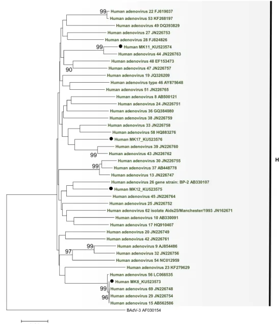
Figure 1.3. Phylogenetic tree of the adenovirus hexon gene loop 1 of HAdV-E 0.2 94 99 96 99
Human adenovirus E isolate NHRC90339_EF371058
Bovine adenovirus 3_AF030154
Human adenovirus E_AY487947
Human adenovirus type 4 vaccine strain_AY594254 Human adenovirus type 4 strain RI-67_AY594253
100
Human adenovirus type 4 strain GZ01_KF006344 Human adenovirus type 4 strain NHRC3_AY599837
Human adenovirus type 4 strain NHRC42606_AY599835 Gorilla gorilla adenovirus E1_KM886330
Simian adenovirus 39_FJ025924.Ch Simian adenovirus 30_FJ025920.Ch
Chimpanzee adenovirus Y25_JN254802 Pan troglodites schweinfurthii adenovirus 13_JN163983 Simian adenovirus 23 strain ATCC VR-592_AY530877.Ch
Simian adenovirus 37.2_FJ025919.Bo
Simian adenovirus 37.1_FJ025921.Ch
HAdV-E
Simian adenovirus 28 2 FJ025915.Go Gorilla gorilla adenovirus B20 KM886326
Gorilla gorilla adenovirus B22 KM886328 Simian adenovirus 47 FJ025929.Go
Gorilla gorilla adenovirus B17 KM886317 Gorilla gorilla adenovirus B13 KM886309 Gorilla gorilla adenovirus B12 KM886308 Gorilla gorilla adenovirus B18 KM886319 Gorilla gorilla adenovirus B11 KM886307 Gorilla gorilla adenovirus B16 KM886315 Simian adenovirus 27 1 FJ025909.Ch
Simian adenovirus -46 FJ025930.Go Simian adenovirus 27.1 KM886316 Simian adenovirus 27.1 KM886318 Gorilla gorilla adenovirus B14 KM886311
Simian adenovirus 29 FJ025916.Ch
Gorilla gorilla adenovirus B15 KM886314 Simian adenovirus 28 1 FJ025914.Ch Simian adenovirus 27 2 FJ025928.Go Simian adenovirus 32 FJ025911.Ch
Simian adenovirus 41 1 FJ025913.Go Simian adenovirus 41 2 FJ025927.Go
Human adenovirus type 3 strain NHRC 1276 AY599836 Human adenovirus 7 KP670861
Human adenovirus 21 KJ364592 Human adenovirus type 50 AY737798 Human adenovirus type 16 AY601636
Gorilla gorilla adenovirus B19 KM886325 Gorilla gorilla adenovirus B21 KM886327 Human mastadenvoris B isolate 6588 KM659150.Go
Human mastadenvoris B isolate 6575 KM659142.Go
Human adenovirus 55 KC857701 Human adenovirus 14 JN032132 Human adenovirus type 34 AY737797 Human adenovirus type 35 AC 000019 Human adenovirus type 11 strain Slobitski AF532578 Human adenovirus type 4 vaccine strain AY594254 Human adenovirus type 4 strain RI-67 AY594253 Human adenovirus E AY487947 Human adenovirus E isolate NHRC90339 EF371058
Human adenovirus E X74508
Human adenovirus type 4 strain NHRC 42606 AY599835 Human adenovirus 4 strain GZ01 KF006344 Human adenovirus type 4 strain NHRC 3 AY599837
Pan troglodites adenovirus1 Gorilla gorilla adenovirus E1 KM886330 Simian adenovirus 26 FJ025923.Ch
Chimpanzee adenovirus Y25 JN254802 Simian adenovirus 39 FJ025924.Ch Simian adenovirus 38 FJ025922.Ch Simian adenovirus 25.2 FJ025918.Ch Simian adenovirus 25 2 FJ025918.Ch Simian adenovirus 24 strain ATCC VR-593 AY530878.Ch Simian adenovirus 36 FJ025917.Ch
Simian adenovirus 23 strain ATCC VR-592 AY530877.Ch Simian adenovirus 37.1 FJ025921.Ch Simian adenovirus 30 FJ025920.Ch
Simian adenovirus 37.2 FJ025919.Bo
Human adenovirus type 26 EF153474 Human adenovirus 33 JN226758 Human adenovirus 39 JN226760 Human adenovirus 45 JN226764 Human adenovirus 43 JN226762 Human adenovirus 69 JN226748 Human adenovirus 47 JN226757 Human MK17_KU523571 Human adenovirus 44 JN226763 Human adenovirus 56 LC066535
HAdV D strain human/DEU/IAI-1/2005/53 FJ169625 Human adenovirus 8 strain: Trim variant AB746853 Human adenovirus type 48 EF153473 Human adenovirus 58 HQ883276 Human adenovirus 15 AB562586 Human adenovirus 29 JN226754
Simian adenovirus -48 isolate AJ75 HQ241818.Cy
Simian adenovirus 3 strain ATCC VR-1449 AY598782.Rh
Human adenovirus 6 FJ349096 Human adenovirus 2 AC 000007 Human adenovirus 5 AC 000008 Human adenovirus type 1 AC 000017 Human adenovirus type 1 (HAdV- C) AF534906
Human MK1_KU523570
Gorilla gorilla adenovirus C14 KM886321 Gorilla gorilla adenovirus C16 KM886323 Gorilla gorilla adenovirus C17 KM886324 Gorilla gorilla adenovirus 1
Gorilla gorilla adenovirus C15 KM886322 Gorilla gorilla adenovirus C10 KM886310 Gorilla gorilla adenovirus C12 KM886313 Gorilla gorilla adenovirus C17 KM886329 Gorilla gorilla adenovirus C11 KM886312 Simian adenovirus 43 FJ025900.Go
Gorilla gorilla adenovirus C13 KM886320 Simian adenovirus -45 FJ025901.Go
Human adenovirus 18 GU191019 Human adenovirus type 31 AM749299 Human adenovirus 12 X73487
Pan troglodites adenovirus 5
Human adenovirus -40 NC 001454
Simian adenovirus -7 DQ792570.Rh ATCC
Human adenovirus -52 isolate T03-2244 DQ923122
Simian adenovirus 1 strain ATCC VR-195 AY771780
Bovine adenovirus 3_AF030154 99 99 99 94 96 99 99 98 99 99 97 99 99 99 99 99 99 99 97 99 99 99 99 99 91 98 0.05 HAdV-B HAdV-E HAdV-D HAdV-A HAdV-C SAdV-A HAdV-F HAdV-G Outgroup
Fig. 1.5. Phylogenetic tree of the partial sequence of the hexon gene
Bovine adenovirus 3_AF030154
100 100 99 95 100 98 92 0.05
Human mastadenovirus B isolate 6560 KM659138 Human mastadenovirus B isolate 6674 KM659157
100 100 100 100 97 98 98 100 100 100 100 100 100 99 94 94 99 0.05
Human adenovirus 22 FJ619037 Human adenovirus 53 KF268197 Human adenovirus 49 DQ393829 Human adenovirus 27 JN226753 Human adenovirus 28 FJ824826 Human MK11_KU523574 Human adenovirus 44 JN226763 Human adenovirus 48 EF153473 Human adenovirus 47 JN226757 Human adenovirus 19 JQ326209
Human adenovirus type 46 AY875648 Human adenovirus 51 JN226765
Human adenovirus 8 AB500121 Human adenovirus 24 JN226751 Human adenovirus 36 GQ384080 Human adenovirus 38 JN226759 Human adenovirus 33 JN226758 Human adenovirus 58 HQ883276 Human MK17_KU523576 Human adenovirus 39 JN226760 Human adenovirus 43 JN226762 Human adenovirus 30 JN226755 Human adenovirus 37 AB448778 Human adenovirus 13 JN226747
Human adenovirus 26 gene strain: BP-2 AB330107 Human MK12_KU523575
Human adenovirus 45 JN226764 Human adenovirus 25 JN226752
Human adenovirus 62 isolate Aids25/Manchester/1993 JN162671 Human adenovirus 10 AB330091
Human adenovirus 17 HQ910407 Human adenovirus 20 JN226749 Human adenovirus 42 JN226761
Human adenovirus 9 AJ854486 Human adenovirus 32 JN226756 Human adenovirus 54 NC012959 Human adenovirus 23 KF279629 Human adenovirus 56 LC066535 Human MK8_KU523573 Human adenovirus 69 JN226748 Human adenovirus 29 JN226754 Human adenovirus 15 AB562586
BAdV-3 AF030154 99 96 99 99 99 99 99 97 90 0.05
Figure 1.6. Phylogenetic tree of partial hexon of HAdV-D
Abstract
Bocaparvoviruses have been studied extensively owing to their ability to cause respiratory illness or gastroenteritis in humans. Some bocaparvoviruses have been detected in non-human primates (gorillas and chimpanzees), but the diversity and evolution of these viruses are not fully understood. In this study,
wild western lowland gorillas in MDNP in Gabon to investigate the presence of bocaparvoviruses. Using a combination of pan-bocaparvovirus PCR
. To my knowledge, this is the first report showing the presence of a non-human primate bocaparovirus within Primate
bocaparvovirus 2.
III.1. Introduction
Bocaparvoviruses belong to the genus Bocaparvovirus of the sub-family
Parvovirinae and the family Parvoviridae. Currently, 12 species have been reported in
the genus: Carnivore bocaparvovirus 1 3, Pinniped bocaparvovirus 1 and 2, Primate
bocaparvovirus 1 and 2, and Ungulate bocaparvovirus 1 5 (Cotmore et al., 2014; ICTV,
2016). Further viruses, genetically related, recently described have not been classified yet (Guo et al., 2016; Lanave et al., 2015; Lau et al., 2017; Yang et al., 2016). Members of
Primate bocaparvovirus 1 and 2 are known to infect human and non-human primate hosts
2010b, 2009; Sharp et al., 2010). The first primate bocaparvovirus was detected in humans in 2005 from pooled nasopharyngeal aspirate specimens by large-scale molecular virus screening (Allander et al., 2005). Since then, many molecular epidemiological studies have suggested that human bocaviruses (HBOV) are associated with respiratory or gastrointestinal illnesses (Ahn et al., 2014; Arthur et al., 2009; Jin et al., 2011; Lu et al., 2015; Medici et al., 2012; Nunes et al., 2014; Phan et al., 2012). Additionally, some bocaparvoviruses have been detected from non-human primates, such as gorillas (Kapoor et al., 2010a, Sharp et al., 2010) and chimpanzees Sharp et al., 2010). Currently, Primate bocaparvovirus 1 can be classified into four genotypes, including human and non-human primate bocaparoviruses: HBOV1, HBOV3, gorilla bocavirus 1 (GBOV1), and primate bocaparvovirus 1 isolate CPZh2 (Allander et al., l., 2010a, 2010b). Primate bocaparvovirus 2 includes only two genotypes: HBOV2 and HBOV4 (Cotmore et al., 2014; Kapoor et al., 2009, 2010b; Khamrin et al., 2013).
Bocaparvoviruses are small non-enveloped viruses with a linear single-stranded DNA genome of approximately 4.9 5.5 kb. The viral genome contains three major open reading frames (ORFs); the left ORF encodes a non-structural protein (NS1) involved in replication (Allander et al., 2005), the right ORF encodes the viral capsid proteins (VP1 and VP2), and the middle ORF is a unique feature of bocaparvoviruses in the family
Parvoviridae and encodes the highly phosphorylated non-structural protein NP1.
Although primate bocaparvoviruses have been detected in diseased and healthy gorillas
and chimpanzees , the clinical significance or
symptoms in apes is unknown thus far. Additionnally, bocaparvovirus-like genes have been found in healthy free-ranging gorillas (prevalence: 36%) and chimpanzees (prevalence: 73%) (Sharp et al., 2010); However, the diversity and evolution of bocaparvoviruses infecting wild apes remain poorly understood.
In this study, I have investigated the bocaparvovirus infection in wild western lowland gorillas (Gorilla gorilla gorilla) in MDNP, Gabon. I have detected a novel bocaparovovirus genotype and characterized the nearly complete genome of two novel gorilla bocaparvoviruses that form a single cluster within the species Primate bocaparvovirus 2.
III.2. Material and methods
III.2.1. Sample collection and DNA extraction
Between November and December 2014, 107 fresh fecal samples were collected
from wild western lowland gorillas subjected to habituation in MDNP, Gabon (Ando et
al., 2008). During the sample collection period, the target gorilla group included one adult
male or silverback (estimated age, >13 years old), four adult females (estimated age, >10
years old), two sub-adults (estimated age, >6 years old), eight juveniles (estimated age,
4 6 years old), and three infants (estimated age, <3 years old) (Table 2.1).
Opportunistically, other individual wild gorillas sharing the same home range with the
target group were sampled. Gorillas were followed daily as part of the habituation process
and fecal samples were collected on trails when following the animals. To detect
bocaparvoviruses, few grams of the feces were taken using a sterile plastic stick and
preserved in 5 ml of RNAlater (Ambion, Austin, TX, USA). In addition, for individual
dual g the
surface of each feces, which contains gorilla-derived tissues, were scratched using a
EDTA, 100 mM Tris-HCl, and 10 mM NaCl) until DNA extraction. The following
information: date, time, GPS coordinates, and fecal diameter were recorded upon
collection. To prevent cross contamination, fecal collection were performed using
disposable plastic sticks and protective gloves were used once. DNA was extracted from
both samples using the QIAamp DNA Stool Mini Kit (QIAGEN, Hilden, Germany),
following t
III.2.2. Individual genetic identification
To identify gorilla individuals, multiplex PCR was performed using QIAGEN Multiplex PCR Kits (QIAGEN), as described by (Inoue et al., 2013), with host DNA extracted from the swab samples of feces from gorillas. One primer set was used for six autosomal microsatellite loci (D7s817, D1s550, vWF, D1s2130, D7s794, and D6s1056 (Inoue et al., 2013). Genotype data for target individuals were already available and genetic variation at these six loci was sufficient to identify individuals (Inoue et al., unpublished results). After PCR to amplify these microsatellite loci, genotypes were determined using Peak Scanner V1.0 (Applied Biosystems, Foster City, CA, USA).
III.2.3. PCR detection of bocaparvovirus
The samples were tested for the presence of bocaparvovirus DNA by hemi-nested