Tuberculous Cold Abscess in the Axillary Lymph Nodes
Takuhiro YOSHIDA , Ko‑ichi TAZAWA, Eriko KASUGA Akihiko YOSHIZAWA and Shu‑ichi IK E D A
1) Department of Medicine (Neurology and Rheumatology), Shinshu University School of Medicine 2) Department of Laboratory Medicine, Shinshu University Hospital
Key words:tuberculous lymphadenitis, pericarditis, cold abscess, axillary lymph node
A 56‑year‑old woman was admitted to the regional medical center suffering from dyspnea.She was diagnosed with tuberculous pericarditis and was treated with antituberculous drugs (INH,RFP,
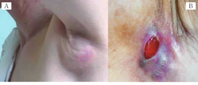
EB, and PZA). One month later, her cardiac condi- tion was ameliorated and she was discharged. Sub- sequently EB and PZA were stopped. Two months later, she started to suffer from painful swelling of the left axillary lymph nodes.Although her cardiac condition was stable, the size of the lymph nodes was gradually enlarged (Fig. 1 A). CT and FDG‑
PET showed a few translucent lymph nodes (Fig.2 A) and there was no sign of other malignancy.
Because the swelling of the lymph nodes progressed in spite of the amelioration of pericarditis, the etiology was assumed to be neoplastic or autoim-
mune in nature. She was therefore referred to our hospital.On admission,she was afebrile and had no symptoms except for the swelling of a left axillary lymph node, which was approximately 4 cm in diameter,elastic and partially reddish.Blood exam-
ination revealed a slight inflammatory reaction (CRP 0.14 mg/dl, ESR 13 mm/hr). Contrast enhan- ced CT showed a consistent low density area sur- rounded by a ring‑enhancing lesion (Fig. 2 B). The tumorʼs elasticity and CT findings suggested that the swelling was an abscess and EB was resumed.
The abscess was located next to the skin and was on the verge of collapse. In case of collapse, there was the risk of nosocomial transmission, so we selected needle aspiration. Aspirated fluid revealed yellow pus (Fig. 3 A) which contained acid‑fast bacteria (Fig. 3 B), though the subsequent culture revealed no growth of those.A diagnosis of tubercu-
lous cold abscess was made. Two weeks after the aspiration,the abscess swelled and collapsed.It was surgically resected and she received an additional antituberculous medication (SM)because the lesions seemed to extend into neighboring lymph nodes in spite of medication with INH, RFP and EB. Three months after the surgical procedure, the abscess swelled again and broke forming a fistula (Fig.1 B),
with the histology typical of a granuloma (Fig.3 C).
With daily lavage through the fistula and continuous use of antituberculous drugs,her tuberculous lymph node lesions were completely cured 6 months after the last collapse.Although the lymph nodes are the most common sites of extrapulmonary tuberculo-
sis ,tuberculous cold abscess in lymph nodes is less frequent in Japan .This case suggests that tubercu-
lous lymphadenitis is an important differential diag- nosis in examining lymph node swelling and that its progression is not always parallel to the exacerba-
tion of other tuberculous disease conditions.
Corresponding author:Takuhiro Yoshida
Department of Medicine (Neurogy and Rheumatology),
Shinshu University School of Medicine, 3‑1‑1 Asahi, Matsumoto, Nagano 390‑8621 Japan
257
No. 5, 2012
Shinshu Med J, 60⑸ :257〜259, 2012
Fig. 2 FDG‑PET (A)and CT (B)showing a few lymph nodes in the left axilla.
Fig. 3 The aspirated yellow pus (A) which contained acid‑fast bacteria (B). C : Histology of the collapsed lymph node, which was typical of granuloma.
A B
A
B
C
Fig. 1
A :Swelling of the left axillary lymph node.
B :A fistula formed three months after the surgical procedure.
A B
258 Shinshu Med J Vol. 60
Yoshida.Tazawa.Kasuga et al.
References
1) Ilgazli A,Boyaci H,Basyigit I,Yildiz F :Extrapulmonary tuberculosis:clinical and epidemiologic spectrum of 636 cases. Arch Med Res 35:435‑441, 2004
2) Aoki M :Tuberculosis in the world, and in Japan. Kekkaku 81:623‑629,2006 (in Japanese,Abstract in English)
(2012. 3.16 received;2012. 6.13 accepted)
259 No. 5, 2012
Tuberculous cold abscess in the axillary lymph nodes