Protective Effects of Fucoidan Against Interleukin-1β-induced Inflammation in SW982 Human Synovial Cells
Masayuki ARAI1,2), Mayumi TSUJI1), Hiromichi TSUCHIYA1), Mai MURAYAMA1), Mizuka OGAWA1), Haruka YANAKI1),
Ryoma HAYATAKE1) and Katsuji OGUCHI1)
Abstract : Fucoidan, a natural sulfated polysaccharide found in the extracellular matrix of brown algae, is rich in L-fucose and sulfate. Fucoidan has a variety of biological actions, including anti-oxidative, anti-coagulative, anti-cancer, and anti- inflammatory activity. However, the cellular molecular mechanism underlying the anti-inflammatory effects of fucoidan remains poorly understood. Rheumatoid arthritis (RA) is a chronic inflammatory disease that causes synovitis and pro- gressive joint destruction. Interleukin (IL)-1β, one of the important mediators involved in the pathogenesis of RA, is known to activate various intracellular signaling pathways. Therefore, in the present study we investigated the inhibitory effects of fucoidan on IL-1β-induced inflammation in human synovial (SW982)
cells. SW982 cells were pretreated with fucoidan (100μg / mL) for 1 h before cotreatment with 5 ng / mL IL-1β plus fucoidan for periods ranging from 20 min to 24 h. Levels of the proinflammatory mediators IL-6, tumor necrosis factor-α, and cyclooxygenase-2 were then determined. We also assayed translocation of nuclear factor (NF)-κB into the nucleus and activation of mitogen-activated protein kinase
(MAPK). Significant increases in the production of proinflammatory mediators were observed from 6 to 24 h of IL-1β treatment. The translocation of NF-κB into the nucleus peaked after approximately 6 h incubation. After 20 min incuba- tion, IL-1β activated c-jun N-terminal kinase and p38 MAPK in SW982 cells. This effect was ameliorated by the coincubation of cells with fucoidan. These results suggest that fucoidan exerts its anti-inflammatory effect by regulating the gene expression of proinflammatory mediators by suppressing the activity of transcription factors and MAPK. Thus, fucoidan may have therapeutic potential for the treat- ment of RA.
Key words : fucoidan, IL-1β-induced inflammation, SW982 cells, NF-κB, MAPK, rheumatoid arthritis
Introduction
Fucoidan, a sulfate polysaccharide complex from brown seaweed such as Laminaria japonica and Cladosiphon okamuranus, which are widely distributed in many countries, has been used Original
1)Department of Pharmacology, Showa University School of Medicine, 1—5-8 Hatanodai, Shinagawa-ku, Tokyo 142-8555, Japan.
2)Department of Orthopaedic Surgery Showa University School of Medicine.
therapeutically in Oriental medicine for a considerable period of time1). Fucoidan has many biological actions, including anticoagulant2), antithrombotic3), antiviral4), antitumor5), and antioxidant properties6). An anti-inflammatory action of fucoidan has been recently reported in mammalian cells7, 8) and inflammatory animal models9), but the cellular molecular mechanism underlying this action is poorly understood.
Rheumatoid arthritis (RA) is a chronic inflammatory disease of the joint synovium that leads to bone erosion, deformation, and joint destruction10, 11). Activation of T cells, synovial cells, and macrophages results in the overproduction of cytokines and growth factors, triggering proliferative synovitis and arthritis. Ultimately, this leads to destruction of the articular cartilage and bone.
A number of genes are activated as part of the inflammatory response. Nuclear factor (NF)- κB is an important transcription control factor involved in the inflammatory response that is typically present in the cytoplasm as a result of binding to an inhibitory protein (IκB). Upon activation by an inflammatory stimulus, IκB is rapidly phosphorylated and ubiquitinated, and then broken down by proteasomes. Degradation of IκB frees NF-κB and causes it to move to the nucleus, where it transcriptionally induces the expression of inflammation-related genes12). Exogenous stimuli also cause an upstream kinase of mitogen-activated protein kinase
(MAPK), namely MAPK kinase (MKK) 3 / 6, to phosphorylate p38 MAPK, ultimately activating transcription control factors such as activator protein-1 (AP-1)13). Proinflammatory cytokines
(e.g. tumor necrosis factor (TNF)-α, interleukin (IL)-1, IL-6, and IL-8), cyclooxygenase (COX)- 2, prostaglandin E2(PGE2), and matrix metalloproteinases (MMPs ; MMP-1, MMP-9, and MMP- 13) are induced via the MAPK signaling pathway14).
In the present study, we investigated whether fucoidan has protective effects against the inflammation mediated by NF-κB and MAPK activity in human synovial (SW982) cells following stimulation with IL-1β.
Materials and methods Drugs and reagents
Fucoidan, fetal bovine serum (FBS), phenylmethylsulfonyl fluoride, and protease inhibitor cocktail were purchased from Sigma-Aldrich (St. Louis, MO, USA); IL-1β and Dulbeccoʼs modified Eagleʼs medium (DMEM) were purchased from Wako (Osaka, Japan); penicillin G sodium, streptomycin sulfate, and amphotericin B were obtained from Invitrogen (Carlsbad, CA, USA). All other chemicals used in the study were of the purest grade available commercially.
Cell culture
The human synovial SW982 cell line was obtained from American Tissue Culture Collection
(HTB-93 ; Manassas, VA, USA). SW982 cells were cultured and grown in DMEM containing 10% FBS and maintained in a humidified atmosphere of 5% CO2 and 95% air at 37℃. For studies on the effects of fucoidan, SW982 cells were incubated in DMEM with or without
(control) 5 ng / mL IL-1β for times ranging from 20 min to 24 h.
Elevated IL-6 levels have been detected in patients with RA, as well as in patients with inflammatory arthritis and traumatic arthritis. To determine the concentration of IL-1β to use in the present series of experiments, we first undertook preliminary experiments to determine IL-6 production by SW982 cells following 48 h incubation with different concentrations of IL-1β(1, 5, and 10 ng / mL). After 48 h incubation with 0, 1, 5, and 10 ng / mL IL-1β, IL-6 levels in the culture medium were 0.77 0.16, 1.24 0.10, 2.10 0.24, and 2.45 0.34 ng / mL, respectively. On the basis of these results, which indicated that significant increases in IL-6 were not observed with concentrations <5 ng / mL IL-1β(P<0.05), we decided to use 5 ng / mL IL-1β in subsequent experiments to induce an inflammatory response in SW982 cells. To evaluate the anti-inflammatory effects of fucoidan, SW982 cells were pretreated with 100μg / mL fucoidan for 1 h before the addition of 5 ng / mL IL-1β to the culture medium and further culture in the presence of IL-1β plus fucoidan for times ranging from 20 min to 24 h. All treatments were performed under sterile conditions.
Measurement of cytokines and COX-2
As described above, SW982 cells were pretreated with 100μg / mL fucoidan for 1 h, followed by cotreatment with 5 ng / mL IL-1β plus 100μg / mL fucoidan and culture for a further 24 h.
After 24 h culture, concentrations of IL-6, TNF-α, and COX-2 in the culture supernatant were determined using commercially available ELISA kits. IL-6 and TNF-α levels were measured using a Human sIL-6 Instant Elisa Kit and TNF-α Instant ELISA, respectively (Affymetrix, Santa Clara, CA, USA), whereas COX-2 levels were measured using an enzyme immunoassay kit (Human COX-2 Assay Kit ; Immuno-Biological Laboratories, Gunma, Japan).
Detection of NF-κB activation
To monitor the nuclear translocation of p65, SW982 cells were treated with IL-1β for 24 h with or without 1 h pretreatment with 100μg / mL fucoidan. Cells were harvested and the nuclei were extracted using a commercially available nuclear extraction kit (Imgenex, San Diego, CA, USA). For quantitative determination of the relative increase of p65 translocation into the nucleus, an NF-κB / p65 ActivElisa kit (Imgenex) was used.
Detection of c-Jun N-terminal kinase, p38 MAPK and Extracellular Signal-regulated Kinase1 / 2 To examine the role of MAPK in IL-1β-induced inflammation in SW982 cells, we evaluated the phosphorylation of c-jun N-terminal kinase (JNK), p38 MAPK and Extracellular Signal- regulated Kinase (ERK) 1 / 2 using the Cell-Based JNK (Thr183/ Tyr185) ELISA kit, Cell-Based p38 MAPK (Thr180/ Tyr182) ELISA kit and Cell-Based ERK1 / 2 (Thr202/ Tyr204) ELISA kit (Ray Biotech, Norcross, GA, USA), respectively. SW982 cells were incubated with or without 100μ g / mL fucoidan pretreatment for 1 h, followed by culture in the presence of 5 ng / mL IL-1β for times ranging from 20 min to 24 h. Cells were fixed and blocked as per the manufacturerʼs instructions and then incubated for 2 h at 20℃ with anti-phosphorylated (p-) JNK (Thr183/ Tyr185), anti-JNK (primary antibody), anti-p-p38 (Thr180/ Tyr182), anti-p38 (primary antibody), anti-
p-ERK1 / 2 (Thr202/ Tyr204), or anti-ERK1 / 2 (primary antibody). After incubation, cells were washed with wash buffer before being incubated for 1 h at 20℃ with horseradish peroxidase- conjugated anti-mouse IgG (secondary antibody). Finally, cells were washed with wash buffer, and 3,3',5,5'-tetramethylbenzidine (TMB) substrate solution was added, resulting in the development of color in proportion to the amount of protein. The reaction was stopped by the addition of Stop Solution from the assay kit, resulting in a change of color from blue to yellow.
The intensity of the color was measured at 450 nm.
Cell viability assay
The SW982 cells divided into the control, IL-1β-, fucoidan-, and IL-1β+fucoidan-treated groups were incubated for 6 h. Each group was stained with Count and Viability Reagent
(EMD Millipore, Billerica, MA, USA), and the viability was analyzed on Muse Cell Analyzer
(EMD Millipore, Billerica, MA, USA).
Statistical analysis
Each experiment was repeated three times. Data are presented as the mean SEM.
Comparisons of the effects of various treatments with untreated control cells were performed with one-way ANOVA and Dunnettʼs post hoc test. One-sided P<0.05 was considered significant.
Results
Effects of fucoidan on IL-1β-induced increases in cytokine and COX-2 production
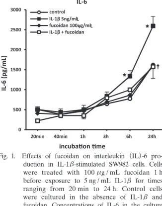
To determine whether fucoidan suppressed the production of proinflammatory mediators (IL- 6, TNF-α, COX-2) in IL-1β-stimulated SW982 cells, cells were pretreated with 100μg / mL fucoidan before being cocultured in the presence of 5 ng / mL IL-1β+fucoidan for a further 24 h. As shown in Fig. 1, IL-1β alone (5 ng / mL) induced significant IL-6 production compared with control cells. Pretreatment of cells with 100μg / mL fucoidan significantly reduced IL-1β- stimulated IL-6 production.
The effects of fucoidan on TNF-α were also evaluated. As shown in Fig. 2, IL-1β induced significant release of TNF-α by SW982 cells compared with the untreated control after 24 h culture. This IL-1β-induced increase in TNF-α production by SW982 cells was inhibited by 1 h pretreatment with 100μg / mL fucoidan. Together, these observations suggest that fucoidan pretreatment can significantly suppress IL-1β-mediated release of proinflammatory mediators from SW982 cells.
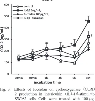
In the present study, 6 h exposure of SW982 cells to IL-1β resulted in the induction of COX- 2 compared with the untreated control group (Fig. 3). Fucoidan pretreatment significantly reduced IL-1β-induced COX-2 production.
The data presented in Figs. 1〜3 indicate that fucoidan may be able to regulate proinflammatory cytokine production in SW982 cells.
The suppression of cytokine and COX-2 production by fucoidan may have been due to
cytotoxic effects of fucoidan in IL-1β-stimulated SW982 cells. To exclude this possibility, the viability of SW982 cells was evaluated. Treatment of cells with 100μg / mL fucoidan, alone or in combination with IL-1β, had no significant effect on cell viability. Specifically, the percentage of living cells in the control, IL-1β-, fucoidan-, and IL-1β+fucoidan-treated groups was 94.89 0.66%, 93.01 0.37%, 95.64 0.37%, and 94.58 0.09%, respectively. These results confirm that the inhibition of cytokine and COX-2 production by fucoidan in IL-1β-stimulated SW982 cells is not due to cytotoxic effects of fucoidan. Similarly, another study has reported recently that 100μg / mL fucoidan treatment, with or without lipopolysaccharide (LPS), does not significantly affect cell viability15).
Effects of fucoidan on translocation of NF-κB / p65 into the nucleus of IL-1β-stimulated SW982 cells
NF-κB is an important transcription factor that regulates the gene expression of proinflammatory mediators ; thus, in the present study we investigated whether fucoidan has any effect on NF-κB activity by examining IL-1β-induced translocation of NF-κB / p65 into the nucleus. As shown in Fig. 4, 1 h exposure of SW982 cells to IL-1β resulted in a marked increase in the translocation of NF-κB / p65 compared with control. However, 1 h pretreatment of cells with fucoidan
Fig. 1. Effects of fucoidan on interleukin (IL)-6 pro- duction in IL-1β-stimulated SW982 cells. Cells were treated with 100μg / mL fucoidan 1 h before exposure to 5 ng / mL IL-1β for times ranging from 20 min to 24 h. Control cells were cultured in the absence of IL-1β and fucoidan. Concentrations of IL-6 in the culture medium were determined using commercially available ELISA kits. Data are the mean SEM. P<0.05 compared with untreated control cells ; †P<0.05 compared with IL-1β-treated cells.
Fig. 2. Effects of fucoidan on tumor necrosis factor
(TNF)-α production in interleukin (IL)-1β- stimulated SW982 cells. Cells were treated with 100μg / mL fucoidan 1 h before exposure to 5 ng / mL IL-1β for times ranging from 20 min to 24 h. Control cells were cultured in the absence of IL-1β and fucoidan. Con- centrations of TNF-α in the culture medium were determined using commercially available ELISA kits. Data are the mean SEM. P<
0.05 compared with untreated control cells ;
†P<0.05 compared with IL-1β-treated cells.
inhibited the IL-1β-induced translocation of NF-κB / p65 into the nucleus. On the basis of these results, fucoidan may inhibit NF-κB activation in SW982 cells by suppressing IκB degradation and hence the nuclear translocation of NF-κB.
Effects of fucoidan on the phosphorylation of MAPKs in IL-1β-stimulated SW982 cells
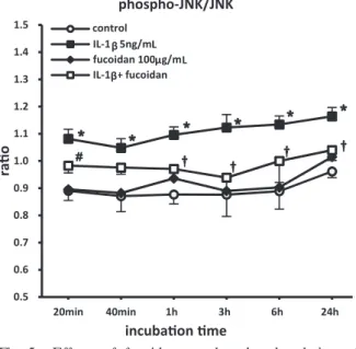
The phosphorylation of ERK, JNK, and p38 MAPK were compared in cells treated with or without IL-1β(5 ng / mL) in the presence or absence of 100μg / mL fucoidan pretreatment.
There was no significant difference in ERK phosphorylation among the different groups
(data not shown). However, there was significant upregulation of JNK and p38 MAPK phosphorylation in IL-1β-treated cells, which was significantly inhibited by fucoidan pretreatment
(Figs. 5, 6). Discussion
Fucoidan is a natural sulfated polysaccharide extracted from brown seaweed. Several studies have demonstrated an anti-inflammatory action of fucoidan. In inflammatory animal models,
Fig. 3. Effects of fucoidan on cyclooxygenase (COX)- 2 production in interleukin (IL)-1β-stimulated SW982 cells. Cells were treated with 100μg / mL fucoidan 1 h before exposure to 5 ng / mL IL-1β for times ranging from 20 min to 24 h.
Control cells were cultured in the absence of IL-1β and fucoidan. Concentrations of COX- 2 in the culture medium were determined using commercially available ELISA kits. Data are the mean SEM. P<0.05 compared with untreated control cells ; †P<0.05 compared with IL-1β-treated cells.
Fig. 4. Effects of fucoidan on translocation of nuclear factor (NF)-κB / p65 into the nucleus of interleukin (IL)-1β-stimulated SW982 cells.
Cells were treated with 100μg / mL fucoidan 1 h before exposure to 5 ng / mL IL-1β for times ranging from 20 min to 24 h. Control cells were cultured in the absence of IL-1β and fucoidan. Cells were harvested and nuclei were extracted using a nuclear extraction kit
(Imgenex). For quantitative determination of the relative increase in p65 translocation into the nucleus, an NF-κB / p65 ELISA kit was used. Data are the mean SEM. P<
0.05 compared with untreated control cells ;
†P<0.05 compared with IL-1β-treated cells.
fucoidan has been shown to inhibit leukocyte recruitment16) and to have neuroprotective effects against β-amyloid-induced neurotoxicity17). Fucoidan has also been reported to inhibit the synthesis of PGE2 and COX-2 in LPS-stimulated microglia15). However, the pharmacological effects of fucoidan on synovial cells have not been reported. Therefore, in the present study we investigated the anti-inflammatory action of fucoidan in IL-1β-treated synovial cells. Our findings demonstrate that fucoidan reduces the production of inflammatory cytokines and COX-2 by IL-1β-stimulated synovial cells. In addition, we demonstrated that the actions of fucoidan are related to the inhibition of NF-κB and MAPK activation.
Cytokines are proteins with a molecular mass of approximately 10—50 kDa ; they have various physiological activities. In the body, a cytokine network is formed by the interactions between numerous cytokines that have different effects. Of these, inflammatory cytokines have an important role in the pathology of RA as inflammatory mediators.
The synovial response in patients with RA is characterized by increased levels of proinflammatory cytokines (TNF-α, IL-1β, IL-6, etc.). These cytokines are known to play an important role in the pathogenesis of RA18). In the present study we investigated the action of fucoidan on the
Fig. 5. Effects of fucoidan on the phosphorylation of c-Jun N-terminal kinase (JNK) in interleukin
(IL)-1β-stimulated SW982 cells. Phosphorylation of JNK was determined using a commercially available ELISA kit. Cells were treated with 100μg / mL fucoidan 1 h before exposure to 5 ng / mL IL-1β for times ranging from 20 min to 24 h. Cells were processed according to the instructions in the kit, and the intensity of the color was measured at 450 nm. Data are the mean SEM. P<0.05 compared with untreated control cells ; †P<0.05 compared with IL-1β- treated cells.
Fig. 6. Effects of fucoidan on the phosphorylation of p38 mitogen-activated protein kinase (MAPK)
in interleukin (IL)-1β-stimulated SW982 cells.
Phosphorylation of p38 MAPK was determined using a commercially available ELISA kit.
Cells were treated with 100μg / mL fucoidan 1 h before exposure to 5 ng / mL IL-1β for times ranging from 20 min to 24 h. Cells were processed according to the instructions in the kit, and the intensity of the color was measured at 450 nm. Data are the mean SEM. P<0.05 compared with untreated control cells ; †P<0.05 compared with IL-1β-treated cells.
production of proinflammatory cytokines (TNF-α and IL-6) by human synovial (SW982) cells.
TNF-α has been implicated in the erosive changes to cartilage and bone associated with early joint swelling, such as in chronic arthritis19). IL-6 is a cytokine with a wide range of biological activities in immunomodulation, hematopoiesis, inflammation, and tumorigenesis. It is known that IL-6 can increase serum c-globulin and rheumatoid factor levels20). High IL-6 levels are observed in serum and synovial fluid samples from patients at the onset of chronic RA21). In the present study, fucoidan treatment significantly inhibited IL-1β-induced TNF-α and IL-6 production by SW982 cells, proving that fucoidan can control the expression of proinflammatory cytokines in synovial cells.
IL-1β, one of the cytokines that acts as a mediator of intercellular communication, was used in the present study to induce an inflammatory response in SW982 cells. IL-1 plays an important role in inflammatory responses, the prevention of infection, and other aspects of the immune response. By binding to IL-1 receptors expressed on the cell membrane, IL-1β induces intracellular signaling, with the activation of MAPKs and the transcription factor NF-κB then inducing the expression of cytokines and chemokines.
In the present study, we investigated whether the anti-inflammatory action of fucoidan in IL- 1β-stimulated SW982 cells involves modulation of MAPKs activity. The findings demonstrate that fucoidan is a strong inhibitor of IL-1β-stimulated MAPK (p38 MAPK and JNK) activation in SW982 cells (Figs. 5, 6) and suggest that the anti-inflammatory action of fucoidan is due to inhibition of the MAPKs signaling pathway.
MAPKs play an important role in inflammatory signaling in many cells and have been shown to be the principal tyrosine phosphorylated proteins in human synovial cells following IL-1β stimulation22). JNK and p38 MAPK activate AP-122), which is a transcription regulating factor, and induce the expression of inflammatory mediator proteins. IL-1β-induced activation of JNK results in the increased expression of cytokines that are inflammatory mediators. In human breast cancer cells, IL-1β treatment is followed by a marked increase in p38 MAPK activation, which peaks early, as well as an increase in the levels of COX-2, an inflammatory mediator23). In addition, a reduction in MAPK activation and production of inflammatory cytokines (e.g. IL-1 β, IL-1, and TNF-α) by fucoidan has been reported in mouse macrophage cells24).
In synovial cells, JNK and p38 MAPK may be the most important kinases activated by proinflammatory cytokines. This is supported by recent studies showing that inhibition of JNK activation is involved in arthropathy and remodeling in arthritic animal models25). As such, the inhibition of JNK activation by fucoidan would be important in inhibiting inflammatory mediator expression induced by IL-1β in synovial cells.
NF-κB is one of the most important transcriptional control factors, and activation of NF-κB protein expression plays an important role in inflammation because it induces the transcription of proinflammatory genes. Upon activation, NF-κB forms a complex with inhibitory IκB proteins in the cytoplasm and is then suppressed. Thereafter, the IκB phosphorylation response leads to ubiquitination and degradation12). IκB degradation then frees the NF-κB and causes it to move to the nucleus, where it induces the transcription of inflammation-related genes.
In the present study, fucoidan significantly inhibited IκB degradation and the IL-1β-induced nuclear translocation of NF-κB / p65 in SW982 cells (Fig. 4). These results suggest that the downregulation of proinflammatory mediators by fucoidan is due to inhibition of both MAPK activation and the NF-κB pathway.
In conclusion, we have demonstrated in the present study that fucoidan inhibits translocation of the transcription factor NF-κB and activation of MAPK (which is a transcription factor adjustment enzyme), in addition to suppressing the synthesis of COX-2 and proinflammatory cytokines by synovial cells following IL-1β stimulation. The data suggest that fucoidan could be a therapeutic drug for the treatment of inflammatory diseases, including RA.
Conflict of interest
The authors declare no conflict of interest.
References
1) Li B, Lu F, Wei X, et al. Fucoidan : structure and bioactivity. Molecules. 2008;13:1671-1695.
2) Irhimeh MR, Fitton JH, Lowenthal RM. Pilot clinical study to evaluate the anticoagulant activity of fucoidan.
Blood Coagul Fibrinolysis. 2009;20:607-610.
3) Millet J, Jouault SC, Mauray S, et al. Antithrombotic and anticoagulant activities of a low molecular weight fucoi- dan by the subcutaneous route. Thromb Haemost. 1999;81:391-395.
4) Baba M, Snoeck R, Pauwels R, et al. Sulfated polysaccharides are potent and selective inhibitors of various enveloped viruses, including herpes simplex virus, cytomegalovirus, vesicular stomatitis virus, and human immunode- ficiency virus. Antimicrob Agents Chemother. 1988;32:1742-1745.
5) Alekseyenko TV, Zhanayeva SY, Venediktova AA, et al. Antitumor and antimetastatic activity of fucoidan, a sulfat- ed polysaccharide isolated from the Okhotsk Sea Fucus evanescens brown alga. Bull Exp Biol Med. 2007;143:730- 732.
6) Wang J, Zhang Q, Zhang Z, et al. Potential antioxidant and anticoagulant capacity of low molecular weight fucoi- dan fractions extracted from Laminaria japonica. Int J Biol Macromol. 2010;46:6-12.
7) Yang JW, Yoon SY, Oh SJ, et al. Bifunctional effects of fucoidan on the expression of inducible nitric oxide syn- thase. Biochem Biophys Res Commun. 2006;346:345-350.
8) Matsumoto S, Nagaoka M, Hara T, et al. Fucoidan derived from Cladosiphon okamuranus Tokida ameliorates murine chronic colitis through the down-regulation of interleukin-6 production on colonic epithelial cells. Clin Exp Immunol. 2004;136:432-439.
9) Saito A, Yoneda M, Yokohama S, et al. Fucoidan prevents concanavalin A-induced liver injury through induction of endogenous IL-10 in mice. Hepatol Res. 2006;35:190-198.
10) Lipsky PE. Rheumatoid arthritis. In Isselbacher KJ, Braunwald E, Wilson JD, et al. eds. Harrisonʼs principles of internal medicine. 13th ed. New York: McGraw Hill; 1994. pp 1648-1655.
11) Dhaouadi T, Sfar I, Abelmoula L, et al. Role of immune system, apoptosis and angiogenesis in pathogenesis of rheumatoid arthritis and joint destruction, a systematic review. Tunis Med. 2007;85:991-998.
12) Mankan AK, Lawless MW, Gray SG, et al. NF-kappaB regulation : the nuclear response. J Cell Mol Med.
2009;13:631-643.
13) Ichijo H. From receptors to stress-activated MAP kinases. Oncogene. 1999;18:6087-6093.
14) Lee JC, Laydon JT, McDonnell PC, et al. A protein kinase involved in the regulation of inflammatory cytokine biosynthesis. Nature. 1994;372:739-746.
15) Park HY, Han MH, Park C, et al. Anti-inflammatory effects of fucoidan through inhibition of NF-κB, MAPK and
Akt activation in lipopolysaccharide-induced BV2 microglia cells. Food Chem Toxicol. 2011;49:1745-1752.
16) Cumashi A, Ushakova NA, Preobrazhenskaya ME, et al. A comparative study of the anti-inflammatory, antico- agulant, antiangiogenic, and antiadhesive activities of nine different fucoidans from brown seaweeds. Glycobiology.
2007;17:541-552.
17) Jhamandas JH, Wie MB, Harris K, et al. Fucoidan inhibits cellular and neurotoxic effects of beta-amyloid (a beta)
in rat cholinergic basal forebrain neurons. Eur J Neurosci. 2005;21:2649-2659.
18) Tanaka Y. The role of chemokines and adhesion molecules in the pathogenesis of rheumatoid arthritis. Drugs Today (Barc). 2001;37:477-484.
19) Warren JS. Interleukins and tumor necrosis factor in inflammation. Crit Rev Clin Lab Sci. 1990;28:37-59.
20) Hirano T. The biology of interleukin-6. Chem Immunol. 1992;51:153-180.
21) Tan PL, Farmiloe S, Yeoman S, et al. Expression of the interleukin 6 gene in rheumatoid synovial fibroblasts. J Rheumatol. 1990;17:1608-1612.
22) Barchowsky A, Frleta D, Vincenti MP. Integration of the NF-kappaB and mitogen-activated protein kinase / AP-1 pathways at the collagenase-1 promoter: divergence of IL-1 and TNF-dependent signal transduction in rabbit primary synovial fibroblasts. Cytokine. 2000;12:1469-1479.
23) Park EJ, Kwon TK. Rottlerin enhances IL-1beta-induced COX-2 expression through sustained p38 MAPK activa- tion in MDA-MB-231 human breast cancer cells. Exp Mol Med. 2011;43:669-675.
24) Kim KJ, Yoon KY, Lee BY. Low molecular weight fucoidan from the sporophyll of Undaria pinnatifida suppresses inflammation by promoting the inhibition of mitogen-activated protein kinases and oxidative stress in RAW264.7 cells. Fitoterapia. 2012;83:1628-1635.
25) Han Z, Boyle DL, Chang L, et al. c-Jun N-terminal kinase is required for metalloproteinase expression and joint destruction in inflammatory arthritis. J Clin Invest. 2001;108:73-81.
[Received January 16, 2014 : Accepted January 27, 2014]