Journal of Dermatology Original Article
1
2
The expression of cell adhesion molecule 1 and its splicing variants in
3
Sézary cells and cell lines from cutaneous T-cell lymphoma
4
5
Mari Yamaguchi1, Shin Morizane1*, Toshihisa Hamada1, Tomoko Miyake1, Makoto
6
Sugaya2, Hiroaki Iwata3, Kazuyasu Fujii4, Rie Haramoto-Shiratsuki5, Yuki Nakagawa1,
7
Mayumi Miura6, Koichi Ohshima6, Kazuhiro Morishita7, Takahide Takahashi8,
8
Masahide Imada8,9, Ken Okada8, Jiro Uehara10, Junko Sowa-Osako11 and Keiji
9
Iwatsuki1
10
11
1Departments of Dermatology, Okayama University Graduate School of Medicine,
12
Dentistry and Pharmaceutical Sciences, Okayama, Japan
13
2Department of Dermatology, Faculty of Medicine, University of Tokyo, Tokyo, Japan
14
3Department of Dermatology, Hokkaido University Graduate School of Medicine,
15
Sapporo, Japan
16
4Department of Dermatology, Kagoshima University Graduate School of Medical and
17
Dental Sciences, Kagoshima, Japan
18
5Department of Dermatology, Shimane University Faculty of Medicine, Izumo, Japan
19
6Department of Pathology, Kurume University School of Medicine, Kurume, Japan
20
7Division of Tumor and Cellular Biochemistry, Department of Medical Sciences,
21
Faculty of Medicine, University of Miyazaki, Miyazaki, Japan
22
8Division of Medical Support, Okayama University Hospital, Okayama, Japan
23
9Central Clinical Laboratory, Kawasaki Medical School Hospital, Okayama, Japan
24
10Department of Dermatology, Asahikawa Medical University, Asahikawa, Japan
25
11Department of Dermatology, Osaka City University Graduate School of Medicine,
26
Osaka, Japan
27
28
*Address correspondence to
29
Keiji Iwatsuki, M.D., Ph.D.
30
Department of Dermatology, Okayama University Graduate School of Medicine,
31
Dentistry and Pharmaceutical Sciences. 2-5-1, Shikata-cho, Kita-ku, Okayama, 700-
32
8558, Japan
33
Phone: +81-86-235-7282, Fax: +81-86-235-7283
34
E-mail: [email protected]
35
36
Short title
37
CADM1 expression in Sézary syndrome
38 39
Abbreviations
40
cell adhesion molecule-1 (CADM1), tumor suppressor lung cancer-1 (TSLC1), Sézary
41
syndrome (SS), mycosis fungoides (MF), adult T-cell leukemia/lymphoma (ATLL),
42
anaplastic large cell lymphoma (ALCL), C-C chemokine receptor type 4 (CCR4),
43
human T-cell leukemia virus 1 (HTLV-1), peripheral blood mononuclear cell (PBMC),
44
cutaneous T-cell lymphoma (CTCL), diffuse large B-cell lymphoma (DLBCL), enzyme-
45
linked immunosorbent assay (ELISA), reverse transcriptase-polymerase chain reaction
46
(RT-PCR)
47
48
49
Abstract; 242 words (limit: 250 words)
50
Main article; 3087 words (limit: 6,000 words)
51 52
ABSTRACT
53
Cell adhesion molecule 1 (CADM1) is aberrantly expressed by T-cell neoplasms such as
54
adult T-cell leukemia/lymphoma (ATLL) and mycosis fungoides (MF). We studied the
55
expression of CADM1 and its splicing variants in Sézary syndrome (SS), MF, other
56
cutaneous T-cell lymphoma (CTCL), and cell lines derived from T- and B-cell
57
lymphomas. Soluble CADM1 was measured in the patients’ sera. CADM1+ cells in the
58
blood and skin lesions were examined by flow cytometry and immunostaining,
59
respectively. Soluble CADM1 was measured by ELISA, and the splicing variants of
60
CADM1 transcripts were determined by reverse transcriptase-polymerase chain
61
reaction, followed by sequencing. As a result, circulating CADM1+ cells were
62
significantly increased in 7 of 10 patients with SS, ranging from 7.9% to 74.5% of the
63
CD3+CD4+ fractions (median; 33.7%) (cut off value; 6.5%). The percentages of
64
CADM1+ cells were usually less than those of circulating Sézary cells. CADM1 was
65
expressed, to various degrees, in 6 of 9 T-cell lines derived from SS, MF, ATLL, and
66
anaplastic large cell lymphoma (ALCL), but negative in B-cell lymphoma-derived cell
67
lines. CADM1+ cells were present in the skin infiltrates of MF, SS, ATLL and ALCL.
68
Serum levels of soluble CADM1 were not significantly elevated in SS/MF. Three major
69
splicing variants of CADM1 expressed by neoplastic T cells contained different
70
combinations of the exons 7, 8, 9 and 11, including a putative oncogenic variant
71
composed of exons 7-8-9-11. In conclusion, CADM1 is frequently expressed in Sézary
72
cells and cell lines from CTCL.
73
74
Keywords
75
CADM1, Mycosis fungoides, Sézary syndrome, splicing variant, T-cell lines,
76 77
INTRODUCTION
78
Cell adhesion molecule 1 (CADM1), a member of the immunoglobulin superfamily of
79
cell adhesion molecules (IgCAM) encoded on chromosome 11q23.2, has been
80
designated with a variety of different names because of its multiple functions: TSLC1
81
(tumor suppressor in non-small cell lung cancer 1) ,1 IGSF4 (immunoglobulin
82
superfamily 4),2 RA175 mRNA,3 SynCAM (synaptic cell adhesion molecule),4 and
83
Necl-2 (nectin-like molecules).5 CADM1 expression has been observed in the human
84
lung, brain, testis, and various epithelial tissues including skin, and has been shown to
85
function in cell-cell adhesion through the homophilic binding of its ectodomains
86
between the adjacent cells.6
87
The absence of CADM1 expression was first shown to be a prognostic indicator in
88
non-small cell lung cancer,1 and it was subsequently shown that methylation of the
89
CADM1 promoter inhibits CADM1 expression in various cancers.7 In contrast, small
90
cell lung cancer expressed a unique CADM1 splicing variant composed of exons 7-8-9-
91
11.8 It has been shown that this splicing has the ability to enhance tumorigenesis,
92
whereas other splicing variants lacking exon 9 may function as tumor suppressors.7,8
93
Although no expression of CADM1 mRNA was detected in normal CD4+ T-cells,
94
molecular and flow cytometric studies revealed the expression of surface CADM1 in
95
adult T-cell leukemia/lymphoma (ATLL) cells. 9,10 Therefore, CADM1 may play dual
96
roles in human oncogenesis: as a tumor suppressor in epithelial cancers, and as an
97
oncoprotein in small cell lung cancer and T-cell malignancies such as ATLL. In cases of
98
ATLL, it has been reported that the presence of circulating CADM1+ T-cells with a
99
CD7dim+/CD7- phenotype is associated with overt or progressive disease. 10
100
In the present study, we detected the expression of CADM1 in leukemic cells of
101
patients with Sézary syndrome (SS) and in infiltrating cells in cutaneous lesions of SS
102
and mycosis fungoides (MF). Since ADAM10 (a disintegrin and metalloproteinases
103
10) cleaves the ectodomain of CADM1 to form soluble CADM1 11, we measured
104
soluble CADM1 in the patients’ sera, and examined the possibility of being a biomarker
105
for SS, MF, and other CTCL. We determined the splicing variant of CADM1
106
expressed by circulating Sézary cells, T-cell lines and cultured human epidermal
107
keratinocytes.
108
Here, we report that CADM1 was expressed by various types of CTCL including
109
SS, MF, ATLL, and cell lines derived from T-cell lymphomas. We detected three major
110
splicing variants of CADM1 in SS, one of which was a putative oncogenic variant as
111
observed in small cell lung cancer.
112
113
MATERIALS AND METHODS
114
Patient’s samples
115
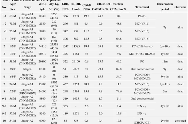
PBMCs were obtained from ten SS patients whose clinical data are summarized in Table
116
1. In one SS patient (case 1), four blood samples were obtained in different occasions:
117
before and during the treatment with oral etoposide, 100 mg/week for 7 years. And in
118
another one (case 9), three samples were collected before and during the treatment with
119
interferon-γ. Control PBMCs were also obtained from 25 patients with MF, three with
120
non-MF/SS cutaneous T-cell lymphomas (CTCL), two with diffuse large B-cell
121
lymphoma (DLBCL), eight with inflammatory skin diseases with erythroderma, and 19
122
healthy volunteers.
123
Cutaneous biopsy specimens were obtained from patients with SS (n=3), MF(n=8),
124
ATLL (n=3) and ALCL (n=5) for diagnostic use, and the rest of them were used for
125
immunostaining. Serum samples from patients with SS (n=7), MF (n=21), ATLL (n=6),
126
ALCL (n=6), and healthy volunteers (n=69) were used for the measurement of soluble
127
CADM1 by ELISA. The present study was approved by the Institutional Review Board
128
(IRB) of Okayama University Hospital (No. 1802-006 and Genome No. 319). Informed
129
consent was obtained from all blood and tissue donors according to the Helsinki
130
Declaration.
131
132
Cell lines
133
We used thee MF/SS cell lines (Hut78, Myla and MJ), one ATL cell line (TL-SU), one
134
non-MF/SS cutaneous T-cell lymphoma line (HH), four ALCL cell lines (SU-DHL-1,
135
Karpas299, SR786, and SUP-M2), and six B-cell lines (Raji, Akata, N83-1, BJ-AB, IB4, 136
and LCL-TT).
137
138
Flow cytometric analysis
139
In addition to our routine panel of conjugated antibodies for flow cytometry, including
140
anti-CD3, CD4, CD7, CD8, CD25, CD30, CD45 and HLA-DR antibodies (Beckman
141
Coulter Inc., CA, U.S.A.), PBMCs were stained with phycoerythrin (PE)-conjugated
142
anti-TSLC1/CADM1 antibody (polyclonal, rabbit, bs-6026R) and allophycocyanin
143
(APC)-conjugated anti-CCR4 antibody (mouse, L291H4). Navios instrument was used
144
for all multicolor flow cytometry, and data were analyzed using Kaluza software
145
(Beckman Coulter Inc., CA, U.S.A.).
146
147
Cell activation by CD3/CD28 stimulation
148
PBMCs from healthy volunteers were stimulated with CD3/CD28-coating beads
149
(Thermo Scientific Inc., MA, U.S.A.) according to the manufacturer’s protocol. After 48
150
h stimulation, the PBMCs were processed for flow cytometry as described above.
151
152
Immunohistochemistry
153
Formalin-fixed, paraffin-embedded tissue sections were used for immunostaining. After
154
deparaffinization and peroxidase-blocking, the sections were stained with mouse
155
monoclonal anti-human CD194 (CCR4) antibody (Becton, Dickinson and Company
156
Inc., NJ, U.S.A.), or with chicken monoclonal anti-SynCAM/TSLC1/CADM1 antibody
157
(MBL, Nagoya, Japan). Slides were then incubated with the ChemMate Envision
158
polymer (DAKO Japan, Tokyo) or with biotinylated anti-chicken IgY (Immuno HRP
159
DAB kit, Immuno Bio Science Corp., WA, U.S.A.). The target proteins were detected
160
using diaminobenzidine tetrahydrochloride (DAB) solution.
161
162
Reverse transcriptase-polymerase chain reaction (RT-PCR) and sequencing
163
Total RNA was extracted from cell lines and converted to complementary DNA. The
164
PCR was carried out for 33 cycles of denaturation at 94°C, annealing at 64°C, and
165
extension at 72°C. The primer sequences used were as follows: CADM1 forward, 5’-
166
GTGATGGTAACTTGGGTGAGAGTC-3’; CADM1 reverse, 5’-
167
CCAGAATGATGAGCAAGCACAG-3’. The PCR products were fractionated on 2%
168
agarose gels and visualized by ethidium bromide staining.The products of target bands
169
from gel were sequenced using an Applied Biosystems 310 Genetic Analyzer (Applied
170
Biosystems, CA, U.S.A.). The results were read by ApE (v2.0.49) (free software by M.
171
Wayne Davis), and compared with the database registered in NCBI BLAST (Basic
172
Local Alignment Search Tool).
173
174
Enzyme-linked immunosorbent assay (ELISA)
175
Soluble forms of CADM1 in patients’ sera and the culture supernatants were measured
176
by a sandwich ELISA using phage anti-human CADM1 antibody (Institute for
177
Antibodies Co., Ltd., Nagoya, Japan; phage 035-212) for the capture antibody, and
178
peroxidase-labeled anti-CADM1 antibody (MBL Co., Ltd., Nagoya, Japan; chicken IgY,
179
Clone3E1) for the second antibody. For quantification of soluble CADM1, calibration
180
curve was made using a standard sample with known concentration (the recombinant
181
protein in soluble form from CADM1-transfected HEK293 cells).
182
183
Statistical analysis
184
Statistical analyses were conducted with GRAPHPAD PRISM, version 4.03 (GraphPad,
185
La Jolla, CA, U.S.A.) and IBM SPSS Statistics 22.0 (IBM, Tokyo, Japan). Mann-
186
Whitney U test and Spearman rank correlation coefficient were used for statistical
187
analysis; P-values < 0.05 were considered significant.
188 189
RESULTS
190
CADM1 expression in circulating Sézary cells
191
Flow cytometric analysis revealed a background level of CADM1+ cells in the PBMCs,
192
ranging from 0.0% to 4.5% (mean: 1.66±1.62%) in the healthy subjects, and from 0.1%
193
to 2.4% (mean: 0.97±1.39%) in the disease control group with non-lymphoma skin
194
disorder. Based on these data, a significant increase of CADM1+ cells was considered
195
to be an increase to 6.5% or more of the CD3+CD4+ fraction (the mean + 3SD in the
196
normal individuals: n=19).
197
Ten SS patients were enrolled in the present study: all patients met the B2 criteria
198
for SS upon initial diagnosis, i.e., ≥1,000 L-1 Sézary cells with positive clones, or
199
either CD4/CD8 ≥10, CD4+CD7- cells ≥40% or CD4+CD26- cells ≥30%.(12) Ten
200
blood samples obtained from the ten SS patients for the first examination contained
201
CADM1+ cells that accounted for 0.4% to 74.5% (median; 22.4%) of the CD3+CD4+
202
cell fraction (Table 1, Fig. 1a, b). Of the 15 samples tested, seven samples from seven
203
patients (sample No. 1-1, 2, 3, 4, 5, 6 and 7 in Table 1) exhibited significantly increased
204
percentages (>6.5%) of CADM1+ cells in the CD3+CD4+ fraction, ranging from 7.9%
205
to 74.5% (median; 33.7%) (Fig. 1a, c). The remaining eight samples (No. 1-2, 1-3, 1-4,
206
8, 9-1, 9-2, 9-3, and 10) obtained from four patients with SS showed a background level
207
(<6.5%) of CADM1+ cells, ranging from 0.4% to 4.8% of the CD3+CD4+ fraction.
208
209
CADM1 expression in other cutaneous lymphomas, activated T-cells, and
210
inflammatory skin diseases
211
In 24 of 25 patients with MF, the percentages of CADM1+ cells were not significantly
212
increased (<6.5% CADM1+ cells) in the PBMCs (Fig. 1b). The one exceptional patient
213
with MF, stage IVB, had a slightly increased CD4/CD8 ratio (3.3%) with 8.8%
214
CADM1+ cells in the CD3+CD4+ fraction, although no abnormal T cells were 215
detectable in blood smear or by flow cytometry. No significant increase of CADM1+
216
cells was observed in the PBMCs from two patients with DLBCL, eight patients with
217
inflammatory skin diseases, including atopic dermatitis (n=6) and generalized drug
218
eruption (n=2), or in the 19 healthy volunteers (Fig. 1b).
219
220
CD3/CD28-stimulated normal T-cells showed no increase of CADM1+ cells in the
221
CD3+ fraction (n=12, mean 2.9±1.8%) (p=0.0091, Mann-Whitney U test) (Fig. 2a),
222
although HLA-DR and CD25 were induced after stimulation (Fig. 2b). Therefore,
223
CADM1 was not inducible in T-cells by CD3/CD28 stimulation.
224
Among the cell lines examined, CADM1+ cells were observed in the T-cell lines
225
ranging from 1.3% to 22.4% (n=9, mean 11.0±8.3%) (Fig. 2a, c). Six of 9 T-cell lines
226
contained CADM1+ cells over the cut-off value (6.5%), including all three T-cell lines
227
derived from MF/SS (Myla, MJ and Hut78), one from ATLL (TL-SU), one from non-
228
MF/SS CTCL (HH), and one from ALCL (SR-786) (Fig. 2a). The remaining three T-cell
229
lines derived from ALCL contained CADM1+ cells below the cut-off value. No increase
230
of CADM1+ cells was observed in all B-cell lines examined (n=6, mean 0.87±0.95%)
231
(p=0.0016) (Fig. 2a). Therefore, CADM1 was expressed in some neoplastic T-cell lines
232
selectively, and not induced in the neoplastic B-cell lines or CD3/CD28-stimulated T
233
cells.
234
235
A phenotype of CADM1+ Sézary cells
236
In addition to cytological examination in the blood smear, the percentages of Sézary
237
cells was estimated by two indicators in the present study: CD3+CD4+CD7dim+/CD7-
238
cells, and CD3+CD4+CD26dim+/CD26- cells by flow cytometry. The percentages of
239
CADM1+ cells did not always correspond to those of Sézary cells determined by
240
cytological criteria, CD4+CD7dim+/CD7- cells or CD4+CD26dim+/CD26- cells (Fig.
241
1c). In our series of patients (n=10), the percentages of CD4+CD7dim+/CD7- cells were
242
higher than those of CADM1+ cells, except for a few patients (case 3 in Table 1).
243
Since CCR4 is usually expressed by Sézary cells, we compared the co-expression of
244
CADM1 and CCR4 in the CD3+CD4+CD7dim+/CD7- fractions. In all six patients
245
(eight samples) studied, the percentages of CADM1+ cells were lower than those of
246
CCR4+ cells: 0.3% (case 1-3 and 1-4), 49.0% (case 4), 39.5% (case 5) 16.4% (case6),
247
50.2% (case 8), 5.1% (case 9-2) and 21.8% (case 9-3) of the CCR4+ cells, respectively.
248
249
CADM1+ cells in skin lesions of cutaneous T-cell lymphomas
250
In the cutaneous lesions of MF/SS at stages IIA (n=1), IIB (n=4), IIIA (n=2), IVA1 (n=2) 251
and IVA2 (n=2), CADM1+ cells were present in the dermal and epidermal infiltrates to 252
various degrees (Fig. 3). Cutaneous lesions of ATLL (n=3) and primary cutaneous ALCL 253
(n=5) also contained CADM1+ cell in the infiltrates (Fig. 3). Since neoplastic T cells of 254
MF, SS and ATLL are known to express CCR4, we examined the ratios of CADM1+
255
cells among CCR4+ cells in the skin lesions, excluding the inflammatory cell infiltrates 256
as much as possible. Similar to the cases of ATLL (n=3), the numbers of CADM1+ cells 257
were less than those of CCD4+ cells in the skin lesions of SS (n=3) and MF (n=8), 258
except for one MF case (Fig. 3, Fig. S1). There was no clear difference in CADM1 259
expression by the stages of illness. By contrast, atypical lymphoid cells of the primary 260
cutaneous ALCL (n=5), which is usually not related to CCR4 expression, were positive 261
for CADM1 without CCR4 expression to various degrees. When compared with the 262
percentages of CADM1+ cells between the skin infiltrates and peripheral blood in the 263
same patient with SS (case 1 in Table 1), only a small percentage of CADM1+ cells 264
(approximately 18%) was observed among CCR4+ cells in the dermis, although 74.5%
265
of CADM1+ cells were present in the CD3+CD4+ fraction in the blood.
266
267
Splicing variants of CADM1 mRNA
268
The RT-PCR amplification revealed that cDNA generated from mRNA extracted from a
269
Sézary cell line (Hut78) contained three major PCR products with the respective
270
molecular sizes of 368, 332, and 257 bp, respectively. A direct sequencing study
271
revealed that the three PCR products were composed of exons 7, 8, 9 and 11 of CADM1
272
(368-bp product), exons 7,8 and 11 (332-bp product), and exons 7 and 11 (257-bp
273
product) (NCBI, BLAST data) (Fig. 4a-c).
274
Cultured normal human keratinocytes also showed three PCR products
275
corresponding to those observed in Sézary cells (Fig. 4b, c). Therefore, one Sézary cell
276
line (Hut78) and cultured normal human keratinocytes expressed the same combination
277
of exons of CADM1. Freshly isolated Sézary cells (cases 4 and 6 in Table 1) expressed
278
the two major isoforms composed of exons 7, 8, 9 and 11, and exons 7, 8, and 11,
279
respectively. Other T-cell lines from MF, ATLL and ALCL also expressed splicing
280
isoforms with a different combination of the above-mentioned three variants (Fig. 4b).
281
No CADM1 mRNA was expressed in the three B-cell lymphoma cell lines examined
282
(N83-1, BJ-AB and Raji).
283
284
Comparison of CADM1 expression with hematological markers and clinical courses
285
When CADM1 expression was compared with hematological markers having
286
prognostic significance, such as lactate dehydrogenase (LDH) and soluble IL-2
287
receptors (sIL-2R), there were weak correlations between the percentages of CADM1+
288
cells in the peripheral blood and the serum levels of LDH or soluble IL-2 receptor (sIL-
289
2R) (Fig. 5).
290
In our series of ten patients with SS, four of five patients whose CADM1+ cells
291
made up more than 20% (median value of CADM1+ cells; 22.4%) of the CD3+CD4+
292
fraction died of SS-related complications in a 7-year follow-up period (Fig. S2).
293
294
Soluble form of CADM1 in patients’ sera
295
In order to evaluate the shedding of CADM1 ectodomains, we measured soluble
296
CADM1 in the sera of SS patients by ELISA. Two of six serum samples from ATLL
297
patients contained extremely high levels of soluble CADM1, i.e., 2913.6 and 2132.4 ng
298
mL-1, but the other four ATLL samples and five samples obtained from patients with SS
299
at different interval (cases 1, 2 and 3 in Table 1) showed low concentrations of soluble
300
CADM1 (below 400 ng mL-1) (Fig. 6). No difference was observed in the serum levels
301
of soluble CADM1 between SS and the other CTCL excluding ATLL (196.8±89.5 vs.
302
198.2±92.2 ng mL-1).
303 304
Discussion
305
Similar to the previous observations in overt ATLL,13 we herein detected circulating
306
CADM1+ cells in a group of patients with SS. Neoplastic cells of ATLL and SS share a
307
cytological profile: a convoluted or flower-like nucleus and a CD3+CD4+CCR4+
308
phenotype. In addition to these cytological similarities, our observations indicate that
309
CADM1 expression is also shared by both cell types. The expression of CADM1,
310
however, was not specific for leukemic cells of ATLL, SS and MF because its
311
expression was detected in infiltrating cells in ALCL and cell line cells from CTCL. The
312
expression of CADM1 was not restricted to the type 2 helper T cells (Th2) or regulatory
313
T cells (Treg), both of which are positive for CCR4, but could be induced in the other
314
cell types negative for CCR4, as shown in cases of ALCL. It is intriguing to note that
315
the B-cell lines used for the present study did not express CADM1.
316
The detection of a CADM1+CD7dim+/CD7- fraction has been used to predict the
317
progressive form of ATLL.9,13 In our small series of SS patients, CADM1 expression
318
was observed in patients with progressive SS: four of five SS patients harboring
319
CADM1+ cells that made up over 20% of the CD3+CD4+ fraction died of progressive
320
SS within 1.5 year after the appearance of more than 20% of CADM1+ cells. Similar to
321
our observations, recent reports have described that CADM1 is expressed in the
322
cutaneous infiltrates of MF, and its expression might be related to the poor prognosis.
323
14,15 Therefore, further cohort study is required to address whether CADM1 expression
324
is a marker of progressive MF and SS.
325
Our study indicates that the expression of CADM1 is not a simple activation marker,
326
because CD3/CD28 stimulation of normal human lymphocytes induced CD25 and
327
HLA-DR expression, but did not induce CADM1. It has been postulated that CADM1
328
expression is induced by HTLV-1-encoded gene products such as Tax and HBZ in
329
ATLL. 13,16 But this scenario cannot explain the fact that CADM1 is expressed by
330
Sézary cells without HTLV-1 infection.
331
Sézary cells are characterized by the expression of CCR4 and the loss or diminished
332
expression of CD7 and/or CD26.17 Our present flow cytometric study revealed that
333
the percentages of CADM1+ cells did not necessarily correspond to those of Sézary
334
cells determined by cytological criteria, or a CD4+CD7dim+/CD7- or a
335
CD4+CD26dim+/CD26- phenotype.
336
In addition to the loss or diminished expression of CD7 and CD26, recent studies
337
described expression of CD158k/ KIR3DL2, CD164,and the central memory T-cell
338
phenotype (CD27+CD45RA- CD45RO+) as characteristic features of Sézary cells.18-21
339
Various novel gene alterations have been reported in Sézary cells: MYC gain and MNT
340
loss, up-regulation of DNM3, TWIST1, EPHA4 and PLS3, and down-regulation of
341
STAT4.22 Whole exome and RNA sequencing revealed a complex genomic landscape of
342
somatic copy number variations and fusion genes possibly related to the pathogenesis of
343
SS.23
344
Concerning the oncogenic isoform of CADM1, Kikuchi et al. reported that normal
345
human lung cDNA reveals a single major isoform composed of the exons 7-8 -11 (the
346
332 bp in Fig. 4a), whereas small cell lung cancer expresses another splicing variant
347
containing the exons 7-8-9-11 (the 368 bp in Fig. 4a). The authors’ experimental data
348
suggest that this variant is associated with the malignant features of small cell lung
349
carcinoma, as is observed in progressive ATLL8. In our study, Sézary cells and T-cell
350
lines including Hut78, TL-SU and SUP-M2 expressed the 368bp variant composed of
351
exons 7-8-9-11 to various degrees.
352
Our study indicates that cultured normal human keratinocytes showed the three
353
major splicing variants of CADM1, identical to those observed in Sézary cells.
354
Furthermore, a previous report has described that the extracellular domains of CADM1
355
interact with integrin 64 in hemidesmosomes24. These observations suggest the
356
possible involvement of CADM1 in epidermotropic infiltration of neoplastic T-cells via
357
the homophilic and heterophilic binding of CADM1 to epidermal keratinocytes and
358
basement membrane zone.
359
In conclusion, CADM1 is frequently expressed by neoplastic cells in SS and MF,
360
and T-cell lines from CTCL. The pathogenic properties of CADM1 in the progression of
361
CTCL remain to be answered.
362 363
Acknowledgments
364
We thank Michinori Aoe, Department of Laboratory Medicine, Okayama University
365
Hospital, Okayama, and Hiroko Katayama, Departments of Dermatology, Okayama
366
University Graduate School of Medicine, Dentistry and Pharmaceutical Sciences,
367
Okayama, for support in this project.
368
369
Conflict of Interest
370
The authors state no conflict of interest.
371
Funding: None
372
373
374
References
375
1. Kuramochi M, Fukuhara H, Nobukuni T, et al. TSLC1 is a tumor-suppressor gene 376
in human non-small-cell lung cancer. Nature genetics. 2001;27(4):427-30.
377
2. Gomyo H, Arai Y, Tanigami A, et al. A 2-Mb sequence-ready contig map and a 378
novel immunoglobulin superfamily gene IGSF4 in the LOH region of chromosome 11q23.2.
379
Genomics. 1999;62(2):139-46.
380
3. Urase K, Soyama A, Fujita E, Momoi T. Expression of RA175 mRNA, a new 381
member of the immunoglobulin superfamily, in developing mouse brain. Neuroreport.
382
2001;12(15):3217-21.
383
4. Biederer T, Sara Y, Mozhayeva M, et al. SynCAM, a synaptic adhesion molecule 384
that drives synapse assembly. Science (New York, NY). 2002;297(5586):1525-31.
385
5. Shingai T, Ikeda W, Kakunaga S, et al. Implications of nectin-like molecule- 386
2/IGSF4/RA175/SgIGSF/TSLC1/SynCAM1 in cell-cell adhesion and transmembrane protein 387
localization in epithelial cells. The Journal of biological chemistry. 2003;278(37):35421-7.
388
6. Masuda M, Yageta M, Fukuhara H, et al. The tumor suppressor protein TSLC1 is 389
involved in cell-cell adhesion. The Journal of biological chemistry. 2002;277(34):31014-9.
390
7. Murakami Y. Involvement of a cell adhesion molecule, TSLC1/IGSF4, in human 391
oncogenesis. Cancer Sci. 2005;96(9):543-52.
392
8. Kikuchi S, Iwai M, Sakurai-Yageta M, et al. Expression of a splicing variant of the 393
CADM1 specific to small cell lung cancer. Cancer Sci. 2012;103(6):1051-7.
394
9. Sasaki H, Nishikata I, Shiraga T, et al. Overexpression of a cell adhesion molecule, 395
TSLC1, as a possible molecular marker for acute-type adult T-cell leukemia. Blood.
396
2005;105(3):1204-13.
397
10. Kobayashi S, Nakano K, Watanabe E, et al. CADM1 expression and stepwise 398
downregulation of CD7 are closely associated with clonal expansion of HTLV-I-infected cells 399
in adult T-cell leukemia/lymphoma. Clinical cancer research : an official journal of the 400
American Association for Cancer Research. 2014;20(11):2851-61.
401
11. Mimae T, Hagiyama M, Inoue T, et al. Increased ectodomain shedding of lung 402
epithelial cell adhesion molecule 1 as a cause of increased alveolar cell apoptosis in 403
emphysema. Thorax. 2014;69(3):223-31.
404
12. Olsen EA, Whittaker S, Kim YH, et al. Clinical end points and response criteria in 405
mycosis fungoides and Sezary syndrome: a consensus statement of the International 406
Society for Cutaneous Lymphomas, the United States Cutaneous Lymphoma Consortium, 407
and the Cutaneous Lymphoma Task Force of the European Organisation for Research and 408
Treatment of Cancer. Journal of clinical oncology : official journal of the American Society of 409
Clinical Oncology. 2011;29(18):2598-607.
410
13. Nakahata S, Saito Y, Marutsuka K, et al. Clinical significance of 411
CADM1/TSLC1/IgSF4 expression in adult T-cell leukemia/lymphoma. Leukemia.
412
2012;26(6):1238-46.
413
14. Mashima E, Sawada Y, Yamaguchi T, et al. A high expression of cell adhesion 414
molecule 1 (CADM1) is an unfavorable prognostic factor in mycosis fungoides. Clinical 415
immunology (Orlando, Fla). 2018;193:121-2.
416
15. Yuki A, Shinkuma S, Hayashi R, et al. CADM1 is a diagnostic marker in early- 417
stage mycosis fungoides: Multicenter study of 58 cases. Journal of the American Academy of 418
Dermatology. 2018.
419
16. Pujari R, Hunte R, Thomas R, et al. Human T-cell leukemia virus type 1 (HTLV-1) 420
tax requires CADM1/TSLC1 for inactivation of the NF-kappaB inhibitor A20 and 421
constitutive NF-kappaB signaling. PLoS pathogens. 2015;11(3):e1004721.
422
17. Kim EJ, Hess S, Richardson SK, et al. Immunopathogenesis and therapy of 423
cutaneous T cell lymphoma. The Journal of clinical investigation. 2005;115(4):798-812.
424
18. Bagot M, Moretta A, Sivori S, et al. CD4(+) cutaneous T-cell lymphoma cells 425
express the p140-killer cell immunoglobulin-like receptor. Blood. 2001;97(5):1388-91.
426
19. Campbell JJ, Clark RA, Watanabe R, Kupper TS. Sezary syndrome and mycosis 427
fungoides arise from distinct T-cell subsets: a biologic rationale for their distinct clinical 428
behaviors. Blood. 2010;116(5):767-71.
429
20. Poszepczynska-Guigne E, Schiavon V, D'Incan M, et al. CD158k/KIR3DL2 is a new 430
phenotypic marker of Sezary cells: relevance for the diagnosis and follow-up of Sezary 431
syndrome. The Journal of investigative dermatology. 2004;122(3):820-3.
432
21. Wysocka M, Kossenkov AV, Benoit BM, et al. CD164 and FCRL3 are highly 433
expressed on CD4+CD26- T cells in Sezary syndrome patients. The Journal of investigative 434
dermatology. 2014;134(1):229-36.
435
22. Boonk SE, Zoutman WH, Marie-Cardine A, et al. Evaluation of Immunophenotypic 436
and Molecular Biomarkers for Sezary Syndrome Using Standard Operating Procedures: A 437
Multicenter Study of 59 Patients. The Journal of investigative dermatology.
438
2016;136(7):1364-72.
439
23. Prasad A, Rabionet R, Espinet B, et al. Identification of Gene Mutations and 440
Fusion Genes in Patients with Sezary Syndrome. The Journal of investigative dermatology.
441
2016;136(7):1490-9.
442
24. Mizutani K, Kawano S, Minami A, Waseda M, Ikeda W, Takai Y. Interaction of 443
nectin-like molecule 2 with integrin alpha6beta4 and inhibition of disassembly of integrin 444
alpha6beta4 from hemidesmosomes. The Journal of biological chemistry.
445
2011;286(42):36667-76.
446
SUPPORTING INFRMATION
447
Figure S1. CADM1+ cells in the skin lesions of MF/SS, ATLL, and ALCL (all
448
data)
449
450
Figure S2. Outcomes of SS patients during the observation periods, and the
451
absolute numbers of CADM1+ cells on diagnosis
452 453
FIGURE LEGENDS
454
Figure 1. CADM1+ cells in peripheral blood
455
(a) CADM1+ cells in the CD3+CD4+ fraction of PBMCs were analyzed by flow
456
cytometry. (b) The percentages of CADM1+ cells of the CD3+CD4+ fraction in
457
PBMCs are shown. (c) The absolute numbers of CADM1+ cells the CD3+CD4+
458
fraction in PBMCs are shown.
459
460
Figure 2. CADM1 expression by T-cell line cells and activated PBMCs
461
(a) The CADM1+ cells were significantly higher in T cell lines than those in the B-cell
462
ones (p=0.0016) and the activated PBMCs (p=0.0091). (Mann-Whitney U test) (b)
463
CD3/CD28-activated PBMCs from healthy controls begin to express activation markers
464
such as CD25 and/or HLA-DR, but do not express CADM1. (c) CADM1 expression in
465
MF/SS-derived cell lines (Myla, Hut78 and MJ), and non-MF/SS CTCL-derived one
466
(HH).
467
468
Figure 3. CADM1+ cells in the skin lesions of MF/SS, ATLL, and ALCL
469
The representative cases show that CADM1+ cells are present among CCR4+ cells in
470
the dermal and epidermal infiltrates in MF/SS and ATLL. The expression of CADM1+
471
cells are also observed in the skin infiltrates of primary cutaneous ALCL. (See all the
472
data of immunostaining in Fig. S1.)
473
474
Figure 4. Splicing variants of CADM1 expressed by Sezary cells, T-cell lines, and
475
normal human epidermal keratinocytes
476
(a) Possible splicing variants of CADM1 mRNA (Kikuchi et al, 2012). (b) One Sézary
477
cell line (Hut78) and cultured normal human keratinocytes (NHEK) express the
478
identical splicing variants of CADM1. Other T-cell lines also express splicing isoforms
479
with a different combination. No CADM1 mRNA was expressed in B-cell lymphoma
480
cell lines (N83-1, BJ-AB and Raji). (c) Three major PCR products expressed by a SS
481
cell line (Hut78) are composed of exons 7, 8, 9 and 11 of CADM1 for the 368 bp, exons
482
7,8 and 11 for the 332bp, and exons 7 and 11 for the 257 bp.(NBCI, BLAST)
483
484
Figure 5. Comparison of CADM1 expression with hematologic markers, LDH and
485
sIL-2R
486
(a) The percentages of CADM1+ cells in the peripheral blood tend to correlate with the
487
serum levels of LDH or sIL-2R. (Spearman’s rank correlation coefficient) (b)
488
Comparison of hematological markers between the alive and dead cases (Mann-
489
Whitney U test). All four SS patients with fatal outcome in the follow-up period had
490
more than 20% of CADM1+ cells (See Figure S2).
491
492
Figure 6. Serum levels of soluble form of CADM1
493
Two of six serum samples from ATLL patients contained extremely high levels of
494
soluble CADM1, but other samples including 5 serum samples from patients with
495
Sézary syndrome showed low concentrations of soluble CADM1. n.s.: not significant.
496
(Mann-Whitney U test)
497 498
Table 1. Clinical data of the SS patients 499
500
501 502
Age/ WBC, Aty-Ly, LDH, sIL-2R, Observation
Sex /μL /μL (%) IU/L U/mL CADM1+ % CD7+dim % period
Stage4A1 3256
(T4N1M0B2) (40.5)
Stage4A1 232
(T4N1M0B2) (9.0)
Stage4A1 56
(T4N1M0B2) (1.5)
Stage4A1 167
(T4N1M0B2) (4.0)
Stage4A1 23538
(T4N3M0B2) (43.5)
Stage4A2 3434
(T4N3M0B2) (65.6)
Stage4A1 11024
(T4N3M0B2) (53.0)
0 (0)
0 (0) 9302 (58.5)
Stage4A2 3472
(T4N3M0BX) (14)
Stage4A2 1063
(T4NxM0B2) (12)
Stage4A2 522
(T4NxM0B2) (7.5)
Stage4A2 1153
(T4NxM0B2) (13.5)
120 (2.0)
Abbreviations: WBC, white blood cell; Aty-Ly, atypical lymphocyte; LDH, lactate dehydrogenase; sIL-2R; soluble interleukin-2 receptor; CD, cluster of differentiation; CADM, cell adhesion molecule; Photo., phototherapy; MC, monochemotherapy; PC, polychemotherapy; VP16, etoposide; HDACi, histone deacetylase inhibitor; IFN -γ, intravenous interferon-γ; C, cyclophosphamide; H, doxorubicin (hydroxydaunomycin); O, vincristine; P, prednisolone; I, ifosfamide; C, carboplatin; E, etoposide; y, year(s); m, month(s).
Table 1. Clinical characteristics of SS patients
10 54/M Stage4A2 6000 88 838 0.6 0.4 17.8 PC
(CHOP, ICE) censored
9-1 56/M 8860 319 1033 9.6 1.7 5.1
MC (VP16) dead
dead
7 74/M Stage4A1 15900 452 2753 28.7 7.9 11.1
8 72/F 24000 298 3584 15.4 4.8 74.6 PC (CHOP) 5m
MC (HDACi)
2y+11m Oral corticosteroid 9y dead 6 64/F Stage4A2
(T4N3M0B2) 5410 380 413 3.9 15.3 34.7 PC (CHOP)
MC (HDACi) alive
5 89/F Stage4 12000 511 7077 98 29.4 82.8
5y+1m dead
4 58/M 20800 322 26100 0.6 33.7 49.2 11m dead
3 76/F 20510 375 1184 98 38 9.6 MC (VP16 / HDACi)
PC
1y+2m
2 62/F 54110 1347 11385 18.4 65.1 83.8 PC (CHP-based) 3y+10m dead
48.8 MC (VP16)
1-3 75/M 3700 342 737 11.2 0.5 55.6 MC (VP16)
7y alive
No. Stage CD4/8
ratio
CD3+CD4+ fraction
Treatment Outcome
1-1 69/M 8040 366 1739 19.3 74.5 84 Photo.
1-2 75/M 2580 296 481 6.4 0.9
1-4 76/F 4170 306 582 13.5 0.5 64.8 MC (VP16)
2y+6m 4y+1m alive
9-3 57/M 8540 180 1271 21 2.0 17.8 IFN -γ
Oral corticosteroid
9-2 56/M 6960 365 - 2.6 2.2 1.4 IFN -γ
Figure 1.
503
504 505
Figure 2.
506
507 508
Figure 3.
509
510 511
Figure 4.
512
513 514
Figure 5.
515
516 517
Figure 6.
518
519 520
Figure S1.
521
522 523
Figure S2.
524
525