INTRODUCTION
The serine/arginine-rich splicing factor (SRSF)
family comprises at least 12 conserved and struc-turally related RNA-binding proteins (SRSF1 to SRSF12) (1). SRSF3 (also known as SRp20) binds
cis-acting RNA elements through an N-terminal
RNA recognition motif (RRM), and a C-terminal re-gion enriched in Ser-Arg dipeptides (SR domain) mediates interactions with other proteins for regu-lation of both constitutive and alternative splicing of pre-mRNA (2). In addition, several lines of recent
ORIGINAL
Truncated serine/arginine-rich splicing factor 3
accelerates cell growth through up-regulating c-Jun
expression
Shizuka Kano
1, 2, Kensei Nishida
1, Chihiro Nishiyama
2, Yoko Akaike
1, Keisuke Kajita
1,
Ken Kurokawa
1, Kiyoshi Masuda
1, Yuki Kuwano
1, Toshihito Tanahashi
1,
and Kazuhito Rokutan
11
Department of Stress Science, Institute of Health Biosciences, the University of Tokushima Graduate School, Tokushima, Japan ;2
Student Lab, the University of Tokushima Faculty of Medicine, Tokushima, Japan
Abstract : Serine/arginine-rich splicing factor 3 (SRSF3), a member of the SRSF family, plays a wide-ranging role in gene expression. The human SRSF3 gene generates a major mRNA isoform encoding a functional, full-length protein and a PTC-containing isoform (SRSF3-PTC ). The latter is expected to be degraded through the nonsense-mediated mRNA decay system. However, it was reported that SRSF3-PTC mRNA was produced un-der stressful conditions and translated into a truncated SRSF3 protein (SRSF3-TR). To disclose unknown functions of SRSF3-TR, we established Flp-In-293 cells stably express-ing SRSF3-TR. The SRSF3-TR-expressexpress-ing cells increased mRNA and protein levels of posi-tive regulators for G1 to S phase transition (cyclin D1, cyclin D3, CDC25A, and E2F1) and accelerated their growth. c-Jun is required for progression through the G1 phase, the mechanism by which involves transcriptional control of the cyclin D1 gene. We also found that the JUN promoter activity was significantly increased in the Flp-In-293 cells stably expressing SRSF3-TR, compared with mock-transfected control cells. The SRSF3-TR-expressing cells increased c-Jun and Sp-1 levels, which are important for the positive autoregulation and basal transcription of JUN , respectively. Our results suggest that stress-inducible SRSF3-TR may participate in the acceleration of cell growth through fa-cilitating c-Jun-mediated G1 progression under stressful conditions. J. Med. Invest. 60 : 228-235, August, 2013
Keywords :truncated SRSF3 protein, oxidative stress, cell growth, c-Jun
Received for publication April 19, 2013 ; accepted April 26, 2013.
Address correspondence and reprint requests to Kensei Nishida, MD, PhD, Department of Stress Science, Institute of Health Biosciences, the University of Tokushima Graduate School, 3 18 15 Kuramoto cho, Tokushima 770 8503, Japan and Fax : +81 -88 - 633 - 9008.
evidence suggest that SR protein family also regu-lates RNA elongation (3), transport (4), and transla-tion (5), suggesting a wide-ranging role in gene ex-pression. In addition, SRSF3 is overexpressed in hu-man ovarian cancer, and its knockdown results in apoptosis of ovarian cancer cells (6). Recently, we have shown that reduction of SRSF3 causes G1 cell cycle arrest and apoptosis in colon cancer cells (7). These findings suggest that SRSF3 may be one of the essential factors for regulation of cell fate.
The human SRSF3 gene is composed of 7 ex-ons and generates two mRNA isoforms. Like other SRSF family member genes, the human SRSF3 gene contains an element of extreme sequence conser-vation, termed “ultraconserved element” (UCE).
SRSF3 contains a 577-bp UCE that includes entire
exon 4, and this exon 4 has multiple premature termination codons (PTCs). A major SRSF3 mRNA isoform encoding a functional full-length SRSF3 protein (SRSF3-FL) excludes this exon 4. Alterna-tive splicing of SRSF3 pre-mRNA produces another mRNA isoform consisting of entire 7 exons. This PTC-containing SRSF3 isoform (SRSF3-PTC ) is considered to be degraded through the nonsense-mediated mRNA decay (NMD) system, a surveil-lance mechanism that decomposes PTC-containing mRNAs. However, it was reported that SRSF3-PTC mRNA was translated into a truncated SRSF3 pro-tein (SRSF3-TR) partially lacking RS domain in the murine B-cell lymphoma K46 cells (8). We also found that colon cancer cells (HCT116) induced SRSF3-TR in response to sodium arsenite (unpub-lished observations). At present, however, the bio-logical function of SRSF3-TR is unknown.
In this study, we established Flp-In-293 cells stably expressing SRSF3-TR, and showed that SRSF3-TR facilitated cell growth through up-regu-lating c-Jun expression.
MATERIALS AND METHODS
Cell culture and cell countingFlp-In-293 cells (Invitrogen, Carlsbad, CA) were maintained in high-glucose Dulbecco’s modified Eagle’s medium (Nacalai tesque, Kyoto, Japan) sup-plemented with 10% (v/v) heat-inactivated fetal calf serum, 2 mM l-glutamine, 100 U/ml penicillin, and 100μg/ml streptomycin in a humidified atmosphere of 5% CO2 in air. Numbers of growing cells were
counted manually using a hematocytometer.
Plasmid construction
Using a cDNA library prepared from a human colon cancer cell line (HCT116), the human
SRSF3-PTC mRNA (Ensembl Transcript ID :
ENST00000477442) were amplified by PCR using a primer set : 5’-AAAAAAGGATCCATGCATCGT-GATTCCTGTCCATTG-3’ (forward, BamHI site is underlined) and 5’-AAAAAAGATATCCTATTTCC-TTTCATTTGACCTAGA-3’ (reverse, EcoRV site is underlined). The amplified product was cloned into the mammalian expression vector pCMV-3Tag (pCMV-3Tag/SRSF3-TR ; Stratagene, La Jolla, CA). FLAG was appended to the NH2termini of SRSF3.
Subsequently, PCR was performed using the pCMV-3Tag/SRSF3-TR plasmid as the template and the following primer set : 5’-AAAAAAGGTACCGCCA-CCATGGATTACAAGGA-3’ (forward, KpnI site is underlined) and the same reverse primer used for amplification of SRSF3-PTC mRNA. The amplified products were cloned into the pcDNA5/FRT vector (pcDNA5/FRT-3xFLAG-SRSF3-TR). All constructs were confirmed to have the expected sequence by DNA sequencing. The plasmid was transfected us-ing FuGENE HD (Promega, Madison, WI) accord-ing to the manufacturer’s instructions.
Establishment of a cell line stably expressing SRSF3-TR
Flp-In-293 cells were co-transfected with the pcDNA5/FRT-3xFLAG-SRSF3-TR vector and a Flp recombinase expression plasmid pOG44 accord-ing to the manufacturer’s protocol of the Flp-In sys-tem (Invitrogen). A single colony resistant to hygro-mycin B (Invitrogen) was selected and subcultured. Selection of positive colonies (293/SRFS3-TR) was performed by Western blotting using an anti-SRSF3 antibody. Flp-In-293 cells were transfected with pcDNA5/FRT-3xFLAG-mock and pOG44 vectors (293/mock cells) and used as a control.
Quantitative real-time reverse transcription-PCR (qPCR)
Total RNA was extracted from 293/SRSF3-TR or 293/mock cells using an RNAiso plus (Takara, Otsu, Japan). One microgram of isolated RNA was reverse-transcribed using ReverTra Ace reverse transcriptase (TOYOBO, Osaka, Japan). Primer sequences are shown in Table 1. Target mRNA levels were measured using SYBR green master mix and the 7500 real-time system (Applied Biosys-tems, Foster City, CA). Glyceraldehyde 3-phosphate
dehydrogenase (GAPDH) mRNA and 18S were
meas-ured as internal controls for normalization. Us-ing theΔΔCt method, data are presented as fold changes in mRNA levels relative to those in 293/ mock cells.
Western blotting
Whole-cell lysates were prepared in RIPA buffer (10 mM Tris-HCl, pH 7.4 ; 150 mM NaCl ; 1 mM EDTA ; 1 mM DTT ; 0.1% sodium dodecyl sulfate ; 1% Nonidet P-40) containing a protease and phos-phatase inhibitor cocktail (Sigma-Aldrich, St. Louis, MO). The extracted proteins were separated by so-dium dodecyl sulfate-polyacrylamide gel electropho-resis and transferred to a polyvinylidene difluoride membrane (BioRad, Hercules, CA). After blocking with 5% non-fat dry milk, the membranes were incu-bated overnight at 4!!with a mouse monoclonal SRSF3 (1 : 1000 dilution, Sigma-Aldrich), anti-FLAG (1 : 1000, Sigma-Aldrich), anti-cyclin D1 (1 : 1000, Cell Signaling Tech., Danvers, MA), anti-cyclin D3 (1 : 1000, Cell Signaling Tech.), anti-cy-clin E (1 : 1000, Santa Cruz Biotech., Santa Cruz, CA), anti-CDC25A (1 : 1000, Cell Signaling Tech.), anti-E2F1 (1 : 1000, Cell Signaling Tech.), anti-c-Jun (1 : 1000 ; Cell Signaling Tech.), anti-Sp1 (1 : 1000, Sigma-Aldrich), or anti-GAPDH (1 : 5000 ;
Santa Cruz Biotech.) antibody. Following incuba-tion with an appropriate secondary antibody for 1 h at room temperature, bound antibodies were de-tected with an ECL or ECL Prime Western Blot-ting Detection System (GE Healthcare, Piscataway, NJ). The membrane was re-blotted using an anti-GAPDH antibody as a loading control.
Promoter activity assay
The 5’ flank of the human JUN gene was cloned into the pGL3-basic luciferase reporter vector (Promega). In brief, the first PCR was performed using the human genomic DNA as a template. The
JUN proximal promoter region (from -1,266 to +170
bp) was amplified using the following primer set : 5’-AAAAAGGTACCTTACATGTGTCTAGCGCTTC-3’ (forward) and 5’-AAAAACTCGAGGGGAGCCA-CAGGCGCTAGCTCT-3’ (reverse). Subsequently, the amplified product was used as a template to gen-erate deletion constructs consisting of the -798 to +170 bp, -331 to +170 bp, and -32 to +170 bp re-gions using the same reverse primer and one of the following forward primers : 5’-AAAAAGGTACCTC-TCGGCTTCTACGAGCAGC-3’ for -798 to +170 bp, 5’-AAAAAGGTACCCCAAGACGTCAGCCCACAA-TG-3’ for -331 to +170 bp and 5’-AAAAAGGTACC-AGCCAATGGGAAGGCCTTG-3’ for -32 to +170 bp. The amplified products were subcloned into the pGL3-basic vector using KpnI and SacI restriction sites. Using FuGENE HD (Promega), pGL-3 lu-ciferase constructs (100 ng) and pRL-CMV vector (50 ng) were co-transfected into 293/mock or 293/ SRFS3-TR cells (7.5 x 10⁴cells) growing on 24-well
plates. Twenty-four hours after the transfection, cells were harvested, and the firefly and renilla luciferase activities were measured using the Dual-Luciferase Reporter Assay System (Promega).
RESULTS
Effect of SRSF3-TR on cell growth
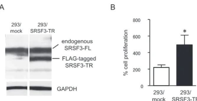
To disclose novel function(s) resided in a trun-cated SRSF3 (SRSF3-TR), we established a cell line stably expressing FLAG-tagged SRSF3-TR us-ing the Flp-In system (Invitrogen). After transfec-tion of Flp-In-293 cells with the vectors encoding 3xFLAG-SRSF3-TR and Flp recombinase, a posi-tive clone (293/SRSF3-TR) was selected. The 293/ SRSF3-TR cells stably expressed similar amounts of both endogenous SRSF3-FL and FLAG-tagged SRSF3-TR (Figure 1A). The stable expression of
Table 1. Primer sets used for quantitative real - time RT- PCR (qPCR)
Targets Primer Sequences (5’ - 3’)
SRSF3 - PTC forward TCCACCTCGTCGCAGAGTCACCATC reverse TCATGTGAAACGACACCAGCCAAGC CCND1 forward GAAGATCGTCGCCACCTG reverse GACCTCCTCCTCGCACTTCT CCND3 forward GCTTACTGGATGCTGGAGGTA reverse AAGACAGGTAGCGATCCAGGT
CCNE1 forward GGCCAAAATCGACAGGAC reverse GGGTCTGCACAGACTGCAT
CDC25A forward ATCTCTTCACACAGAGGCAGAA reverse CCCTGGTTCACTGCTATCTCTT
E2F1 forward TCCAAGAACCACATCCAGTG reverse CTGGGTCAACCCCTCAAG
JUN forward CAGGTGGCACAGCTTAAACA reverse GTTTGCAACTGCTGCGTTAG
GAPDH forward AGCCACATCGCTCAGACAC reverse GCCCAATACGACCAAATCC
18S forward CCCTATCAACTTTCGATGGTAGTCG reverse CCAATGGATCCTCGTTAAAGGATTT
SRSF3-TR did not change endogenous SRSF3-FL levels (Figure 1A). Unexpectedly, SRSF3-TR - ex-pressing cells showed significantly accelerated growth, compared with control 293/mock cells (Figure 1B).
Effects of SRSF3-TR on expression of genes crucial for G1 to S phase transition
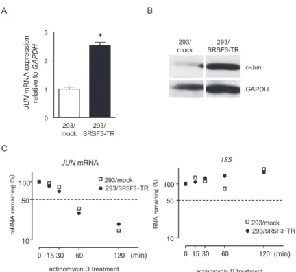
In a previous study, we showed that SRSF3 knock-down caused G1 cell cycle arrest through inhibi-tion of cyclin D/CDK4/6 activainhibi-tion and reducinhibi-tion of E2F1 expression (7). Based on the data, we com-pared mRNA and protein levels of cyclins (D1, D3, and E1), E2F1, and CDC25A between 293/SRSF3-TR and 293/mock cells. As shown in Figure 2, SRSF3-TR-expressing cells significantly up-regu-lated expression of CCND1, CCND3, CCNE1, E2F1, and CDC25A mRNAs (Figure 2A) and increased amounts of cyclin D1, cyclin D3, CDC25A, and E2F1 (Figure 2B). The first kinase complexes to be-come active in G1 phase are cyclin D/CDK4/6 that initiate phosphorylation of retinoblastoma (RB) proteins and release E2F1 from the RB-repressor complex, permitting transcription essential for the G1/S transition. Induction of cyclin D1 is important for the activation of cyclin D/CDK4/6. We, there-fore, focused on the mechanism how SRSF3-TR increased expression of cyclin D1. Several lines of evidence suggest that c-Jun is one of the crucial fac-tor for cyclin D1 expression (9, 10). In fact, 293/ SRSF3-TR cells increased JUN mRNA (Figure 3A) and c-Jun protein (Figure 3B) levels, compared with 293/mock cells. We also confirmed that the stable expression of SRSF3-TR did not change the stability of JUN mRNA, suggesting that SRSF3-TR might activate transcription of the JUN gene. Effects of SRSF3-TR on JUN promoter activity
To test whether SRSF3-TR regulated c-Jun ex-pression at the transcriptional level, we cloned the 5’-flank of JUN (from -1,266 to +170 bp), and 293/ SRSF-TR or 293/mock cells were transfected with a luciferase reporter construct containing -1,266/ +170, -798/+170, -331/+170, or -32/+170 bp re-gion of the human JUN promoter (Figure 5A). As shown in Figure 4A, 293/SRSF3-TR cells signifi-cantly increased the luciferase reporter activity of the -1,266/+170 and -798/+170 bp regions of the
JUN promoter, compared with 293/mock cells. The
-331/+170 bp construct partially reduced the pro-moter activity in 293/SRSF3-TR cells, and the -32/ +170 bp region almost completely lost the promoter
activity (Figure 4A). The -331/-32 bp region con-tains two AP-1-binding sites (-190 to -183 and -71 to -64) (Figure 4C), and the proximal AP-1-binding
Figure 2. Expression of G1 to S phase transition regulators in 293/SRSF3 - TR cells.
(A) Levels of CCND1, CCND3, CCNE1, CDC25A, and E2F1 mRNAs in 293/SRSF3 - TR cells were measured by qPCR us-ing GAPDH mRNA as an endogenous quantity control. Val-ues (means!SD, n = 3) are expressed as fold changes, com-pared with those of 293/mock cells. *P!0.05 by ANOVA and Bonferroni test. (B) Amounts of cyclins (D1, D3, and E1), CDC25A, and E2F1 proteins in these cells were measured by Western blotting using GAPDH as a loading control.
Figure 1. Cell proliferation of 293/SRSF3 - TR cells.
(A) Flp - In - 293 cells stably expressing FLAG tagged - SRSF3 - TR (293/SRSF3 - TR) or mock (293/mock) were established using the Flp In system as described in Materials and Methods. Whole -cell lysates were prepared from them and subjected to Western blotting using the SRSF3 antibody. GAPDH was served as a load-ing control. (B) Numbers of growload-ing cells in 35 mm cloture dishes were counted on day 4 after plating. Values represent means!SD, n = 3. *P!0.05 by ANOVA and Bonferroni test.
Figure 3. Up - regulation of c- Jun expression in 293/SRSF3 - TR cells.
(A) Levels of JUN mRNA in 293/SRSF3 - TR cells were measured by qPCR using GAPDH mRNA as an endogenous quantity control. Val-ues (means!SD, n = 4) are expressed as fold changes, compared with those of 293/mock cells. *P!0.05 by ANOVA and Bonferroni test. (B) Amounts of c- Jun proteins were measured by Western blotting using GAPDH as a loading control. (C) To compare the stability of JUN mRNA between 293/mock ( ) and 293/SRSF3 - TR (") cells, both cells were incubated with 2.5 μg/ml actinomycin D for the indicated times. Amounts of JUN mRNA remained were measured by qPCR. Amounts of 18S were also measured as a control.
Figure 4. JUN promoter activity in 293/SRSF3 - TR cells.
(A) Luciferase reporter plasmids (pGL3) driven by 1,266 -, 798 -, 331 -, 32 - bp JUN promoter fragments were transfected into 293/ SRSF3 - TR and 293/mock cells. After transfection for 24 h, these cells were harvested, and luciferase activity was measured using the Dual - Luciferase Reporter Assay System. Values are expressed as fold changes (means!SD, n = 6). *P!0.05 by ANOVA and Bonferroni test. (B) Amounts of Sp - 1 proteins were measured by Western blotting using GAPDH as a loading control. (C) Nucleo-tide sequence of the 5’-flanking region of the human JUN gene. Putative binding sites for Sp-1, CTF and AP-1 are indicated with underlines. Sp - 1 predicted binding sites between - 798 and - 332 bp based on the JASPER database (relative profile score threshold 85%) are shown by a square. Putative transcription initiation site is indicated by a bent arrow.
site is responsible for trans-activation of the JUN promoter by its gene product (11). Sp-1 is also known as a crucial transcription factor for JUN (12), and several Sp-1-binding sites are distributed in both -331/-32 bp and -798/-332 bp regions. More-over, 293/SRSF3-TR significantly induced Sp-1 (Figure 4B), suggesting that multiple cis elements distributed in the -798/-32 bp region might be in-volved in the SRSF3-TR-mediated JUN promoter activity.
DISCUSSION
Alternative splicing is important for generation of protein isoform diversity and for regulation of gene expression program. At the same time, how-ever, up to one-third of alternative splicing of hu-man genes generates PTC-containing mRNAs (13). All SRSF family member genes produce PTC - con-taining splice variants, which is coincided with the presence of UCEs (14). To avoid production of the C-terminally truncated proteins which may have potential dominant-negative or gain-of-function, PTC-containing mRNAs are expected to be de-graded by NMD. However, several lines of evidence have shown that NMD activity is often inhibited by stresses, such as hypoxia, amino acid starvation, and generation of reactive oxygen species (15-17). Moreover, it has been shown that the impairment of NMD is involved in the oncogenic mechanism (18). In fact, several cancer cell lines (HCT116, RKO, and HeLa cells) constitutively expressed sig-nificant amounts of the SRSF1 PTC variant. For example, HCT116 cells expressed the SRSF1 PTC variant nearly equivalent to the major isoform (data not shown). These PTC variants are believed not to be translatable, whereas SRSF3-TR was docu-mented to be translated from SRSF3-PTC mRNA (8). We also found that treatment of HCT116 cells with sodium arsenite induced SRSF3-TR possibly through inhibiting NMD (unpublished observa-tions). The inclusion of exon 4 to mature SRSF3 mRNA was reported to occur under serum starva-tion (8), and the biological significance of exon 4-inclusion is considered to be an auto-regulatory feedback loop by SRSF3 itself (19).
To uncover unknown functions of SRSF3-TR, we established a cell line stably expressing SRSF3-TR using the Flp-In system, and found that SRSF3-TR-expressing cells (293/SRSF3-TR) significantly ac-celerated their growth, compared with control cells
(293/mock). As to the mechanism for the acceler-ated cell growth, 293/SRSF3-TR significantly in-creased mRNA and protein levels of essential regu-lators for the G1 to S phase transition, which in-cluded cyclin D1, cyclin D3, CDC25A, and E2F1. c-Jun is required for progression through the G1 phase, the mechanism by which involves transcrip-tional control of the cyclin D1 gene (CCND1). We also confirmed that SRSF3-TR-expressing cells sig-nificantly increased expression of JUN mRNA and induced c-Jun protein without any growth stimulus. c-Jun positively regulates cell proliferation through the induction of CCND1 transcription and the re-pression of tumor suppressor gene exre-pression and function (20). The regulatory elements crucial for the human CCND1 transcription contain two AP-1 binding sites and several AP-1 proteins including c-Jun and c-FOS were shown to bind these sites and suggested to stimulate CCND1 transcription (21-23). Along with this line, 293/SRSF3-TR cells exhibited a significantly augmented promoter activ-ity of JUN . The proto-oncogene JUN is positively autoregulated by its product, c-Jun/AP-1 (11). It was reported that two AP-1-binding sites (-182 and -64) of the JUN promoter were important for basal activity and dexamethasone-mediated repression of the JUN promoter (24). The proximal -71 to -64 bp region is responsible for the autoregulation of the
JUN promoter activity (11). Our luciferase reporter
assay also indicated the importance of the -331 to -32 bp region for both basal and SRSF3-TR-depend-ent activities of the JUN promoter. At the same time, however, our results implicated that there might be SRSF3-TR responsive element(s) in the -798 to -332 bp region. In addition to c-Jun, Sp-1 plays an essential role for basal expression of JUN (12). The -331/-32 bp region of the JUN promoter contains one Sp-1-binding site, and several putative Sp-1-binding sites are scattered in the -798 to -332 bp region. Moreover, Sp-1 was induced in 293/ SRSF3-TR cells. Thus, multiple trans factors and cis elements appear to be involved in the SRSF3-TR-mediated transcription of JUN . Further studies are needed to elucidate the mechanism how SRSF3-TR regulates c-Jun expression.
Although the mechanism how SRSF3-TR regu-lates c-Jun expression remains to be elucidated, our results suggest that a truncated SRSF3 derived from a PTC-containing splice variant of the SRSF3 gene may have a mitogenic property. NMD was often in-hibited under certain conditions such as hypoxia and oxidative stress. Cancer tissues are exposed
to similar microenviromental conditions, and it has been shown that the impairment of NMD is in-volved in the oncogenic mechanism (18). In such situations, SRSF3-TR could be induced and might participate in abnormal cell growth. Further studies are needed to fully elucidate the molecular mecha-nism for the SRSF3-TR-mediated acceleration of cell growth.
CONFLICTS OF INTEREST
The authors declare no conflict of interest.
ACKNOWLEDGEMENTS
This work was funded by a Grants-in-Aid for Scientific Research from the Ministry of Education, Culture, Sports, Science and Technology, Japan (#12016603 to K.N.).
REFERENCES
1. Manley JL, Krainer AR : A rational nomencla-ture for serine/arginine-rich protein splicing factors (SR proteins). Genes Dev 24 : 1073-1074, 2010
2. Chen M, Manley JL : Mechanisms of alternative splicing regulation : insights from molecular and genomics approaches : Nat Rev Mol Cell Biol 10 : 741-754, 2009
3. Lin S, Coutinho-Mansfield G, Wang D, Pandit S, Fu X-D : The splicing factor SC35 has an ac-tive role in transcriptional elongation. Nat Struct Mol Biol 15 : 819-826, 2008
4. Sapra AK, Anko ML, Grishina I, Lorenz M, Pabis M, Poser I, Rollins J, Weiland EM, Neugebauer KM : SR protein family members display diverse activities in the formation of nas-cent and mature mRNPs in vivo. Mol Cell 34 : 179-190, 2009
5. Sanford JR, Gray NK, Beckmann K, Caceres JF : A novel role for shuttling SR proteins in mRNA translation. Genes Dev 18 : 755-768, 2004
6. He X, Arslan AD, Pool MD, Ho TT, Darcy KM, Coon JS, Beck WT : Knockdown of splicing fac-tor SRp20 causes apoptosis in ovarian cancer cells and its expression is associated with ma-lignancy of epithelial ovarian cancer. Oncogene
30 : 356-365, 2011
7. Kurokawa K, Akaike Y, Masuda K, Kuwano Y, Nishida K, Yamagishi N, Kajita K, Tanahashi T, Rokutan K : Downregulation of serine/arginine-rich splicing factor 3 induces G1 cell cycle ar-rest and apoptosis in colon cancer cells. On-cogene 2013 (in press)
8. Jumaa H, Guenet JL, Nielsen PJ : Regulated ex-pression and RNA processing of transcripts from the Srp20 splicing factor gene during the cell cycle. Mol Cell Biol 17 : 3116-3124, 1997 9. Schreiber M, Kolbus A, Piu F, Szabowski A,
Mohle-Steinlein U, Tian J, Karin M, Angel P, Wagner EF : Control of cell cycle progression by c-Jun is p53 dependent. Genes Dev 13 : 607-619, 1999
10. Wisdom R, Johnson RS, Moore C : c-Jun regu-lates cell cycle progression and apoptosis by distinct mechanisms. EMBO J 18 : 188-197, 1999
11. Angel P, Hattori K, Smeal T, Karin M : The jun proto-oncogene is positively autoregulated by its product, Jun/AP-1. Cell 55 : 875-885, 1988 12. Han TH, Lamph WW, Prywes R : Mapping of
epidermal growth factor-, serum-, and phorbol ester-responsive sequence elements in the c-jun promoter. Mol Cell Biol 12 : 4472-4477, 1992
13. Lewis BP, Green RE, Brenner SE : Evidence for the widespread coupling of alternative splic-ing and nonsense-mediated mRNA decay in humans. Proc Natl Acad Sci U S A 100 : 189-192, 2003
14. Lareau LF, Inada M, Green RE, Wengrod JC, Brenner SE : Unproductive splicing of SR genes associated with highly conserved and ultra-conserved DNA elements. Nature 446 : 926-929, 2007
15. Gardner LB : Hypoxic inhibition of nonsense-mediated RNA decay regulates gene expression and the integrated stress response : Mol Cell Biol 28 : 3729-3741, 2008
16. Mendell JT, Sharifi NA, Meyers JL, Martinez-Murillo F, Dietz HC : Nonsense surveillance regulates expression of diverse classes of mam-malian transcripts and mutes genomic noise. Nat Genet 36 : 1073-1078, 2004
17. Wang D, Zavadil J, Martin L, Parisi F, Friedman E, Levy D, Harding H, Ron D, Gardner LB : Inhibition of nonsense-mediated RNA decay by the tumor microenvironment promotes tu-morigenesis. Mol Cell Biol 31 : 3670-3680, 2011
18. Gardner LB : Nonsense-mediated RNA decay regulation by cellular stress : implications for tumorigenesis. Mol Cancer Res 8 : 295-308, 2010
19. Jumaa H, Nielsen PJ : Regulation of SRp20 exon 4 splicing. Biochim Biophys Acta 1494 : 137-143, 2000
20. Shaulian E, Karin M : AP-1 in cell proliferation and survival. Oncogene 20 : 2390-2400, 2001 21. Bakiri L, Lallemand D, Bossy-Wetzel E, Yaniv
M : Cell cycle-dependent variations in c-Jun and JunB phosphorylation : a role in the control of cyclin D1 expression. EMBO J 19 : 2056-2068, 2000
22. Beier F, Lee RJ, Taylor AC, Pestell RG, LuValle
P : Identification of the cyclin D1 gene as a tar-get of activating transcription factor 2 in chon-drocytes. Proc Natl Acad Sci U S A 96 : 1433-1438, 1999
23. Brown JR, Nigh E, Lee RJ, Ye H, Thompson MA, Saudou F, Pestell RG, Greenberg ME : Fos family members induce cell cycle entry by activating cyclin D1. Mol Cell Biol 18 : 5609-5619, 1998
24. Wei P, Inamdar N, Vedeckis WV : Transrepres-sion of c-jun gene expresTransrepres-sion by the glucocor-ticoid receptor requires both AP-1 sites in the c-jun promoter. Mol Endocrinol 12 : 1322-1333, 1998