Investigation of lymphatic network and cancer
metastasis
著者
Shao Lenan
学位授与機関
Tohoku University
学位授与番号
11301乙第9297号
URL
http://hdl.handle.net/10097/00096759
Investigation of lymphatic network and cancer
metastasis
Lenan Shao
Doctoral Program in Biomedical Engineering
Submitted to the Graduate School of Biomedical
Engineering
in Partial Fulfillment of the Requirements
for the Degree of Philosophy in Biomedical Engineering
at
INVESTIGATION OF LYMPHATIC NETWORK AND
CANCER METASTASIS
This dissertation is dedicated to Prof. Tetsuya Kodama and Dr. Shiro Mori, who taught me the significance of pre-clinical research and the ways to approach the study of cancer lymphatic metastasis. I am grateful to both of them for providing me with the opportunity to learn from the experience of pre-clinical research and grow from being a theatre surgeon to an academic surgeon.
i
TABLE OF CONTENTS
Acknowledgements ... v
List of abbreviations ... vii
Abstract ... ix
CHAPTER 1
Introduction ... 1
1.1. Cancer metastasis ... 2
1.1.1. Lymphatic metastasis of cancer ... 4
1.1.2. Hematogenous metastasis of cancer ... 7
1.1.3. Iatrogenic metastasis of cancer ... 11
1.1.4. Mechanisms of cancer metastasis ... 12
1.2. Lymphatic network ... 14
1.2.1. What is the lymphatic system? ... 14
1.2.2. Research methods used to investigate the lymphatic system 15
1.2.3. Embryology of the lymphatic system ... 16
1.2.4. Composition of the lymphatic system ... 17
1.2.5. Primary functions of the lymphatic system ... 20
1.3. The lymphatic network and cancer metastasis ... 21
1.3.1. The sentinel lymph node and cancer metastasis ... 23
1.3.2. Biopsy of the sentinel lymph node ... 24
1.4. Biopsy of tumor-positive sentinel lymph nodes: does it promote
cancer metastasis ... 26
1.4.1. Hypothesis ... 27
1.4.2. Rationale of the hypothesis ... 27
1.4.3. Hypothesis evaluation and consequences ... 28
1.4.4. Concluding remarks ... 29
1.5. Thesis objectives ... 29
CHAPTER 2
Lymphatic mapping of mice with systemic lympho-proliferative
disorder: usefulness as an inter-lymph node metastasis model of cancer
... 33
ii
2.1. Summary ... 34
2.2. Introduction ... 34
2.3. Materials and Methods ... 36
2.3.1. Mice ... 36
2.3.2. Characteristics of MXH10/Mo/lpr mice ... 37
2.3.3. LN mapping ... 37
2.3.4. Emulsion dye casting of peripheral lymph drainage routes ... 38
2.3.5. Fluorescence imaging of peripheral lymph drainage routes .. 38
2.3.6. Cell culture ... 39
2.3.7. Induction of metastasis in the PALN ... 39
2.3.8. Histological analysis ... 40
2.3.9. Statistical analysis ... 41
2.4. Results ... 41
2.4.1. Weight of LNs ... 41
2.4.2. Pathological scores for glomerulonephritis and renal
vasculitis ... 41
2.4.3. Harvesting and mapping of LNs ... 42
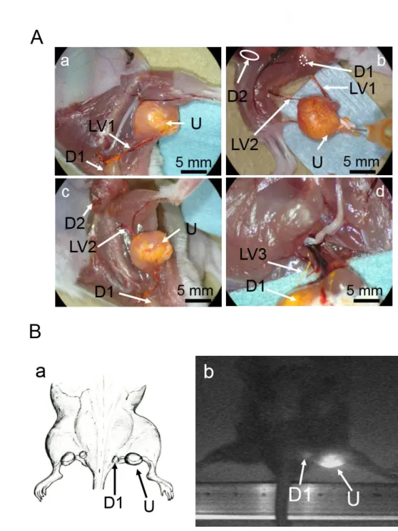
2.4.4. Peripheral lymph drainage routes ... 43
2.4.4.1. LV from the SiLN to the PALN ... 43
2.4.4.2. LV from the superficial salivary LN to the caudal deep
cervical LN... 45
2.4.4.3 LVs from the popliteal LN to both the sciatic LN and the
SiLN ... 45
2.4.5. Induction of metastasis from the SiLN to the PALN ... 50
2.5. Discussion ... 53
CHAPTER 3
Communication between lymphatic and venous systems in mice ... 57
3.1. Summery ... 58
3.2. Introduction ... 59
3.3. Materials and Methods ... 60
3.3.1. Mice ... 60
3.3.2. Identification of the lymphatic and venous systems ... 61
3.3.3. Production of NDB-liposomes ... 61
3.3.4. Visualization of flow in veins ... 61
3.4. Results ... 62
3.4.1. Communication between lymphatic and venous systems in the
axillary and subiliac regions ... 62
iii
3.4.2. Direction of blood flow in the TV ... 67
3.5. Discussion ... 69
CHAPTER 4
Activation of latent metastases in the lung after resection of a
metastatic lymph node in a mouse model of lymph node metastasis ... 73
4.1. Summary ... 74
4.2. Introduction ... 74
4.3. Materials and Methods ... 75
4.3.1. Mice ... 75
4.3.2. Cell culture ... 76
4.3.3. Visualization of flow from the SiLN into the efferent LVs and
thoracoepigastric vein ... 76
4.3.4. Induction of metastasis to the PALN by injection of tumor
cells into the SiLN ... 77
4.3.5. Resection of the SiLN ... 77
4.3.6. Assessment of tumor and metastasis development through
measurement of luciferase activity ... 78
4.3.7. Histological analysis ... 80
4.3.8. Statistical analysis ... 81
4.4. Results ... 82
4.4.1. Ex vivo detection of metastases in the lungs using IVIS ... 82
4.4.2. Histological analysis ... 90
4.5. Discussion ... 92
CHAPTER 5
Conclusions ... 95
Bibliography ... 99
Appendix ... 125
Achievements ... 126
v
Acknowledgements
Completing my PhD degree has probably been one of the greatest challenges of the half-century of my life. Many people have shared the best and worst moments of my doctoral journey. Over the last 5 years, it has been a great privilege to spend several months in the Kodama laboratory at Tohoku University, and its staff members will always remain dear to me. My first debt of gratitude goes first and foremost to my supervisor Professor Tetsuya Kodama, Graduate School of Biomedical Engineering, Tohoku University, for his worldwide vision of cancer science and his constant encouragement and guidance, which was necessary for me to complete my dissertation. Furthermore, he has walked me through all the stages of the experiments, the writing of scientific papers and the assembly of this thesis. Without his consistent and illuminating instruction, this dissertation would not have been possible and would not have reached its present completed form.
I would also like to thank Dr. Shiro Mori, D.D.S., Maxillofacial Surgery Department of Tohoku University Hospital, for his unflagging encouragement and for serving as an academic role model to me as both a cancer metastasis researcher and senior oral and maxillofacial surgeon. He has been a strong and supportive adviser throughout my stay in the Kodama laboratory; his research enthusiasm, continued hard work, commitment and focus always gave me an unending ‘supply of motivation’.
Special thanks are due to my committee members, Professor Yasufumi Sato, Professor Kei Takase, Professor Tetsuaki Kawase and Associate Professor Masakazu Kawashita, for their support, guidance and helpful suggestions. Their guidance has served me well and I owe them my heartfelt appreciation. Special thanks are also expressed to Professor Masato Nose for his sagacity and pertinent pathological discussions in preparing the published papers. Members of the Kodama laboratory also deserve my sincerest thanks; their friendship and assistance has meant more to me than I can ever express in words. I could not have completed my research without their participation and invaluable friendly assistance during this JSPS project.
My friends in China, Japan, the United States and other parts of the world were sources of laughter, joy and support in my voyage of discovery. Special thanks go to Dr.
vi
Rui Chen, Dr. Li Li, Dr. Hitoshi Miyashita, Dr. Yunchao Chen, Dr. Zhenghong Zhu and Dr. Peng Cheng. I am very happy that, in many cases, my friendships with you have extended well beyond our shared time both in China and overseas.
I wish to thank my parents. Their love, spirit and family genes have provided me with both inspiration and motivation. I owe them everything and wish I could show them how much I love and appreciate them. My thanks also to my sisters, brothers, the nursing staff of my parents and their families, and especially to Fei Yao (my wife), whose love, support and encouragement allowed me to finish this long journey. She already has my heart so I will just give her a heartfelt “thanks”. My son Yiwei Shao knows his father well, so I just exchange with him a high five, give him a tight bear hug and tell him his dad is almost ready to finish blackening the other eye of the Daruma! I also want to thank my mother-in-law and father-in-law for their solicitude and unconditional support.
Finally, I would like to dedicate this research to Dr. Weimin Chen and all my dear colleagues in the Oral and Maxillofacial Department of Tongji Hospital; I hope that this research and my endeavors will make you proud.
I am also grateful to the organization that has provided funding to support this Dissertation PhD Program: Japan Society for the Promotion of Science (2011-S-1109) and Chinese Scholarship Council.
Finally, I am sorry and thankful to the mice that gave their lives for this research. I appreciate the support, encouragement and hope from people all over the world. Thanks, my dear friends!
Just as I started this Doctoral Dissertation Program at Tohoku University in 2011, Sendai was hit by an earthquake and tsunami on March 11th. In total, about 20,000
vii
List of abbreviations
AALN accessory axillary lymph node ALND axillary lymph node dissection BM bone marrow
C3H/lpr C3H/HeJ-lpr/lpr
CCR CC chemokine receptor CD31 cluster of differentiation 31 CDUS color Doppler ultrasound
CXCL12 chemokine (C-X-C motif) ligand 12 CXCR4 chemokine (C-X-C motif) receptor 4 CT computed tomography
CTC circulated tumor cell DTC disseminated tumor cell ECM extracellular matrix ECS extracapsular spread EM elastic-Masson FBS fetal bovine serum
FITC fluorescein isothiocyanate
FM3A C3H/He mouse mammary carcinoma cell line FNA fine needle aspiration
FNR false negative rate H&E hematoxylin and eosin HEV high endothelial venules ICG indocyanine green
IHC immunohistochemical analysis IMC invasion-metastasis cascade
IVIS in vivo bioluminescence imaging system LSMC lymphatic smooth muscle cell
LN lymph node
LV lymphatic vessel
viii
MHC major histocompatibility complex MRI magnetic resonance imaging MRL/lpr MRL/MpJ-lpr/lpr
MW molecular weight MXH10/Mo/lpr MXH10/Mo-lpr/lpr NBD nitrobenzoxadiazole
PALN proper axillary lymph node PBS phosphate-buffered saline PDE Photodynamic Eye
PET positron emission tomography SCC squamous cell carcinoma SD standard deviation
SEM standard error of the mean SiLN subiliac lymph node SLN sentinel lymph node
SLNB sentinel lymph node biopsy
TV thoracoepigastric vein
ix
Abstract
The objectives of this thesis were to clarify the distributions of lymph nodes (LNs) and lymph vessels (LVs), the communication between lymphatics and the surrounding veins, and the relationship between cancer metastasis and the lymphatic system in MXH10/Mo/lpr, a lympho-proliferative in-bred mouse, to extrapolate the findings to develop potential new clinical treatments.
In Chapter 1, overviews of cancer metastasis, the lymphatic system and the relationship between them are outlined. An extensive literature review strongly indicated that the lymphatic system facilitates cancer cell dissemination throughout the body, acting in concert with the venous system. It is clear that preclinical models of LN metastasis are indispensable to aid the understanding of the mechanisms involved in tumor cell trafficking, which are fundamental for developing new techniques to diagnose and treat LN metastasis.
However, the identification of LNs and LVs in mice is challenging with conventional imaging modalities, since the LN diameter in normal mice is 1–2 mm. In this thesis, a preclinical model of LN metastasis has been used involving MXH10/Mo-lpr/lpr (MXH10/Mo/lpr) inbred mice, which develop systemic swelling of LNs up to 10 mm in diameter.
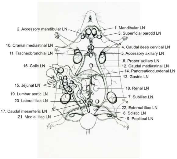
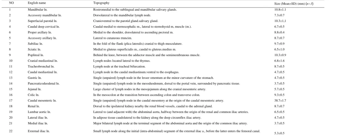
In Chapter 2, the aim was to identify LNs and LVs in MXH10/Mo/lpr mice and establish one of the peripheral inter-LN routes that can induce regional lymphatic cancer metastasis. Twenty-two different LNs were found distributed in the peripheral, thoracic and abdominal regions, and 4 peripheral inter-LN vessels were identified, from the subiliac LN (SiLN) to the proper axillary LN (PALN), the parotid LN to the caudal deep cervical LN, and the popliteal LN to both the sciatic LN and the SiLN. Moreover, peripheral regional metastasis was induced by inoculating FM3A/Luc mouse breast cancer cells into the SiLN. This chapter unveils the anatomy of murine lymphatics to give new insights into the investigation of inter-LN metastasis of cancer, especially the mechanisms involved in the trafficking of cancer cells in inter-LN vessels. The lymphatic anatomy data will be useful in the quest for deepening our understanding of cancer cell trafficking and the mechanisms involved in cancer metastasis. The results
x
also show, for the first time, that the MXH10/Mo/lpr mouse strain is an 'investigator-friendly' and reliable model of peripheral inter-LN cancer metastasis.
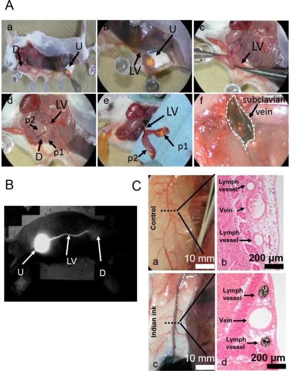
In Chapter 3, the communication pathways between the lymphatic and venous systems are clarified, with a focus on the anatomy of these communication routes in the axillary and subiliac regions. The communication pathways between the lymphatic and venous systems in the axillary and subiliac regions of mice were unequivocally identified. The efferent LVs of the PALN were demonstrated to communicate with the subclavian vein. Furthermore, it was shown that the thoracoepigastric vein (TV), which connects the subclavian vein and inferior vena cava, runs adjacent to the SiLN and PALN, and receives venous blood from these LNs routed through small branches. The direction of blood flow in the TV occurred in two directions in the intermediate region between the PALN and SiLN; one to the subclavian vein, the other to the inferior vena cava. This research reveals the anatomy of the communication between the lymphatic and venous systems in the axillary and subiliac regions of the mouse, and provides further lymphatic-venous anatomy data relevant to the investigation of the trafficking routes of cancer cells in preclinical mouse models. The bi-directional flow of blood in the TV between the PALN and SiLN gives insight into tumor cell trafficking from regional areas to the whole body. It is proposed that the final form of LN metastasis should be recognized as “LN-mediated hematogenous metastasis” based on lympho-venous communication.
In Chapter 4, the surgical and non-surgical outcomes of an implanted LN tumor were explored, with a focus on regional cancer and distant metastasis. The tumor-bearing SiLN was resected to simulate clinical dissection of the sentinel lymph node (SLN). It was found that resection of a tumor-bearing SiLN enhanced lung metastasis in the mouse model. Bioluminescence imaging revealed that metastatic tumor cells in the down-stream LN continued to grow after the resection of the up-stream tumor-bearing SiLN, and that the probability of metastasis to the lungs was increased when the interval between SiLN inoculation and resection was reduced. Furthermore, histological analysis demonstrated that latent cancer cells in the lungs were stimulated to grow after resection of the SiLN. Fluorescence imaging indicated that the route of tumor cell dissemination from the SiLN to the lungs was via the venous system enveloping the SiLN. This part of the thesis confirmed two trafficking routes in the SiLN region: one towards the PALN via the LV, which is related to PALN
xi
lymphogenous metastasis, and the other towards the venous system via the TV, which is related to hematogenous metastasis to distant sites. The resection of a SiLN inoculated with tumor cells led to an accelerated growth of metastatic tumor cells in the lungs and ipsilateral PALN. This result is a timely reminder of the clinical risk of iatrogenic induction of regional and distant cancer metastases. This phenomenon provides new insights into the concept of “LN-mediated hematogenous metastasis” and is the starting point for tracing the activation process of distant dormant cancer cells.
In Chapter 5, it is concluded that the lymphatics can facilitate cancer cell dissemination, and that this is fundamental to the occurrence of cancer metastasis. Presently, two contrasting models are used to explain the formation of distant organ metastases: a lymphatic-independent hematogenous model and a lymphatic-dependent sequential model. The most crucial issue to resolve is whether metastatic cells come to a halt in LNs or continue to disseminate throughout the body by usurping either LN vascular vessels or efferent LVs to colonize distant organs via the blood circulation. To date, the detailed mechanisms involved in this important process remain a mystery. However, there is now little doubt that the interplay between tumors and the lymphatic system represents the main route used by solid cancers to spread.
Based on a study of MXH10/Mo/lpr mice, this dissertation shows the topography of 22 LNs and 4 peripheral lymphatic drainage routes, which can be used to explore peripheral lymphatic cancer metastasis in a preclinical setting. In addition, the thesis has clarified the communications between the lymphatic and venous systems in the axillary and subiliac regions, and revealed that the LV running from the SiLN to the PALN is capable of draining tumor cells. Moreover, the research in this thesis has also shown that lung metastatic foci could be rapidly activated following resection of a tumor-bearing SiLN. It is anticipated that this mouse model will be useful for studying LV imaging, lymphatic trafficking kinetics and the mechanisms of tumor-lymphatic system interplay during both regional and distant metastasis, with the aim of translating this basic research into enhanced clinical diagnosis and novel drug delivery systems.
1
CHAPTER 1
2
1.1. Cancer metastasis
Little was known about cancer until 1829 when the French surgeon, J.C.A. Récamier (1774-1852), gave the first clear reference to metastasis (“métastase”) in his book on cancer [1, 2]. At present, the accepted concept is that cancer begins when healthy cells change and grow uncontrollably, forming a mass called a tumor, which can be malignant or benign. A cancerous tumor is malignant when it can grow and spread, that is metastasize to other parts of the body. Metastatic neoplasia can be found in the lymph nodes (LNs) draining the region affected by the tumor. Metastatic lesions in LNs or distant organs can also arise from neoplastic blood-born emboli from tumors that are not located near to the nodes [3]. Metastasis is one of the hallmarks of cancer; however, the mechanisms underlying invasion and metastasis remain incompletely understood [4, 5]. Concepts of the mechanisms underlying the patterns of metastasis to different sites are considered in two hypotheses, namely: 1) the ‘mechanical’ hypothesis, or the physical dissemination of cancer cells from the primary tumor to distant sites, and 2) the ‘seed and soil’ hypothesis, which concerns the adaptation of these cells to foreign tissue that results in successful colonization [6]. The first is based on the anatomy of the routes of tumor cell dissemination, while the second focuses on patterns due to differential arrest and proliferation of cancer cells. For most cancers, the onset of metastasis involves local invasion of the surrounding stroma by individual cells or groups of cells that were originally formed in the primary tumor. After penetrating the lymphatic or vascular channels, the invading tumor cells can grow at the penetration site or break off either as single cells or as small embolic aggregates, to be transported via the circulatory system. Circulating tumor cells are more susceptible to host immune and non-immune defenses, including turbulent blood flow, monocytes, and lymphocytes called natural killer cells. Those tumor cells that survive these defenses usually lodge in the capillary beds of distant organs by adhering to capillary endothelial cells or attaching to basement membrane that is exposed at regions of endothelial cell retraction. Following this process, extravasation can occur, likely through many of the same mechanisms underlying the initial invasion. Growth of the metastasis in the organ parenchyma necessitates the development of a vascular network, evasion of the host immune system and proliferation. After reaching a certain size, the metastasis can give rise to many additional metastases. In this way, a small primary tumor can produce many metastases in a very short period of time.
3
However, only a small number of cells in the primary tumor can undertake all these steps and develop into metastases. The inefficiency of metastasis is accounted for, at least in part, by the challenge of completing the complex steps involved.
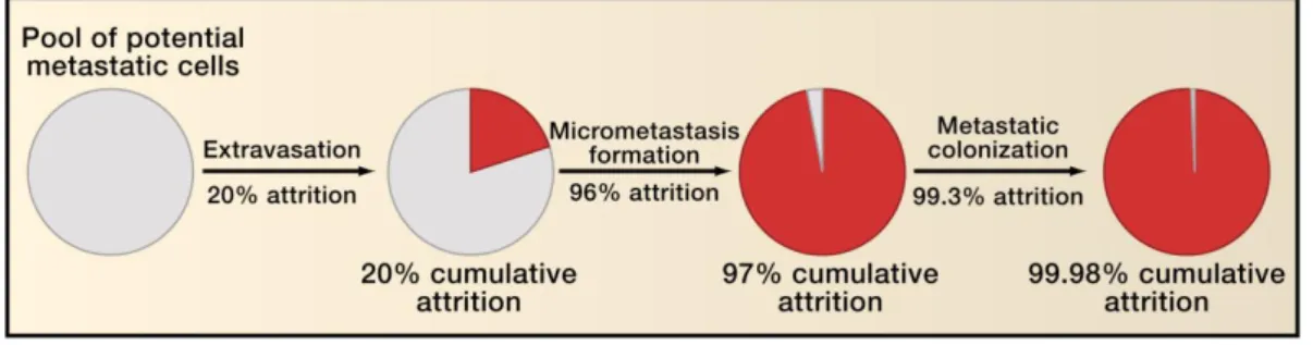
Some of the steps in the invasion-metastasis cascade (IMC) are extremely inefficient. Research using experimental animal models has demonstrated that the rate-limiting step of the IMC is the mechanisms underlying metastatic colonization. Indeed, the attrition rate is usually > 99% of the cells that survive to form micrometastases (Figure 1.1).
Figure 1.1. Inefficiency of the invasion-metastasis cascade. The figure indicates the
approximate proportions of intravenously implanted tumor cells that die as they pass through the various steps of the invasion-metastasis cascade. Figure 1.1 is used with kind permission [7].
The majority of tumor cells that enter the circulation are quickly destroyed (Figure 1.1); hence, the presence of circulating tumor cells does not necessarily predict that metastases will develop. The process of cancer metastasis is still difficult to predict as it contains stochastic elements and has sequential selective and differential proliferation properties. The most fearsome aspect of cancer metastasis is that conventional therapy cannot stop it, which ultimately leads to the death of the patient. Metastatic tumor spread through the blood or lymphatic vessels (LVs) occurs in most forms of cancer, and metastasis to regional LNs is often the single most important factor that predicts the fate of patients with malignant cancer [8]. Although primary tumors can be surgically resected or locally irradiated, it is problematic to apply these treatments to disseminated cancer. In many patients, metastasis may well have occurred before the primary malignant tumor (with the exception of skin cancers) has been diagnosed [9]. Furthermore, at the time of clinical diagnosis of major lesions, malignant tumors and their metastases consist of multiple cell populations that exhibit extensive biological heterogeneity. Such parameters include antigenicity, immunogenicity and rate of growth, karyotype, the cell surface
4
characteristics, susceptibility to cytotoxic drugs and, not least, the ability to metastasize and invade remote organs. This biological heterogeneity makes the development of efficacious chemotherapy extremely challenging [6].
1.1.1. Lymphatic metastasis of cancer
The main reason why people die of cancer is that the cancer cells spread to form daughter tumors, or metastases, in organs vital to life including the lungs, liver and gastrointestinal tract. A route frequently used by cancer cells for dissemination is the lymphatic system. One of the ‘mechanical’ or ‘hemodynamic’ mechanisms of metastasis is based on well-substantiated observations on the anatomical basis of lymphogenous metastatic patterns [10]. The common human cancers spread first by lymphatic metastasis; metastasis to the lymphatic system is the major factor predicting survival in patients with epithelial malignancies. Metastasis along lymphatic pathways occurs in stages, which may usefully be described as: pre-metastatic invasion; approach; penetration; translocation; intra-nodal settling; growth and destruction of the LN; and then subsequent metastasis to further nodes [1, 11]. Unusual patterns of lymphogenous metastasis have been accounted for by perturbations in lymphatic flow, which was suggested by Virchow (1845) and emphasized by von Recklinghausen (1885) as the concept of “retrograde lymphatic embolism” [12, 13].
In the majority of human cancers, the lymphatic vasculature acts as the main conduit for metastasis of tumor cells to local LNs [14-17]. Moreover, the status of the LNs is an important criterion used in the staging, prognosis and treatment of a variety of solid tumor types. However, although it is recognized that tumor-associated LVs play a key role in cancer progression, there is a paucity of information regarding the molecular and cellular mechanisms directing metastasis to the LVs. Tumor cells gain access to the lymphatic vasculature by invading pre-existing LVs at the margin of the tumor or by stimulating lymphangiogenesis [18-20]. It has been reported that malignant tumor cells may make an active contribution to the induction of peri-tumoral and intra-tumoral lymphangiogenesis in some types of cancer [20, 21]. The lymphatic system provides an important conduit for the maintenance of tissue fluid homeostasis and the transportation of macromolecules and immune cells into the circulating blood, and has been implicated in cancer metastasis [22, 23]. Several studies have shown that the expression of vascular
5
endothelial growth factor (VEGF)-C/D stimulates tumor-associated lymphangiogenesis and tumor spread to the LNs [24-30]. Research has indicated that lymphangiogenesis plays a role in both LN metastasis and distant metastasis, and also influences disease-free survival (DFS) [31]. The factors implicated as being relevant to the metastasis of four common types of cancer (breast cancer, colorectal cancer, lung cancer and melanoma) include the expression of growth factors and/or receptors that promote lymphangiogenesis, the tumor lymphatics and lymphatic invasion. In melanoma, the intratumoral lymphatics have been linked to distant metastasis [32], with a poor DFS [33]. In colorectal cancer, high LV density is a predictor of disease recurrence and is associated with LN and distant metastasis [34-36]. In lung cancer, lymphatic invasion is related to an increased risk of regional LN involvement in non-small-cell lung cancer (NSCLC), and is a predictor of distant metastasis, long-term survival of the patient and DFS [37-39]. In breast cancer, the expression of VEGF-C is connected with LN metastasis [40], distant metastasis and a shorter overall survival (OS) time [41, 42]; similar correlations have not been observed in other studies [43, 44]. However, the mechanism involved in the trafficking of tumor cells from LNs to distant organs still remains enigmatic (Figure 1.2).
There is increasing evidence that has highlighted a role for the CC chemokine receptor-7 (CCR7) in cancer metastasis [45-47]. Although peritumoral LVs are believed to be functional, the significance of LVs present within a tumor remains contentious. Several studies have suggested that LVs enable the process of metastasis by providing a conduit for the dissemination of tumor cells [48]. Nevertheless, the mechanisms by which tumor cells cross into the lymphatic system remain unclear, and it is not known whether this occurs via intra-tumor lymphatics or functional lymphatics around the tumor [49]. There is evidence in a melanoma model that cancer cells can induce lymphangiogenesis or some form of lymphatic sprouting in the region of their periphery [50].
6
Figure 1.2. Remodeling of lymphatic vessels in cancer and its contribution to metastasis.
The lymphatic vessels present in adults arise during embryonic development. In cancer, these pre-existing lymphatic vessels undergo various types of remodeling, including lymphangiogenesis (which generates new lymphatics) and lymphatic enlargement (which does not produce new vessels, and can involve proliferative or non-proliferative mechanisms). Tumor lymphangiogenesis and/or lymphatic enlargement (in and around the primary tumor) are stimulated by growth factors that are derived from tumor cells or immune cells in the microenvironment of the tumor. These growth factors also promote lymphangiogenesis of lymph nodes, enlargement of collecting lymphatics that drain the tumor (both proximal and distal to sentinel lymph nodes), and changes in the lymphatic smooth muscle cells (LSMCs) around the collecting lymphatics. These processes are believed to promote the occurrence of metastasis. The mechanism by which tumor cells spread from lymph nodes to distant organs is currently speculative. Figure 1.2 is used with kind permission [31].
In comparison to blood vessels, the LV pathway provides numerous advantages for the invasion and transportation of pre-metastatic cells or embolism, just as it does for white blood cells, such as: 1) a discontinuous basement membrane and loose inter-cellular junctions; 2) an orders of magnitude slower flow rate that promotes cell survival by reducing shear stress; and 3) a 1,000 fold higher lymph concentration of hyaluronic acid, which confers cell protection and pro-survival effects [51]. The lymphatic system has several features suited to the transport of cells throughout the body while maintaining their survival and functions, including long residence times, areas in which flow stagnates, and access to the circulating blood through high
7
endothelial venules. Epithelial tumors (including melanoma, breast cancer, prostate cancer and head and neck cancers) exploit the ability of the lymphatic system to transport cells, and thus spread preferentially via the LVs rather than via a hematogenous route (for which the success rate for metastasis is extremely low, due to the toxicity of the serum, high shear stresses and mechanical trauma). Metastasis may be initially confined to the lymphatic system; however, the presence of discrete extra-nodal metastases indicates embolic spread through the bloodstream [45, 52, 53].
1.1.2. Hematogenous metastasis of cancer
The blood vessels are an important route for cancer metastasis and it is well known that tumor cells can be detected in the blood of patients with cancer in the early stages of tumorigenesis. There are two paradigms of this dissemination: the first is a parallel progression model, which states that early during the formation and growth of a primary tumor (e.g., cancer of the breast), cells are shed by the tumor and then circulate in the bloodstream; the second is a conventional linear progression model, in which the spread of tumor cells that causes the formation of a metastasis is a delayed process in tumor development [52-56].
Newinsights have emerged regarding the pathways that cancer cells take once they have left the primary tumor. Several concepts have been developed concerning the early seeding of metastases in the parallel progression model, the self-seeding of a primary tumor by circulating tumor cells, and the formation in distant organs of premetastatic niches.
One method by which cancer cells can reach distant regions is by transportation in the blood, followed by exiting the vasculature by the process of extravasation [57]. Recent work indicates that some circulating cancer cells that survive might return to the primary tumor rather than extravasate [58]. An alternative view is that tumor cells can enter the lymphatics and be conveyed to regional LNs, where (in principle) they can either adhere and form metastases, reach the bloodstream through high endothelial venules, or pass through the lymphatic system to the bloodstream via anastomoses such as the thoracic duct [59, 60] (Figure 1.3).
Metastatic spread may therefore follow two models that complement each other but place emphasis on different routes of dissemination. In one model (Figure 1.2), cancer
8
cells spread during the early stages of tumor growth from the primary tumor to the LNs or the blood stream. The disseminated tumor cells (DTCs) proliferate and generate solid metastases in LNs, while tumor cells that spread via the blood to distant sites either become dormant or are destroyed. In the later stages, tumor cells spread from the metastases established in the LNs, and can form solid metastases in remote organs. The ability to do this may be acquired when cells are selected in the LN milieu. As a consequence, the metastasis of a tumor to other organs depends on the existence of LN metastases. In the other model (Figure 1.4), cells spread frequently via the blood from the primary tumor to distant regions, where they evolve into explicit metastases without having traversed the LNs [61].
9
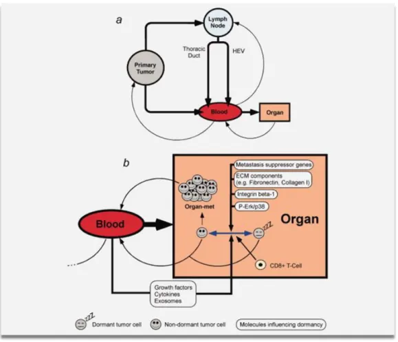
Figure 1.3. Routes for the development of metastasis. (a) Schematic diagram illustrating
the possible pathways that tumor cells could follow as they travel through the body. Cancer cells disseminating from the primary tumor can directly enter the bloodstream. If the cancer cells enter the lymphatic system, they might embed in lymph nodes and develop as lymph node metastases. Alternatively, the tumor cells might enter the bloodstream, for example via the thoracic duct or high endothelial venules (HEV). Disseminated tumor cells that reach the bloodstream and survive might either self-seed the primary tumor, re-enter the lymphatic system or extravasate into organs. Cancer cells spread to the blood by metastases might also contribute to self-seeding. (b) Diagram showing the possible fates of disseminated cancer cells. After the cancer cells have extravasated and invaded vital organs, there are 3 possible outcomes: death, dormancy, or growth as metastases. Several factors, such as those illustrated in Figure 1.3, have been suggested to regulate dormancy. ECM, extracellular matrix. For further details, see main text. The color figure is available online at wileyonlinelibrary.com, and is used here with permission [60].
10
Figure 1.4. Models of metastasis. Tumor cells can spread from the primary site via
lymphatic (red arrows) and hematogenous (blue arrows) routes. Secondary hematogenous dissemination from overt metastases to other distant sites may also occur (black arrows). In the first model, tumor cells disseminate and proliferate in the lymph nodes to form solid metastases, whereas tumor cells at distant sites either die or enter dormancy. At later stages, tumor cells spread from metastases established in the lymph nodes to form secondary metastases at distant sites. This ability may be gained as the cells develop in the environment of the lymph node. As a consequence, the metastasis of the tumor depends on the existence of lymph node metastases. Hematogenous dissemination can occur from the primary tumor, from lymph node metastases or from distant metastases. In the second model, tumor cells disseminate predominantly via the hematogenous route to form metastases at distant sites. This type of dissemination may be seen in patients (such as those with breast cancer) who develop metastases in other organs while the lymph nodes remain tumor free. Hematogenous dissemination appears to begin at the early stages of tumor progression. Figure 1.4 is used with permission [61].
11
Accordingly, the prognosis of patients with carcinoma, even those with small primary tumors, is mainly determined by the hematogenous spread of tumor cells from the primary tumor to more distant organs (such as the bone marrow, liver, lungs or brain) and the subsequent outgrowth of these cells in their new microenvironment [5, 60, 62]. However, DTCs and micro-metastases can remain in a dormant state for several years after complete resection of a primary tumor before giving rise to overt metastases [63]. DTCs derived from such metastases can re-circulate through the bloodstream and colonize other distant organs, giving rise to secondary metastases. Interestingly, animal experiments suggest that DTCs converted into circulating tumor cells (CTCs) can even return to the primary tumor, a process termed “tumor self-seeding” or “cross-seeding,” and give rise to aggressive metastatic variants [58]. It is difficult for CTCs to survive the effects of fast flowing blood and the immune system. The CTCs may try to adhere to platelets to form clumps that give them some protection. This may also help the cancer cells to be filtered out in the next capillary network they come across, so that they can migrate into the surrounding tissues [64]. These CTCs thereby have the potential to contribute to the development of local relapses, although this provocative hypothesis requires support from studies in both animals and human patients with cancer.
1.1.3. Iatrogenic metastasis of cancer
Iatrogenic metastasis of cancer cannot be independent of tumor cell dissemination through vascular vessels. Cancer cells are easily seeded during an operation or clinical diagnostic procedure. For example, primary tumor resection, regional resection of the lymphatics and fine needle aspiration (FNA) are significant opportunities for tumor cells to come into contact with ruptured micro-vessels and lymphatics, which will eventually allow them to gain access to the systemic circulation. These cells may then lodge in organs such as the lung or liver, which are favorable environments that promote the development of the seeded cells into a tumor [65]. Another relevant point is that the majority of patients with colorectal cancer present at a stage when the primary cancer can be resected with curative intent. However, despite the high resectability rate, about 30-50% of these patients subsequently develop metastatic disease. In these patients, neoplastic cells would have been disseminated either before the operation or during surgery to remove the primary cancer. Due to the lack of appropriate detection systems,
12
the extent of pre- and intra-operative hematogenic tumor cell dissemination has not yet been determined [56]. Moreover, there have hitherto been no attempts to differentiate metastasis initiated by invasive medical interventions from the naturally occurring spread of malignancy. The risk of iatrogenic metastasis needs to be readdressed by the use of both animal models and human clinical investigations. It is proposed in this thesis that an animal model of metastasis can be used to investigate the relationship between sentinel LN (SLN) resection and lung cancer metastasis, as this would be a helpful first step toward determining whether the resection of tumor-bearing LNs is a risk factor for the distant dissemination of cancerous cells.
1.1.4. Mechanisms of cancer metastasis
An improved understanding of the biochemical processes underlying cancer metastasis, including the regulation of the mechanisms by which tumor cells invade local tissues and disseminate to distant sites, will facilitate the implementation of better therapies. Over the years, many hypotheses have been put forward to try to describe the inefficiency of the metastatic process, but none of these completely explains current biological and clinical observations [66]. Generally, most cancers must first invade and spread through local vessels at the primary site, allowing intravascular extension and systemic transport. Furthermore, the organ-specific capacity of certain cancer cells to attach, invade and proliferate is poorly understood, and little is known about how the interactions of tumor cells with various host factors determines the fate of the trafficking cells [6, 67]. Investigation of the factors regulating metastasis began in 1889 when Paget examined the postmortem data of women who had died of cancer, and observed a high frequency of metastasis to the ovaries as well as differing incidences of bone metastasis for different primary tumors. Paget deduced that the distribution of metastases to the organs was not simply due to chance, and proposed that metastases develop only when there is compatibility between the “seed” (tumor cells with the ability to metastasize) and the “soil” (organs that are colonized and provide growth advantages to the seeds). There are three principles to the current “seed and soil” hypothesis. First, tumors are biologically heterogeneous; second, the metastatic process is very selective, and favors the growth and survival of a small subpopulation of cells that pre-exist in the heterogeneous parent tumor; and third, the outcome of metastasis is
13
the consequence of numerous interactions between metastatic cells and homeostatic factors [6]. In other words, metastasis is not a random process but is influenced by factors independent of vessel anatomy, blood flow, and the number of cancer cells reaching each organ. Most malignant cancers exploit the homeostatic mechanisms to obtain a growth advantage, with neoplastic angiogenesis as a good illustration of this. The French surgeon, Henry LeDran (1684–1770), first postulated that in its earliest stages, cancer starts as a local disease, before spreading initially to the LNs and then to the bloodstream. He stressed that cancer was a local lesion during the early stages and spread via the lymphatics. LeDran noted that LN involvement made curative treatment far less likely. This theory offered the hope that surgical intervention might be curative if it was performed sufficiently early [68, 69]. Before that time, the widely accepted model had originated in Greek philosophy, and suggested that cancer was the local manifestation of a systemic disease [70]. Around 1840, the German doctor Virchow, who is considered to be the father of pathology, proposed that LNs filter particles from the lymph, and filter out cancer cells as a first line of defense. If the LN ‘filters’ become saturated, they themselves can act as nidi for dissemination of cells to the next line of defense, and eventually to the skeleton and vital organs. Radical mastectomy was introduced in the 19th century on the basis of these proposals, and this therapeutic approach was adopted worldwide until the late 20th century as the default strategy for treating breast cancer. It was at this point that the concept of tumor dormancy with an interrupted growth paradigm was assumed as a working hypothesis to explain the phenomenon of metastasis. It is hypothesized that besides spreading, the primary tumor exerts some kind of homeostatic effect on distant metastases, which results in an inhibition of proliferation and/or an enhancement of apoptosis. Some investigations have shown that most DTCs appear to be in a dormant (i.e., non-proliferating) state [71]. As described in a model proposed for breast cancer [72] as well as other cancers [73], metastatic tumors can either grow continuously or sojourn in two dormant states, named S1 and S2. Orderly transition between these two dormant states eventually results in the progressive appearance of clinical metastases. This supports the idea that the presence of the primary tumor may exert some kind of homeostatic influence on distant metastases by mechanisms that have not yet been elucidated, ultimately causing an inhibition of proliferation and/or an enhancement of apoptosis. It is suggested that before surgery, the primary tumor somewhat restrains the S1→S2 and S2→M
14
transitions, causing the dormancy of single cells and vascular micro-metastases. In contrast, surgical removal of the primary tumor may in itself have a triggering effect. So far, the characteristics of the primary tumor responsible for the early release of cells into the circulatory or lymphatic systems remain unclear. The phenomenon of “tumor cell dormancy,” which refers to the time from tumor cell dissemination until the development of clinically overt metastases, is poorly understood.
1.2. Lymphatic network
1.2.1. What is the lymphatic system?
The lymphatic system is an intricate network of vessels, nodes and tissues, including the spleen and thymus, which circulates lymph, a protein-containing fluid. Lymph slowly leaks out of the blood as it circulates throughout the body. The lymph removes hazardous substances from the blood, and these substances are filtered and entrapped in the LNs. The lymph is transported slowly around the body, and it ultimately re-enters the heart to be circulated once again. Macroscopically, the lymphatic system is a ‘linear’ network of LVs and secondary lymphoid organs, which contrasts with the ‘circular’ arrangement of the blood vascular system. In mammals, the two vascular systems are closely related [74] and together ensure that there is effective circulation: the blood vessels deliver oxygen and nutrients; while the lymphatic system transports fluid and macromolecules from tissues back to the bloodstream for recirculation, and facilitates the function of the protective immune system.
Interstitial fluid is collected by the initial lymphatic capillary plexus, flows from pre-collector LVs to larger collecting LVs, and is returned to the circulating blood via lymphatic trunks or ducts. Collecting LVs are covered with smooth muscle cells and contain luminal valves, which help to maintain the unidirectional flow of lymph. The deep LVs run alongside arteries and veins. The lymphatic system also serves as a route for intestinal lipid absorption and the transportation of lymphocytes and antigen-presenting dendritic cells (the term “dendritic cells” was coined in 1973 by Steinman and Cohn) to regional LNs [75]. Plasma constituents, extravasated white blood cells and particulate matter (such as bacteria) enter the LVs through valve-like openings and travel through a successive number of LNs. Moreover, many malignant
15
tumors take advantage of the lymphatic vasculature for their further dissemination. In summary, the lymphatic system contains a linear network of vessels that transport a fluid, LNs that act as filters to remove impurities and harmful substances, organs such as the spleen that act in the production, storage and regulation of white blood cells, and the bone marrow from which the blood cells originate.
1.2.2. Research methods used to investigate the lymphatic system
Histological preparations of intact organs seldom reveal LVs; hence, various methods have to be employed in order to visualize the macro-anatomy of the lymphatic system. Vivisection in dogs fed appropriate markers was an early method used by Aselli (1627) to demonstrate the lymphatic system. Mercury injection is a notable method that was introduced by Anton Nuck (1692) and used for three centuries by anatomists. Mercury disperses in the luminal structures of cadavers after being injected into the skin. The injected mercury reaches the collecting LVs in the subcutaneous tissue through lymphatic capillaries in the dermis. In the past, technical difficulties existed with regard to distinguishing small veins from LVs without magnification, because the injected mercury can be trapped by both types of vessel [76]. After the use of mercury became obsolete in the early 20th century because of toxicity, Gerota’s method (1896) became a potential replacement [77]. In this technique, a mixture of colored oil paint and ether was injected into the skin. However, due to similarities with the mercury injection method, it also inevitably resulted in the staining of the venous system.
Other methodologies used to investigate the lymphatic system include but are not limited to: lymphatic congestion induced by ligation of the efferent lymphatics or LNs of any given organ; venous congestion leading to dilation of the lymphatics; a combination of venous and lymphatic congestion; ligation of the efferent ducts of certain organs which may lead to lymphatic congestion; examination of pathological edematous material; and in certain cases electron microscopy investigations [78].
Lymph flow can also be observed with the aid of fluorescently labeled macromolecules [79]. This technique is commonly known as ‘micro-lymphangiography’ and can be used for clinical diagnosis [80]. In preclinical studies, quantitative assessments of lymphatic uptake and interstitial mechanical properties (such as
16
hydraulic conductivity and bulk elastic modulus) have been made in the skin of the mouse tail [81]; these parameters are related to lymphatic uptake [82].
In the clinic, the investigation of lymphatic system pathology has been problematic due to the complex anatomy of the lymphatics [83]. Bipedal lymphography [84] is a lymphatic imaging approach introduced several decades ago that enabled radiologists and clinicians to visualize previously undetectable lymphadenopathy. The advance of noninvasive approaches such as lymphoscintigraphy, ultrasound, computed tomography (CT), positron emission tomography (PET), magnetic resonance imaging (MRI) and optical imaging has greatly facilitated the assessment of LNs [85].
Nowadays, emerging and newly evolved modalities such as photoacoustic imaging and nanotechnology are replacing these traditional techniques [86]. The combination of novel modalities with comparatively traditional approaches has guided us towards new developments in the diagnosis and therapeutic monitoring of tumor progression in the lymphatic system, and in targeted drug delivery systems.
1.2.3. Embryology of the lymphatic system
Despite being first described centuries ago, the lymphatic vasculature remained relatively poorly characterized until recently, when several lymphatic-specific molecular markers were identified [87]. During phylogenesis, an independent lymphatic system was first developed by vertebrates [88]. In mammals, LVs originate from embryonic veins to form an independent and unidirectional network of vessels that communicate with blood vessels at a small number of specific sites [89]. Blood vessels are generated during embryonic development through two distinct processes: vasculogenesis and angiogenesis [90]. Vasculogenesis describes the de novo construction of vessels by angioblasts (endothelial precursor cells), while angiogenesis describes the generation of a more elaborate and complex network of vessels through the growth and remodeling of pre-existing blood vessels [91]. LV development occurs secondarily to that of blood vessels. A recent study employing a lineage tracing approach [92] has substantiated the suggestion made by Sabin (1902) more than a century ago, and confirmed that the lymphatic vascular network originates from the embryonic veins. The development of the lymphatic vasculature occurs in a stepwise manner [49], and in the mouse begins in the anterior cardinal vein at around embryonic day 9.75 [92, 93]. In recent years, some of
17
the mechanisms regulating the various stages of this process have been elucidated [49, 87, 91, 94]. Embryologically, the lymphatic system is derived from the mesenchymal clefts that form after the fifth week of gestation, and the LVs develop in close proximity to the veins.
1.2.4. Composition of the lymphatic system
The lymphatic system may be considered to comprise a fluid (lymph), vessels that transport this fluid, and organs that contain lymphoid tissue. The lymphatics are present in the skin and in all viscera except avascular structures (for example, cartilage, hair, nails and epidermis); certain vascularized structures such as the spinal cord, retina and bone marrow also lack LVs. Lymph collector vessels are present on the surface of the dura mater in the epineuria, in peripheral nerves fibers and in the periosteum [95].
Lymph is a fluid that has a composition similar to that of the blood plasma. Lymph originates from blood plasma that has passed through the walls of capillaries at their arterial end; the accumulating interstitial fluid is collected and removed by tiny LVs, and eventually returned to the circulating blood. Once the interstitial fluid has entered the lymphatic capillaries, it is known as lymph. The return of this fluid to the bloodstream helps to prevent edema and maintain normal blood volume and pressure. Lymph fluid consists of chyle, proteins, fat and white blood cells (predominantly lymphocytes).
LVs are found in nearly every organ and blood vessel-containing interstitial tissue and serve as a drainage system for excess fluid and large molecules, or for cells that cannot easily find their way back into venules [96]. The LVs are thin, fragile structures that become thicker as they penetrate deeper into tissues. They consist of three layers: an external (connective tissue) layer, a middle (smooth muscle and elastin) layer and an internal (endothelial cell) layer. Unlike the blood vascular system, LVs only transport fluid away from the tissues. The lymphatic system has six main types of conduit: capillaries, pre-collectors, collecting vessels, LNs, trunks and ducts. The sizes of these range from 10 μm to 2 mm in diameter [82]. The smallest LVs are the microscopic lymphatic capillaries, which arise in the tissue spaces as initial or terminal blind-ended sacs. Lymphatic capillaries exist in all regions of the body, with the exception of the central nervous system, bone marrow, and tissues (such as the epidermis) that lack
18
blood vessels. The lymphatic capillary wall is composed of a single layer containing endothelial cells, typically comprised of one or two non-fenestrated and highly attenuated cells. LVs are anchored to the extracellular matrix by very thin (4–10 nm) fibrillin-containing filaments, which are inserted into the plasma membranes of the endothelial cells. These anchoring filaments prevent the collapse of the vessels when interstitial pressure is elevated [22]. The squamous cells are arranged in an overlapping manner to form a simple one-way valve, which allows fluid to enter the capillary but prevents lymph from leaking out of the vessels. The lymphatic capillaries merge to form LVs. Small LVs combine to form larger tributaries, known as lymphatic trunks, which drain large areas. In turn, the lymphatic trunks merge to form the two lymphatic ducts. The right lymphatic duct drains lymph from the upper right quadrant of the body, and the thoracic duct drains the rest. Similar to veins, these lymphatic conduits have thin walls and contain one-way valves to aid lymph propulsion and prevent retrograde flow. The lymphatic system lacks a central pump (analogous to the heart in the cardiovascular system), thus flow occurs at a low Reynolds number (i.e., without significant inertia) [97]. The pressure gradients that propel the lymph through the vessels arise from contraction of skeletal muscle, respiratory movements, and contraction of smooth muscle in the vessel walls.
Lymphatic organs contain groups of lymphocytes and other cells (such as macrophages) embedded within a network of short, branching connective tissue fibers. The lymphocytes originate (along with other types of blood cell) in the red bone marrow, and are transported to the lymphatic organs by the blood. Lymphocytes make an important contribution to the immune response that aims to destroy invading organisms. On encountering microorganisms or other foreign substances, lymphocytes proliferate within the lymphatic organs and migrate via the bloodstream to the site of the invasion. The lymphatic organs comprise the LNs, tonsils, spleen and thymus.
LNs are bean-shaped structures usually no more than 2.5 cm in length. LNs are widely distributed along lymphatic pathways throughout the body, although they are not found in the central nervous system. The LNs filter the lymph before it is returned to the bloodstream. LNs tend to cluster at three superficial regions on each side of the body: the inguinal nodes in the groin, the axillary nodes in the armpit, and the cervical nodes in the neck. Typically, a LN is encased by a capsule of connective tissue, and is subdivided into compartments known as lymph nodules that are separated by spaces
19
known as lymph sinuses. The lymph nodules contain dense aggregates of lymphocytes and macrophages. The afferent lymphatics, which transport lymph into the LN, enter the node on the convex side at different parts of its periphery. The lymph traverses the lymph sinuses to enter an efferent LV located at an indented region known as the hilum; the efferent LV carries lymph away from the LN.
The three groups of tonsils consist of clusters of lymphatic tissue found beneath the mucous membranes lining the nose, buccal cavity and pharynx. The pharyngeal tonsils are found close to the opening of the nasal cavity into the pharynx. When they become enlarged, these tonsils are known as adenoids and can interfere with breathing. The palatine tonsils are found near the opening of the oral cavity into the pharynx. The lingual tonsils are positioned on the posterior surface of the tongue, near to the opening of the oral cavity into the pharynx. The tonsils contain lymphocytes and macrophages that act to protect against harmful substances and pathogens that access the body through the nose and mouth.
The spleen is found in the upper left cavity of the abdomen, inferior to the diaphragm and posterior to the stomach. The spleen is much larger than a LN (it is the largest lymphatic organ in the body), but is similar to a LN in shape and structure. The spleen is encased in a capsule of connective tissue that extends inward to divide the organ into lobules. The lobules contain two types of tissue, known as white pulp (lymphatic tissue comprised predominantly of lymphocytes arranged around arteries) and red pulp (venous sinuses containing blood and cords of lymphatic cells, such as lymphocytes and macrophages). Blood that enters the spleen via the splenic artery is filtered as it moves through the sinuses and then leaves via the splenic vein. The filtering of blood by the spleen is similar to the filtering of lymph by LNs. Splenic lymphocytes react to pathogens in the blood in order to try to destroy them, and the macrophages then phagocytose the resultant debris (including damaged cells and other large particles). Together with the liver, the spleen also removes old or damaged erythrocytes from circulating blood. Similar to other lymphatic tissues, the spleen generates lymphocytes, particularly when responding to an invading pathogen. In addition, the splenic sinuses act as a reservoir for blood. In pathological situations such as hemorrhage, blood is mobilized from the spleen into the general circulation by the contraction of smooth muscle in the splenic vessel walls and capsule.
20
The thymus comprises two lobes located anterior to the ascending aorta and posterior to the sternum. It is a soft organ that is relatively large in infants and children, but its size begins to decrease after puberty such that it is relatively small in adults. The main physiological role of the thymus is the processing and maturation of specialized cells known as T lymphocytes (T cells). These lymphocytes do not respond to pathogens and foreign agents while in the thymus. However, on maturation, these lymphocytes enter the bloodstream and migrate to other lymphatic organs where they contribute to the immunological defense against disease. The thymus also secretes the hormone thymosin, which induces lymphocyte maturation in other lymphatic organs.
Broadly speaking, the composition of the lymphatic system includes efferent linear vessels and organs. These are integrated with capillaries supported by anchoring filaments in the extracellular matrix, pre-collectors, collecting vessels, trunks and ducts, which are interspersed with LNs and connected to other lymphatic organs such as the tonsils, spleen and thymus.
1.2.5. Primary functions of the lymphatic system
The lymphatic system has three main physiological functions. The first of these is the return of excess interstitial fluid to the blood in order to regulate tissue fluid balance. Approximately 90% of the fluid that diffuses out of a capillary is reabsorbed back into the capillary; the 10% that does not return adds to the interstitial fluid that bathes tissue cells. Small protein molecules can “leak” through the capillary wall to raise the interstitial fluid osmotic pressure, further inhibiting the return of fluid into the capillaries, and promoting the accumulation of fluid in the tissue spaces. If this process were to continue, the blood volume and pressure would fall, and the volume of the interstitial fluid would increase, resulting in edema (swelling). The excess interstitial fluid and proteins are collected by lymphatic capillaries and returned to the venous circulation.
The second physiological role of the lymphatic system is in the absorption of fats and fat-soluble vitamins from the gastrointestinal tract, and the subsequent delivery of these to the venous blood. The mucosal lining of the small intestine has numerous finger-like projections known as villi, in the center of which are blood capillaries and specialized lymphatic capillaries termed lacteals. Most of the nutrients are absorbed into
21
the blood capillaries, but fats and fat-soluble vitamins enter the lacteals. The lymph within the lacteals is known as chyle, and it has a milky appearance due to the high fat content.
The third, perhaps best known, function of the lymphatic system is to act as a holding reservoir for the body’s defenses against invading, disease-causing microorganisms. LNs and other lymphatic organs filter the lymph to remove foreign particles such as microorganisms. The lymphatic organs also house lymphocytes that destroy invading pathogens. It is within these ‘incubators’ that white blood cells proliferate; tumor cells can also proliferate here and disseminate to form metastatic tumors. Thus, the lymphatic system has notable advantages over the blood circulation as a route for drug delivery. In particular, there is great potential for the targeting of immune-modulators, chemotherapeutic drugs and imaging agents to specific LNs, thereby avoiding systemic side effects that limit drug dosages, dilution in the systemic circulation, and degradation by the liver [82].
One can summarize the primary functions of the lymphatic system into two categories: 1) a circulatory role, which includes returning excess tissue fluid to maintain normal blood volume, blood pressure and ionic concentrations, as well as collecting and delivering absorbed lipids from the small intestine to the blood via the left subclavian vein; and 2) a defensive role, which includes filtering out pathogens in LNs, producing and exporting lymphocytes to the vascular system, and the detection of antigens followed by the production of specific antibodies in various lymph organs. In the case of cancer, the lymphatic system carries out its functions of filtering and defense; unfortunately, it can be usurped and used as an incubator and network for tumor cell dissemination.
1.3. The lymphatic network and cancer metastasis
Metastatic tumor spread via the blood or LVs occurs in most types of human cancer, and metastasis to regional LNs is often the most important prognostic factor in patients with carcinoma. For most cancers, local and regional LNs are often the first site of metastasis. The state of the LNs in patients with cancer is critical for diagnosis, staging, treatment and follow-up [8]. The SLN is the first regional LN to which tumor cells metastasize; subsequently, further dissemination may occur to other nodes or into the
22
systemic circulation. The recent introduction of SLN biopsy (SLNB) and its replacement by regional LN excision in patients with melanoma or breast cancer reflects the current understanding of the organized nature of LN metastases [94].
The process of lymphatic metastasis is complex and involves many factors, including gene expression in the cancer and lymphatic epithelium [98], lymphangiogenesis in the tumor and LNs [82], and the location of the primary tumor relative to small LVs [99].
Microarray analyses of gene expression have yielded novel insights into the biology of metastasis [100]. Research has shown that in patients with breast cancer who were free of LN metastases, the clinical outcome could be predicted on the basis of the gene expression profile of their tumor [101]. Other investigations have suggested that gene expression patterns could be used to foresee outcomes in patients with both LN-positive and LN-negative breast tumors [102].
The use of in silico approaches allowed the identification of a gene expression profile in adenocarcinoma metastases that was evident in some primary tumors, and could therefore be exploited to predict which tumors were most likely to become metastatic [98]. Furthermore, several biomarkers have been identified in primary breast tumors, the levels of which were associated with the tendency of a patient to suffer a relapse into metastasis [7, 102]. However, these assays are prognostically informative only in patients with particular subtypes of breast cancer; they are unable to predict increased risk in patients with other subclasses of the disease [103].
Tumor lymphangiogenesis has been reported to exhibit a good correlation with metastasis. VEGF-C [104, 105] was the first growth factor shown to promote angiogenesis as well as lymphangiogenesis [106-108]. Human VEGF-C activates both VEGFR-2 and VEGFR-3 receptors to promote lymphangiogenesis and angiogenesis, whereas the less mature form predominantly activates VEGFR-3 receptors [109] to promote lymphangiogenesis [25, 110]. VEGF-C overexpression in human breast cancer cells (genetically engineered to be fluorescent) transplanted into nude mice led to an increase in the size of peritumoral LVs and a marked enhancement of intratumoral lymphangiogenesis, with no notable influence on tumor angiogenesis. A greater intratumoral LV density was found to be associated with an enhanced incidence of metastasis to regional LNs and the lungs. Intratumoral LV density correlated strongly