Role of Dipeptidyl Peptidase-4 in Atherosclerotic Cardiovascular
Disease in Humans and Animals with Chronic Stress
Limei Piao,
1MD, Yanglong Li,
1MD, Megumi Narisawa,
2MD,
Xionghu Shen,
3MD and Xian Wu Cheng,
1MD
SummaryExposure to psychosocial stress is a risk factor for cardiovascular disease, including vascular atherosclerosis-based cardiovascular disease (ACVD). Dipeptidyl peptidase-4 (DPP-4) is a complex enzyme that acts as a membrane-anchored cell surface exopeptidase. DPP-4 is upregulated in metabolic and inflammatory cardiovascular disorders. DPP-4 exhibits many physiological and pharmacological functions by regulating its ex-tremely abundant substrates, such as glucagon-like peptide-1 (GLP-1). Over the last 10 years, emerging data have demonstrated unexpected roles of DPP-4 in extracellular and intracellular signaling, immune activation, in-flammation, oxidative stress production, cell apoptosis, insulin resistance, and lipid metabolism. This mini-review focuses on recent novel findings in this field, highlighting a DPP-4-mediated regulation of GLP-1-dependent and -inGLP-1-dependent signaling pathways as a potential therapeutic molecular target in treatments of chronic psychological stress-related ACVD in humans and animals.
(Int Heart J 2021; 62: 470-478)
Key words: Vascular senescence, Atherosclerosis, Inflammation, Oxidative stress
C
hronic psychological stress (CPS) is considered arisk factor for vascular aging and atherosclerosis-based cardiovascular disease (ACVD), atherosclerosis-based on
clinical and experimental observations (Table).1) The
im-portance of various psychological stressors as contributors to the initiation and progression of vascular senescence and ACVD has been the focus of concerted research
ef-forts over the past several decades.2-5) For example, the
large-scale case-control INTERHEART trial conducted in 51 countries demonstrated that chronic psychological stressors (e.g., depression, perceived life stress, major life events, and low sense of control) pose an adjusted
2.7-fold enhanced risk of acute myocardial infarction (AMI).6)
Indeed, the contribution of psychological factors (e.g., anxiety and depression) to the increased likelihood of re-current coronary arterial events after coronary artery by-pass grafting and AMI is known, and it is well docu-mented that transient psychological stress may cause
po-tentially fatal arrhythmias and acute cardiovascular
events.7) Over the last 10 years, it has been established
that chronic psychological stressors in modern lifestyles are associated closely with the incidence of hypertension, metabolic syndrome, diabetes mellitus (DM), and
cardio-vascular diseases (CVDs).8) Clinical and laboratory
find-ings from our team and other groups showed that chronic
psychological stressors activate intra- and extracellular pathways (including the hypothalamic-pituitary-adrenal axis and the sympathetic nervous system) by eliciting pathophysiological overactions, resulting in metabolic and
inflammatory cardiovascular disorders (Table).9-12)
How-ever, the precise mechanisms involved in stress-related vascular aging and atherosclerotic lesion formation and progression remain largely uncertain.
A Brief Review of DPP-4
The biological and molecular functions of DPP-4: The
human gene encoding dipeptidyl peptidase-4 (DPP-4) has
been reported to localize to chromosome 2 locus 2q24.2.13)
DPP-4 is a member of a complex gene family, several
members of which nonspecifically truncate many
structure-related peptides, including cytokines,
chemoki-nes, neuropeptides, hormochemoki-nes, and growth factors.14)
Fig-ure 1 shows the gene family of DPP-4-related proteases and their expression of cell types, substrate specificities, working spaces, and functions. The DPP-4 protease family
includes N-acetylated-α-linked acidic dipeptidase I, II, L,
seprase, DPP-1vβ, DPP-6, DPP-8, DPP-9, fibroblast
acti-vation protein α, folate hydrolase, prostate-specific
mem-brane antigen, pteroylpoly-γ-glutamate carboxypeptidase,
From the1
Department of Cardiology, Yanbian University Hospital, Yanji, China, 2
Department of Cardiology, Nagoya University Graduate School of Medicine, Nagoya, Japan and3
Department of Oncology, Yanbian University Hospital, Yanji, China.
This work was supported in part by the Scientific Research Fund of the Chinese Ministry of Education (nos. 81560240 and 81770485).
Address for correspondence: Xian Wu Cheng, MD, Department of Cardiology, Yanbian University Hospital, 1327 Juzijie, Yanji, Jilin, 133000 China. E-mail: xianwu@med.nagoya-u.ac.jp, chengxw0908@163.com or Xionghu Shen, MD, Department of Oncology, Yanbian University Hospital, 1327 Juzijie, Yanji, Jilin, 133000 China. E-mail: ghsxwc0908@yahoo.co.jp
Received for publication March 23, 2020. Revised and accepted November 19, 2020. Released in advance online on J-STAGE May 15, 2021.
doi: 10.1536/ihj.20-181
All rights reserved by the International Heart Journal Association.
Table. Experimental Studies of Chronic Stressors on Vascular Aging, Angiogenesis, and Atherosclerosis Diseases and implications Animals Publication year/journal
Stressor Treatment Mechanism Morphological and
functional alterations Ref.
Angiogenesis BALB/c (2015) Atherosclerosis Immobilized stress (3 weeks) Fluoxetine hydrochloride (18 mg/kg/day) Oxidative stress↓/ VEGF↑/ p-Erk1/2↑ Blood flow↑ Capillary density↑ 5 Metabolic disorder Prothrombosis C57BL/6j (2012) Diabetes Immobilized stress (2 weeks) 7ND Human MCP-1 neu-tralizing antibody Inflammation↓ GTT (–) /ITT↓ PAI-1/TF↓ MCP-1↓ TNF-α/IL-6 ↓ Macrophage infiltration↓ Insulin resistance↓ Prothrombosis↓ 21 HSC activation Atherosclerosis C57BL/6J, ApoE−/− (B6.129P2-Apoetm1Unc/J), (2014) Nature Medicine Chronic variable stress (6 weeks)
Bone marrow niche
Adrβ3 ↑/CXCL12 ↓
Plasma adrenaline/
Noradrenaline↑
Bone marrow lin−c-KithighSca-1high
CD48lowCD150high
HSCs↓
Peripheral blood
neutro-phils↓ Monocytes↓ Leukocytes↓ Atherosclerotic plaque↑ 25 HSC activation C57BL/6j DPP4−/− rats (2017) Journal of American Heart Association Immobilized stress (2 weeks) Anagliptin (30 mg/kg/day) Exenatide (5 μg/kg/day) A specific Adrβ3 inhibitor (L748337: 0.5 mg/kg/day) Brain GLP-1R↑
Bone marrow niche
Adrβ3 ↓/CXCL12 ↑ Plasma adrenaline/ Noradrenaline↓ MMP-2/MMP-9↓ Plasma DPP4↓/APN ↑ Bone marrow lin−c-KithighSca-1high
CD48lowCD150high HSCs↓ Peripheral blood neutrophils↓ Monocytes↓ Leukocytes↓ 26 Metabolic disorder Prothrombosis C57BL/6j (2012) Plos One Immobilized stress (2 weeks) AT1R Antagonist (Irbesartan: 3 or 10 mg/kg/day) Inflammation↓ Angiotensinogen↓ GTT (–) /ITT↓ PAI-1/TF↓ MCP-1 ↓ TNF-α/IL-6 ↓
Free fatty acid↓
GLUT4/IRS-1↑ APN↑ Macrophage infiltration↓ Insulin resistance↓ 27 Metabolic disorder Prothrombosis C57BL/6j (2016) Psychoneuroen-docrinology Immobilized stress (2 weeks) DPP4 inhibitor (alogliptin: 15 or 45 mg/kg/day) Macrophages↓ GTT/ITT↓ 8-OHdG↓ Nox4/MCP-1↓ PAI-1/TF↓ TNF-α/IL-6 ↓ GLUT4/IRS-1↑ Plasma DPP4↓ APN/GLP-1↑ Macrophage infiltration↓ Insulin resistance↓ Prothrombotic state ↓ 29 Metabolic disorder Hyperuricemia C57BL/6j (2017) Scientific Reports Immobilized stress (2 weeks) OX inhibitor (febuxostat: 1 or 5 mg/kg/day)
Plasma and tissue
OX/MDA/XOR activity↓ NADPH oxidase subunit mRNAs ↓ ROS production↓ Mn-SOD mRNA↑ Catalase mRNA↑ Macrophages↓ GTT/ ITT↓ MCP-1↓ PAI-1/TF ↓ TNF-α/IL-6 ↓ GLUT4/IRS-1↑ Macrophage infiltration↓ Hyperuricemia↓ Glucose Dysmetabolism↓ Prothrombotic state↓ 30 Vascular aging Atherosclerosis ApoE−/− mice (KOR/Stm Slc-ApoEshl, BALB/c background) (2017) International Journal of Cardiology Chronic variable stress (12 weeks) DPP4 inhibitor (anagliptin: 30 mg/kg/day) APN neutralizing antibody (450 μg/kg/day) Plasma DPP4↓ APN/GLP-1↑ TLR2-/TLR-4↓ CXCR4/MCP-1↓ NADPH oxidase subunit↓ MMP-2/MMP-9↓ TIMP-1/TIMP-2↓ CatS/CatL/CatK↓ Vascular senescence↓ Neovessel↓ Lipid accumulation↓ Collagen content↑ Elastin broken↓ SMC contents (‒) 31
Diseases and implications
Animals Publication year/journal
Stressor Treatment Mechanism Morphological and
functional alterations Ref.
Vascular aging Angiogenesis Neovascularization C57BL/6j DPP4−/− rats APN−/− mice (2017) Journal of American Heart Association Chronic variable stress (4 weeks) Anagliptin (low: 30 mg/kg/ day; high: 60 mg/ kg/day) Exenatide (5 μg/kg/day) APN neutralizing antibody (450 mg/kg/day) Plasma DPP4↓ Corticosterone↑ GLP-1/APN↑ p-APMKα/Sirt-1 ↑ PPAR-γ/PGC-1α ↑ VEGF↑ MMP-2/MMP-9↓ GLUT4/IRS-1↑ Macrophages↓ Blood flow↑ Capillary density↑ Amputation↑ Inflammation↓ 40 Vascular aging C57BL/6j (2019) Chemico-Biological Interactions Chronic immobilized stress (2 weeks) Anagliptin (30 mg/kg/day) Plasma DPP4↓ eNOS/Sirt1↑ p53/p21/p27↓ gp91phox/p22 phox↓ MMP-2/MMP-9↓ CatS/CatL/CatK↓ Vascular aging↓ 41 Vascular aging Atherosclerosis ApoE−/− mice (KOR/Stm Slc-ApoEshl, BALB/c back-ground) (2017) Atherosclerosis Chronic variable stress (12 weeks) GLP-1 analogue (exenatide: 5 μg/kg/day) Plasma APN/leptin↑ eNOS↑ TLR2-/TLR-4↓ CXCR4/SDF-1↓ gp91phox/p22 phox↓ MMP-2/MMP-9↓ TIMP-1/TIMP-2↓ CatS/CatL/CatK↓ Vascular senescence↓ Neovessel↓ Lipid accumulation↓ Collagen content↑ Elastin broken↓ SMC contents (‒) 42
7ND indicates dominant negative mutation of monocyte chemoattractant protein-1; MCP-1, monocyte chemoattractant protien-1; HSC,
hemato-poietic stem cell; Adrβ3, β3 adrenaline receptor; AT1R, angiotensin II type 1 receptor; VEGF, vascular endothelial growth factor; p-Erk1/2,
phosphate-extracellular signal regulated kinase-1/-2; OX, xanthine oxidase; GTT, glucose tolerance test; ITT, insulin tolerance test; 8-OHdG,
8-hydroxy-2’-deoxyguanosine; PAI-1, plasminogen activator-1; TF, tissue factor; GLUT4, glucose transporter type 4; TNF-α; tumor necrosis
factor-α; IL-6, interleukin-6; GLP-1, glucagon-like peptide-1, DPP-4, dipeptidyl peptidase-4, APN, adiponectin; MDA, lipid peroxidation;
NA-DPH, nicotinamide adenine dinucleotide phosphate; ROS, reactive oxygen species; XOR, xanthine oxidoreductase; Mn-SOD, Mn-superoxide dismutase; SMCs, smooth muscle cells; CatS, cathepsin S; MMP-2, matrix metalloproteinase-2; CXCR4, C-X-C chemokine receptor type 4;
SDF-1, stromal derived factor-1; eNOS, endothelial nitric oxide synthase; p-AMPKα, phospho-AMP-activated protein kinase α; PPAR-γ;
per-oxisome proliferator-activated receptor-γ; PGC-1α, PPAR-γ co-activator; DPP4−/−, DPP4 deficiency; ApoE–/–, apolipoprotein deficiency; APN–/–,
adiponectin deficiency; Ref., reference; (‒), no change; ↑ , increase; and ↓ , decrease or improvement.
Table. Experimental Studies of Chronic Stressors on Vascular Aging, Angiogenesis, and Atherosclerosis (continued)
quiescent cell proline dipeptidase, thymus-specific serine protease, attractin, and other DPP-4 activity and/or
struc-tural homologues.15)
The adenosine deaminase immunoaf-finity chromatography assay showed that soluble CD26/ DPP-4 was responsible for the release of X-pro
dipep-tides.16) Accumulating evidence suggests that the
assess-ment of selectivity of potential clinical candidates may be important to create an optimal safety profile for this new
class of antihyperglycemic agents.17,18)
DPP-4 is a widely expressed glycoprotein that has at-tracted much attention both as a receptor for the Middle Eastern respiratory syndrome virus and for its role in the proteolytic degradation of gastrointestinal hormones, such
as glucose-dependent insulinotropic polypeptide and
glucagon-like peptide-1 (GLP-1).14) DPP-4 consists of
three structural domains: a transmembrane α-helix
do-main, a C-terminal extracellular dodo-main, and an
N-terminal intracellular domain.19) Iwaki-Egawa and
col-leagues reported that the N-terminal intracellular domain
contributes to the enzymatic activity,20)
and it was later demonstrated that the transmembrane region is responsible
for the main enzymatic activity.19)
In addition to the trans-membrane domain, DPP-4 presents as a soluble form, which is the extracellular domain of the peptide
consid-ered to be cleaved from the cell plasma membrane.19)
Since being identified, DPP-4― also known as T-cell activation antigen CD26 or Adenosine deaminase-binding
protein 2 ―was known to be a 766-amino acid serine
exopeptidase belonging to the S9B protein family and to degrade two alanine or X-proline dipeptides from the
N-terminus of polypeptides in the extracellular space.21)
DPP-4 has been reported to be widely expressed on cell surface peptidase, which has a complex biology, participating in the cell membrane-related activation of cell-cell cross-talk, intracellular signal transductions, and the proteolytic activ-ity displayed by the membrane-anchored and soluble
forms of the enzyme.22,23)Over the last 10 years, emerging
findings demonstrated unexpected roles of DPP-4 in intra-cellular signaling, oxidative stress production, lipid me-tabolism, insulin resistance, immune activation, and
in-flammation.24,25) These activities provide a broad range of
molecular functions of the DPP-4 family (Figure 1), with clinical implications for a potential pathophysiological role in metabolic and inflammatory CVDs. Recent data from laboratory and clinical studies highlighted the role of
DPP family members (especially DPP-4) in ACVD.26-28)
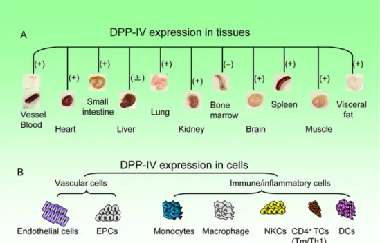
Cell and tissue expression of DPP-4: DPP-4, which is
one of the most potent serine peptidases, is broadly ex-pressed in mammalian tissues, including the kidney, blood vessels, small intestine, lung, liver, brain, spleen, adipose,
Figure 1. The substrate specificities, cell expression, working spaces, and functions of the family of DPP-4-related enzymes. ECs
in-dicates endothelial cells; FAP-α, fibroblast activation protein-α; APP, aminopeptidase P; QPP, quiescent cell praline dipeptidase; PREP,
prolyl endopeptidase; GLP, glucagon-like peptide; PYY, peptide YY; ( + ), working in intra- or extracellular space; (–), not working in intra- or extracellular space; and ?, unknown.
and heart tissues (Figure 2).10) The observations of an
early experimental study using colorimetric enzyme histo-chemistry demonstrated that DPP-4 activity is localized in cardiac venous capillaries. DPP-4 is also expressed on en-dothelial progenitor cells, enen-dothelial cells, some impor-tant immune cells (e.g., monocytes, dendritic cells, natural killer cells, and lymphocytes) and inflammatory cells (i.e.,
macrophages) in various pathological conditions.14,19)As it
is distributed widely, DPP-4 inhibition is a promising ap-proach in various medical fields, such as inflammation regulation, hematopoiesis recovery, and immunomodula-tion, and, of course, in vascular repair and ischemic
ACVD,27,29,30)as highlighted in this review.
DPP-4 Substrates
Numerous neuropeptides, hormones, chemokines,
growth factors, and endocrines contain an alanine or proline at position 2 and are putative DPP-4 substrates. DPP-4 exhibits many physiological and pharmacological
functions by truncating extremely abundant substrates.19)
It is well known that DPP-4 inhibition can enhance insulin secretion and improve glucose tolerance in humans via the
GLP-1-dependent signaling pathway.31)
Synthetic DPP-4 inhibitors were also reported to ameliorate glucose
intoler-ance in Glp1r−/− mice,32) suggesting that DPP-4 exerts its
own biological role, independent of GLP-1. In addition,
DPP-4 degrades a large number of peptide chemokines and hormones in vitro, whereas comparatively limited peptides have been characterized as endogenous
physi-ological substrates for DPP-4 in vivo.14)
Based on DPP-4’s potential catalytic activity in vivo and in vitro, it seems that the proteins/peptides with cleavage sites for DPP-4 could be potential substrates of DPP-4 in various patho-physiological conditions.
Accumulating evidence indicates putative praline or
N-terminal alanine DPP-4 truncation sites in many
chemokines, cytokines, growth factors, and hormones, for
example, interleukin-3 (IL-3), IL-1α, IL-6,
colony-stimulating factor (CSF), stromal cell-derived factor-1α,
granulocyte-CSF, granulocyte macrophage-CSF, erythro-poietin, a number of splice variants of vascular endothelial
growth factor-A, leukemia inhibitory factor,
throm-bopoietin, high-mobility group box 1, and others.19) This
raises the possibility that DPP-4 can modulate ACVD in-itiation and progression through the degradation and modification of these substrate-related factors. Given the potential effects of DPP-4 on ACVD, DPP-4 inhibitors have recently been known as pharmacological targets for ischemic ACVD. In the following sections, we focus on the significance of DPP-4/its inhibitor-mediated cardiovas-cular benefits on vascardiovas-cular inflammatory and metabolic CVDs in animals and humans under CPS.
Figure 2. Dipeptidyl peptidase-4 (DPP-4) is known to be expressed widely in human and animal tissues (e.g., ves-sels, blood, heart, small intestine, lung, kidney, brain, spleen, muscle, and visceral fat) (A) and vascular cells and isch-emic cardiovascular disease (ICVD) -associated cells (B). EPCs indicates endothelial progenitor cells; NKCs, natural killer cells; TCs, T cells; Tm/Th1, T-memory/T-helper; DC, dendritic cells; ( + ), increase in diseased tissues; ( ± ), increase/no changes in diseased tissue; and (–), no detection.
The Impact of CPS on ACVD and Its Mechanisms Stress produces plasma and tissue DPP-4 and GLP-1 imbalance: Individual DPP family members may
partici-pate in inflammatory and metabolic disorders.33,34)The
im-portance of DPP-4 in the initiation and progression of ACVD and the data on DPP-4 inhibition-mediated benefi-cial effects obtained from experimental models, mechanis-tic human studies, and clinical trials are summarized in
two early comprehensive reviews.19,31)
The discovery of incretin-based treatments exhibits a major therapeutic ad-vance in the medical intervention of cardiometabolic dis-orders, and the development of DPP-4 inhibitors as useful antidiabetic drugs was based on the concept that these agents would enhance systematic and tissue glucagon-like peptide-1 (GLP-1) levels, causing an improvement of the
insulinotropic effects of blood sugar.35,36) In addition to
GLP-1-dependent effects on the cardiometabolic risk pro-file, DPP-4 inhibitors provide vascular protective benefi-cial effects by modulating several substrate factor
activi-ties ( e. g. , CSF, stromal cell-derived factor-1α,
granulocyte-CSF, granulocyte macrophage-CSF,
neuropep-tide Y, and high-mobility group box 1).37)A clinical study
reported that individuals with and without DM had in-creased plasma DPP-4 levels and dein-creased plasma GLP-1 levels.38)
In mice and rats, chronic stress increased circu-lating and tissue DPP-4 activities and decreased plasma
and brain GLP-1 levels,2,26,27)
suggesting an imbalance be-tween GLP-1 and DPP-4 as a potential therapeutic target in the management of vascular aging and atherosclerosis in animals under experimental stress conditions.
DPP-4 inhibition attenuated vascular aging and
athe-rosclerosis via the reduction of inflammation and oxi-dative stress production associated with GLP-1-mediated adiponectin production in response to stress:
Although a growing body of evidence indicates that DPP-4 plays an important role in the initiation and progression
of ACVD,19)little is known about the functional relevance
of this exopeptidase as a transmembrane protease in the pathogenesis of stress-related vascular senescence and atherogenesis. Chronic variable stress has been exhibited to produce harmful changes in blood and tissue DPP-4 levels.10,39)
It is well known that inflammation occurs in all stages of atherosclerosis, including initiation, progression, calcification, plaque rupture, and ultimately, thrombotic
complications.40) Data from our research team and those
from other groups clearly revealed that chronic variable stress activated bone-marrow hematopoietic stem cell
pro-liferation via the inactivation of β-adrenergic
receptor-mediated C-X-C motif chemokine 12 (CXCL12) (Table), leading to an increased output of inflammatory monocytes
and neutrophils (Figure 3).9,10) Existing evidence has
con-firmed that stress can increase inflammatory actions in
vascular and adipose tissues.2,4) In vivo, marked increases
in neutrophil and macrophage infiltration and
inflamma-tory chemokine/cytokine expressions (i.e., monocyte
chemoattractant protein-1, osteopontin, toll-like receptor, and CXCR4) and vascular aging were observed in the aortas of stressed mice, and these changes were rectified significantly by DPP-4 inhibitor anagliptin treatment (Fig-ure 3).27)
Accumulating evidence suggests that oxidative stress also plays a critical role in vascular senescence and
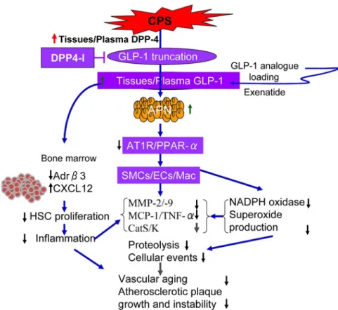
observa-Figure 3. The proposed mechanisms of how GLP-1R activation and DPP-4 inhibition suppress stress-related vascular endothelial senescence and atherosclerotic lesion formation in mice fed a high-fat diet. Stress enhanced the levels of blood DPP-4 levels and decreased blood GLP-1, which decreased adi-pose APN expression and promoted atherosclerotic lesion oxidative stress production, inflammation, and proteolysis, leading to an acceleration of vascular senescence and atherosclerotic lesion formation and its
instability in ApoE−/− mice. CPS indicates chronic psychological stress; DPP-4, dipeptidyl peptidase-4;
APN, adiponectin; PPAR-γ, peroxisome proliferator-activated receptor-γ; Adrβ3, β3 adrenaline receptor;
GLP-1R, GLP-1 receptor; PGC-1α, PPAR-γ co-activator-1α; GLP-1, glucagon-like peptide-1; SMCs,
smooth muscle cells; ECs, endothelial cells; Mac, macrophages; HSC, hematopoietic stem cell; MMP-2, matrix metalloproteinase-2; and CatS, cathepsin S.
tions described herein exhibit that anagliptin mitigated NADPH oxidase component expression (p22phox, p47 phox, p67phox, and gp91phox) and superoxide (O2−) generation. Moreover, the levels of the adiponectin protein and gene were increased in the blood, inguinal, and sub-cutaneous adipose tissues of stressed apoE
lipoprotein-deficient (ApoE)−/−mice, and these changes were reversed
by DPP-4 inhibition.27) In vitro, the GLP-1 analog
ex-enatide increased adiponectin expression in adipose tissue-derived immature adipocytes in a dose-dependent manner,
whereas anagliptin did not affect it.27) Surprisingly,
adi-ponectin depletion with its neutralizing antibody almost completely diminished the anagliptin-mediated vascular
benefits in ApoE−/−mice fed a high-fat diet.27)Adiponectin
was protective against various vascular injuries under con-ditions of stress.42)
These findings thus indicate that an en-hancement of GLP-1 by DPP-4 inhibition may have pro-vided a positive modulation of vascular senescence and atherosclerotic lesion formation through the improvement of adiponectin-induced antioxidative stress production and
anti-inflammation in ApoE−/− mice under our experimental
conditions (Figure 3). This notion was further supported
by the findings of a comparable effect of exenatide on
stress-related vascular harmful changes in ApoE−/−
mice fed a high fat-diet.39)
DPP-4 inhibition prevents stress-related atherosclerotic plaque growth via the reduction of proteolysis:
Accu-mulating evidence of vascular cells have reported that atherosclerosis-associated inflammatory cytokines augment the expression and production of the members of cathep-sin and matrix metalloproteinase (MMP) families from cultured vascular cells (i.e., vascular smooth muscles and endothelial cells), monocyte-derived macrophages, mast cells, and T lymphocytes, and that these inflammatory cy-tokines increase the degradation of extracellular matrix
proteins (collagen and elastin).43) Novel insights into the
actions of these proteases have been made possible by the generation and in-depth analyses of transgenic and knock-out mice.44)
It is well known that both cathepsin and MMP activities modulate neovascularization and vascular remod-eling through the modification, activation, and liberation of cytokines, angiogenic growth factors, cell events (apop-tosis, transmigration, and proliferation),
inhibitors targeting GLP-1 receptor stimulation and DPP-4 activity exhibited a protective effect on the expression and/or activities of proteolytic enzymes [e.g., cathepsin L (CatL), CatS, CatK, matrix metalloproteinase-2 (MMP-2), and MMP-9] and matrix protein metabolism (elastin and collagen) in the lesions of stressed animals fed a high-fat diet.2,27,39)
These therapies also suppressed the levels of
plaque peroxisome proliferator-activated receptor-α
(PPAR-α) and angiotensin II type 1 receptor (AT1R)
pro-teins.27) Both receptor systems with their ligands have
been exhibited to regulate CatS/K and MMP-2/-9 expres-sion by the enhanced productions of oxidative stress and
inflammatory cytokines both in vivo and in vitro.43)In
vi-tro, exenatide suppressed tumor necrosis factor-alpha
ex-pression in cultured macrophages. These findings thus suggest that atherosclerotic lesion development with ne-ovascularization and instability may be attributable to the increase in MMP-2/-9- and CatL/S/K-mediated proteolysis
induced by the stimulation of PPAR-α- and
AT1R-signaling pathway-related oxidative stress production and inflammation in animals under chronic stress conditions (Figure 3).
DPP-4 inhibition and GLP-1 receptor activation at-tenuated atherosclerosis via the modification of lipid metabolism: Previous clinical and basic research studies
indicated that GLP-1 and DPP-4 activities are involved in
lipid metabolism.27,31,39) Biological analyses demonstrated
that anagliptin reduced blood nonesterified fatty acids and
triglycerides in stressed animals,27)and similar results were
found in stressed animals treated with exenatide.39)
Ex-enatide dramatically reduced the foam cell formation of
peripheral blood monocyte-derived macrophages.39)
Clini-cal observations have provided a limited beneficial effect of DPP-4 inhibition on plasma apolipoprotein B-48 and
triglyceride levels.31) Therefore, the improvements in free
fatty acid and triglyceride metabolism may also contribute to the incretin-based glucose-lowering drug-related vascu-lar benefits in mice under stress. Unfortunately, we ob-served that these treatments did not alter plasma levels of “good” and “bad” cholesterols (i.e., high- and low-density lipoprotein cholesterols) in animals under our
experimen-tal stress conditions.27,39) However, clinical research
pro-vides evidence that these preclinical observations are translated into clinical practice.45)
Clinical Trials with DPP-4 Inhibitors in CVD
Incretin-based DPP-4 inhibitors are often the chosen antidiabetic medications that are now most widely used globally. The outcomes of several clinical trials to evaluate the cardiovascular safety of DPP-4 inhibitors have been
reported.46-49)SAVOR-TIMI53 was designed as a
superior-ity trial and failed to meet the prespecified superior out-comes of saxagliptin versus placebo in a high-risk patient population with established vascular diseases and risk fac-tors.46)
In the EXAMINE trial, designed as a safety trial in a high-risk population with post-acute coronary syndrome, the prespecified end point of non-inferiority was met, and alogliptin was non-inferior to the placebo with regard to
cardiovascular outcomes.47) Despite the many preclinical
studies showing the beneficial effects of incretin-related
drugs, most cardiovascular safety trials of incretin-based DPP-4 inhibitors did not show benefits for cardiovascular events. It is important to recognize that cardiovascular safety trials were carried out to meet the US Food and Drug Administration guidance to assess cardiovascular safety of all new antidiabetic drugs; they were not de-signed to assess their benefits for cardiovascular events. Therefore, the long-term potential benefits, as well as the safety, of DPP-4 inhibitors for certain cardiovascular out-comes have not been definitively established, and must be evaluated in more specific and relevant trials. If the need for cardiovascular safety trials is determined based on an individual drug’s safety data during its early development as well as its mechanism of action, resources could be saved when carrying out such clinical trials.
In addition, these negative clinical findings are
incon-sistent with the positive results of animal studies,10,12,26,27)
which demonstrated a DPP-4 inhibitor-mediated cardio-vascular protective effect in animal studies. Although based on their exclusion and inclusion criteria, these large-scare clinical trials recruited huge numbers of par-ticipants with different conditions (e.g., age, sex, body mass index, medical and medication histories, etc.),
espe-cially, ACVD risk factors and its complications.46-49)
In contrast, in animal studies, DPP-4 inhibitors were applied to investigate whether they produce a cardiovascular bene-ficial effect in various special simplified animal models (e. g., an ischemia-induced hindlimb model, a carotid artery ligation with and without cuff-replacement model, a myo-cardial infarction model, or a diet-induced atherosclerosis
model) or model mice (ApoE−/− mice, etc.).14)With the
ex-ception of the vehicle and drugs used, the animal condi-tions (e.g., age, sex, body weight, genetic background, etc.) were controlled to be the same or similar between experimental groups. These factors possibly explain the inconsistent DPP-4 inhibitor-mediated cardiovascular ef-fects between humans and animals. Further studies will be needed to investigate this issue.
Plasma DPP-4, GLP-1, and Adiponectin Levels as Novel Biomarkers for Chronic Stress and Related
CVD Risk
Recently, it was reported that increased circulating DPP-4 and decreased circulating adiponectin and GLP-1 might serve as new useful biomarkers to predict the
pres-ence of stress in animals.10,26) The observations suggest
that among these biological parameters, blood DPP-4 lev-els were more sensitive to chronic stress, and that the noninvasive evaluation of those alterations would be use-ful to assess brain injuries in animals subjected to chronic
stress.10)However, the clinical significance of targeted
hor-mone and exopeptidase changes in the initiation and pro-gression of ACVD associated with modern stressors in hu-mans (including natural disasters, environmental stress, work-related stress, and social anxiety) should be studied as large-scale prospective and/or retrospective cohort stud-ies.
Concluding Remarks
Overall, recent findings from Lei, et al. suggest a po-tential clinically applicable benefit of DPP-4 inhibitors on
vascular senescence and atherogenesis in ApoE−/−
mice
un-der experimental stress conditions,27)
with an effect size
similar to that of GLP-1 analog-based therapies.39)
This protective effect is supported by the finding of a consis-tent effect of genetic inhibition targeting DPP-4 on in-flammation cell production and cytokine expression in the
peripheral blood of rats under chronic stress.10) These
re-sults are consistent with the positive findings of a small clinical trial of a GLP-1 analog or DPP-4 inhibitor as a useful treatment to mitigate atherosclerotic lesion forma-tion and coronary artery events in patients with ACVD
with and without DM.36,50) Nevertheless, given the safety
profile of DPP-4 inhibitors and most current, near-term clinical trial data, these intriguing data make a compelling case for long-term observational studies of GLP-1 analogs and DPP-4 inhibitors on CPS-related ACVD. There are some limitations to these current observations as follows: (1) The absence of preclinical dose dependence when comparing dose administration at 30, 60, and 90 mg/kg/ day; (2) the use of DPP-4 inhibitors in T1DM and T2DM under physiological or psychological stress has not been studied rigorously in long-term, randomized clinical trials; (3) cardiovascular safety endpoints (e.g., chronic heart failure, hospitalization, sudden death) have been studied
rigorously,46)but long-term data may be more challenging
to interpret; and (4) the chronic immobilized stress model used in these studies could not completely mimic human CPS. Thus, it is too hard to fully explore the mechanism of CPS-related CVD and these drug-mediated cardiovas-cular benefits using these animal models.
Disclosure
Conflicts of interest: The authors declare that they have
no conflicts of interest to disclose with respect to this manuscript.
References
1. Lagraauw HM, Kuiper J, Bot I. Acute and chronic psychological stress as risk factors for cardiovascular disease: Insights gained from epidemiological, clinical and experimental studies. Brain Behav Immun 2015; 50: 18-30.
2. Xin M, Jin X, Cui X, et al. Dipeptidyl peptidase-4 inhibition prevents vascular aging in mice under chronic stress: Modula-tion of oxidative stress and inflammaModula-tion. Chem Biol Interact 2019; 314: 108842.
3. Jin X, Jin C, Nakamura K, et al. Increased dipeptidyl peptidase-4 accelerates chronic stress-related thrombosis in a mouse ca-rotid artery model. J Hypertens 2020; 38: 1504-13.
4. Wang H, Meng X, Piao L, et al. Cathepsin S deficiency miti-gated chronic stress-related neointimal hyperplasia in mice. J Am Heart Assoc 2019; 8: e011994.
5. Meng X, Piao L, Wang H, et al. Deficiency of cysteinyl cathep-sin k suppresses the development of experimental intimal hyper-plasia in response to chronic stress. J Hypertens 2020; 38: 1514-24.
6. Rosengren A, Hawken S, Ounpuu S, et al. Association of
psy-chosocial risk factors with risk of acute myocardial infarction in 11119 cases and 13648 controls from 52 countries (the inter-heart study): Case-control study. Lancet 2004; 364: 953-62. 7. Arnold SV, Smolderen KG, Buchanan DM, Li Y, Spertus JA.
Perceived stress in myocardial infarction: Long-term mortality and health status outcomes. J Am Coll Cardiol 2012; 60: 1756-63.
8. Steptoe A, Kivimäki M. Stress and cardiovascular disease. Nat Rev Cardiol 2012; 9: 360-70.
9. Heidt T, Sager HB, Courties G, et al. Chronic variable stress ac-tivates hematopoietic stem cells. Nat Med 2014; 20: 754-8. 10. Zhu E, Hu L, Wu H, et al. Dipeptidyl peptidase-4 regulates
he-matopoietic stem cell activation in response to chronic stress. J Am Heart Assoc 2017; 6.
11. Yisireyili M, Uchida Y, Yamamoto K, et al. Angiotensin receptor blocker irbesartan reduces stress-induced intestinal inflammation via at1a signaling and ace2-dependent mechanism in mice. Brain Behav Immun 2018; 69: 167-79.
12. Yisireyili M, Takeshita K, Hayashi M, et al. Dipeptidyl peptidase- iv inhibitor alogliptin improves stress-induced insulin resistance and prothrombotic state in a murine model. Psycho-neuroendocrinology 2016; 73: 186-95.
13. Abbott CA, Baker E, Sutherland GR, McCaughan GW. Genomic organization, exact localization, and tissue expression of the human CD26 (dipeptidyl peptidase IV) gene. Immunoge-netics 1994; 40: 331-8.
14. Lei Y, Hu L, Yang G, Piao L, Jin M, Cheng X. Dipeptidyl peptidase-iv inhibition for the treatment of cardiovascular disease- recent insights focusing on angiogenesis and neovascu-larization. Circ J 2017; 81: 770-6.
15. Yu DM, Yao TW, Chowdhury S, et al. The dipeptidyl peptidase IV family in cancer and cell biology. FEBS Journal 2010; 277: 1126-44.
16. Durinx C, Lambeir AM, Bosmans E, et al. Molecular characteri-zation of dipeptidyl peptidase activity in serum: Soluble CD26/ dipeptidyl peptidase IV is responsible for the release of x-pro dipeptides. Eur J Biochem 2000; 267: 5608-13.
17. Lankas GR, Leiting B, Roy RS, et al. Dipeptidyl peptidase IV inhibition for the treatment of type 2 diabetes: Potential impor-tance of selectivity over dipeptidyl peptidases 8 and 9. Diabetes 2005; 54: 2988-94.
18. Havale SH, Pal M. Medicinal chemistry approaches to the inhi-bition of dipeptidyl peptidase-4 for the treatment of type 2 dia-betes. Bioorg Med Chem 2009; 17: 1783-802.
19. Zhong J, Maiseyeu A, Davis SN, Rajagopalan S. Dpp4 in cardi-ometabolic disease: Recent insights from the laboratory and clinical trials of dpp4 inhibition. Circ Res 2015; 116: 1491-504. 20. Iwaki-Egawa S, Watanabe Y, Kikuya Y, Fujimoto Y. Dipeptidyl peptidase IV from human serum: Purification, characterization, and N-terminal amino acid sequence. J Biochem 1998; 124: 428-33.
21. Matteucci E, Giampietro O. Dipeptidyl peptidase-4 (CD26): Knowing the function before inhibiting the enzyme. Curr Med Chem 2009; 16: 2943-51.
22. Deacon CF. Metabolism of gip and the contribution of gip to the glucose-lowering properties of dpp-4 inhibitors. Peptides 2020; 125: 170196.
23. Gupta S, Sen U. More than just an enzyme: Dipeptidyl peptidase-4 (dpp-4) and its association with diabetic kidney re-modelling. Pharmacol Res 2019; 147: 104391.
24. Sato H, Kubota N, Kubota T, et al. Anagliptin increases insulin-induced skeletal muscle glucose uptake via an no-dependent mechanism in mice. Diabetologia 2016; 59: 2426-34.
25. Fleenor BS, Ouyang A, Olver TD, et al. Saxagliptin prevents in-creased coronary vascular stiffness in aortic-banded mini swine. Hypertension 2018; 72: 466-75.
26. Piao L, Zhao G, Zhu E, et al. Chronic psychological stress ac-celerates vascular senescence and impairs ischemia-induced ne-ovascularization: The role of dipeptidyl peptidase-4/glucagon-like peptide-1-adiponectin axis. J Am Heart Assoc 2017; 6.
27. Lei Y, Yang G, Hu L, et al. Increased dipeptidyl peptidase-4 ac-celerates diet-related vascular aging and atherosclerosis in apoe-deficient mice under chronic stress. Int J Cardiol 2017; 243: 413-20.
28. Cosenso-Martin LN, Takaoka LY, Vilela-Martin JF. Randomized study comparing vildagliptin vs glibenclamide on glucose vari-ability and endothelial function in patients with type 2 diabetes mellitus and hypertension. Diabetes Metab Syndr Obes 2020; 13: 3221-9.
29. Broxmeyer HE, Hoggatt J, O’Leary HA, et al. Dipeptidylpepti-dase 4 negatively regulates colony-stimulating factor activity and stress hematopoiesis. Nat Med 2012; 18: 1786-96. 30. Higashijima Y, Tanaka T, Yamaguchi J, Tanaka S, Nangaku M.
Anti-inflammatory role of dpp-4 inhibitors in a nondiabetic model of glomerular injury. Am J Physiol Ren Physiol 2015; 308: F878-87.
31. Ferrannini E, DeFronzo RA. Impact of glucose-lowering drugs on cardiovascular disease in type 2 diabetes. Eur Heart J 2015; 36: 2288-96.
32. Marguet D, Baggio L, Kobayashi T, et al. Enhanced insulin se-cretion and improved glucose tolerance in mice lacking CD26. Proc Natl Acad Sci U S A 2000; 97: 6874-9.
33. Kawase H, Bando YK, Nishimura K, Aoyama M, Monji A, Murohara T. A dipeptidyl peptidase-4 inhibitor ameliorates hy-pertensive cardiac remodeling via angiotensin-II/sodium-proton pump exchanger-1 axis. J Mol Cell Cardiol 2016; 98: 37-47. 34. Ghorpade DS, Ozcan L, Zheng Z, et al. Hepatocyte-secreted
dpp4 in obesity promotes adipose inflammation and insulin re-sistance. Nature 2018; 555: 673-7.
35. Ikedo T, Minami M, Kataoka H, et al. Dipeptidyl peptidase-4 inhibitor anagliptin prevents intracranial aneurysm growth by suppressing macrophage infiltration and activation. J Am Heart Assoc 2017; 6.
36. Mita T, Katakami N, Shiraiwa T, et al. Dose-dependent effect of sitagliptin on carotid atherosclerosis in patients with type 2 dia-betes mellitus receiving insulin treatment: A post hoc analysis. Diabetes Ther 2017; 8: 1135-46.
37. Anderluh M, Kocic G, Tomovic K, Kocic R, Deljanin-Ilic M, Smelcerovic A. Cross-talk between the dipeptidyl peptidase-4 and stromal cell-derived factor-1 in stem cell homing and myo-cardial repair: Potential impact of dipeptidyl peptidase-4
inhibi-tors. Pharmacol Ther 2016; 167: 100-7.
38. Yang G, Li Y, Cui L, et al. Increased plasma dipeptidyl peptidase-4 activities in patients with coronary artery disease. PLOS ONE 2016; 11: e0163027.
39. Yang G, Lei Y, Inoue A, et al. Exenatide mitigated diet-induced vascular aging and atherosclerotic plaque growth in apoe-deficient mice under chronic stress. Atherosclerosis 2017; 264: 1-10.
40. Sorci-Thomas MG, Thomas MJ. Microdomains, inflammation, and atherosclerosis. Circ Res 2016; 118: 679-91.
41. Lozhkin A, Vendrov AE, Pan H, Wickline SA, Madamanchi NR, Runge MS. Nadph oxidase 4 regulates vascular inflammation in aging and atherosclerosis. J Mol Cell Cardiol 2017; 102: 10-21. 42. Lim S, Quon MJ, Koh KK. Modulation of adiponectin as a
po-tential therapeutic strategy. Atherosclerosis 2014; 233: 721-8. 43. Wu H, Du Q, Dai Q, Ge J, Cheng X. Cysteine protease
cathep-sins in atherosclerotic cardiovascular diseases. J Atheroscler Thromb 2018; 25: 111-23.
44. Wu H, Cheng XW, Hu L, et al. Cathepsin S activity controls injury-related vascular repair in mice via the tlr2-mediated p38 MAPK and pi3k-akt/p-hdac6 signaling pathway. Arterioscler Thromb Vasc Biol 2016; 36: 1549-57.
45. Moon JY, Woo JS, Seo JW, et al. The dose-dependent organ-specific effects of a dipeptidyl peptidase-4 inhibitor on cardio-vascular complications in a model of type 2 diabetes. PLOS ONE 2016; 11: e0150745.
46. Scirica BM, Bhatt DL, Braunwald E, et al. Saxagliptin and car-diovascular outcomes in patients with type 2 diabetes mellitus. N Engl J Med 2013; 369: 1317-26.
47. White WB, Cannon CP, Heller SR, et al. Alogliptin after acute coronary syndrome in patients with type 2 diabetes. N Engl J Med 2013; 369: 1327-35.
48. Filion KB, Azoulay L, Platt RW, et al. A multicenter observa-tional study of incretin-based drugs and heart failure. N Engl J Med 2016; 374: 1145-54.
49. Green JB, Bethel MA, Armstrong PW, et al. Effect of sitagliptin on cardiovascular outcomes in type 2 diabetes. N Engl J Med 2015; 373: 232-42.
50. Holman RR, Bethel MA, George J, et al. Rationale and design of the exenatide study of cardiovascular event lowering (exscel) trial. Am Heart J 2016; 174: 103-10.