Contents lists available atScienceDirect
Journal of Forensic and Legal Medicine
journal homepage:www.elsevier.com/locate/yjflm
Utility of soluble lectin-like oxidized low-density lipoprotein receptor-1 (sLOX-1) in the postmortem diagnosis of ischemic heart disease
Shojiro Takasu
∗, Sari Matsumoto, Yuko Kanto, Kimiharu Iwadate
Department of Forensic Medicine, The Jikei University School of Medicine, 3-25-8, Nishi-shinbashi, Minato-ku, Tokyo 105-8461, Japan
A R T I C L E I N F O
Keywords:
Forensic Postmortem Biochemical markers Ischemic heart disease sLOX-1
A B S T R A C T
Purpose:Ischemic heart disease (IHD) is a major cause of death in developed countries. Postmortem IHD di- agnosis using biochemical markers is difficult because of the postmortem changes. In the present study, we investigated the utility of soluble lectin-like low-density lipoprotein receptor-1 (sLOX-1) in bodyfluids obtained from forensic autopsy cases.
Methods:We measured pericardialfluid, urine, and serum sLOX-1 levels; these samples were obtained from medicolegal autopsy cases (n = 149, postmortem interval < 72 h), and the utility of these biomarkers post- mortem acute IHD diagnosis was evaluated.
Results:The pericardialfluid and urine of patients with acute IHD had higher sLOX-1 levels (p < .05) compared to the controls. No significant differences were found between the sLOX-1 level and the degree of coronary atherosclerosis, body mass index, and postmortem interval.
Conclusion:sLOX-1 levels in pericardialfluid and urine samples obtained postmortem are useful markers of acute IHD.
1. Introduction
Ischemic heart disease (IHD) is a major cause of sudden death in developed countries. Biochemical markers are central to the clinical diagnosis of IHD. However, the usefulness of biochemical markers for the postmortem diagnosis of IHD in forensic autopsy cases is uncertain because of postmortem changes. In general, the autopsy diagnosis of IHD is achieved through both macroscopic and microscopic findings based on anamnesis, death-related details, or both. Because early-stage pathological consequences of IHD can only be observed 6 h after onset, the diagnosis of IHD in sudden death cases is challenging.1
Several biochemical markers, such as N-terminal pro-brain na- triuretic peptide (NT-proBNP), heart-type fatty acid-binding protein (hFABP), creatine kinase MB (CKMB), myoglobin, cardiac troponin (cTn) T, and cardiac myosin light chain I, are known to increase in patients with IHD.2–16 Previous studies have measured the levels of these biochemical markers in forensic autopsy cases. Although some studies revealed a significant difference in serum cTnT, NT-proBNP, and hFABP levels and pericardialfluid myoglobin, CKMB, and cTnT levels, postmortem elevation of these markers was a major concern.17–29
Atherosclerosis plays a major role in the pathogenesis of IHD.
Oxidized low-density lipoprotein (LDL) is associated with atherogen- esis, and lectin-like oxidized LDL receptor-1 (LOX-1) is the endothelial
receptor of oxidized LDL.30,31LOX-1 is a type II membrane protein that belongs to the C-type lectin family and is overexpressed in the en- dothelial cells, macrophages, and smooth muscle cells of atherosclerotic lesions.30,31 LOX-1 overexpression in the atherosclerotic lesions in blood vessels releases, by a cleavage process, the extracellular domain of this receptor into the blood as soluble LOX-1 (sLOX-1).32,33Tumor necrosis factor alpha-converting enzyme (TACE)/ADAM17, a disin- tegrin metalloproteinase domain-containing protein 10, and inter- leukin-18 are involved in this cleavage process,34–36and sLOX-1 may be associated with plaque vulnerability. Recent studies have reported in- creased serum sLOX-1 levels in patients with acute coronary syndrome (ACS), suggesting that measurement of sLOX-1 may be useful in the clinical diagnosis of acute-phase IHD.37–44Other studies have revealed serum sLOX-1 elevation in patients with obesity, type II diabetes, atherosclerotic conditions, and aortic dissection.45–47
To the best of our knowledge, no studies have investigated the use of sLOX-1 in forensic autopsy cases. In the present study, we measured the level of sLOX-1 not only in serum, but also in pericardialfluid and urine because serum biomarkers are likely to be unstable owing to postmortem changes, and we validated their use in the postmortem diagnosis of IHD.
https://doi.org/10.1016/j.jflm.2018.02.006
Received 11 October 2017; Received in revised form 2 February 2018; Accepted 6 February 2018
∗Corresponding author.
E-mail address:[email protected](S. Takasu).
Journal of Forensic and Legal Medicine 55 (2018) 45–51
Available online 07 February 2018
1752-928X/ © 2018 Elsevier Ltd and Faculty of Forensic and Legal Medicine. All rights reserved.
T
2. Methods 2.1. Case selection
The present study included forensic autopsies performed at the Jikei University School of Medicine, between January 2015 and March 2017.
We examined 70 cases of acute IHD (mean age: 61.9 years; range:
35–87 years) and 79 control cases (mean age: 62.2 years; range: 22–89 years). The diagnosis of acute IHD was based on both macroscopic findings such as coronary stenosis and signs of abrupt death (blood fluidity, organ congestion), and microscopicfindings such as coronary embolism/plaque rupture and myocardial necrosis. In the absence of microscopicfindings, a diagnosis by exclusion of other possible causes of death was made. For all cases, the postmortem interval (PMI) was 72 h or less. The enforcement of cardiopulmonary resuscitation was determined by the medical records presented by the police.
The Ethics Committee of the Jikei University School of Medicine for Biochemical Research approved this study. This research did not re- ceive any specific grant from funding agencies in the public, commer- cial, or not-for-profit sectors.
2.2. Sample collection and laboratory assays
The cardiac blood serum, pericardialfluid, and urine samples ob- tained during autopsies were stored at−80 °C as quickly as possible and were kept frozen until biomarker measurement. sLOX-1 levels were measured by a sandwich chemiluminescence enzyme immunoassay (NK Medico Co., Ltd.) using two kinds of monoclonal antibody against the extracellular domain of LOX-1, that is, B017M and a chicken mono- clonal anti human LOX-1 antibody HUC3-48.
Table 1shows the number of samples. Among the 149 cases in the study, pericardialfluid was obtained in 103 cases, and urine was ob- tained in 82 cases. We measured serum sLOX-1 in 70 acute IHD cases and 79 control cases (23 asphyxia, 11 drowning, 9 congestive heart failure 10 drug intoxications, 9 hypothermia, 8 carbon monoxide in- toxication, 4 head traumas, 2 cervical cord traumas, 2 hyperthermia, and 1 thoracic injury). Pericardialfluid sLOX-1 level was measured in 48 acute IHD cases and 55 control cases (15 asphyxia, 11 drowning, 9 congestive heart failure, 1 drug intoxication, 11 hypothermia, 1 carbon monoxide intoxication, 2 head traumas, 1 cervical cord trauma, 3 hy- perthermia, and 1 thoracic injury), and urine sLOX-1 level was mea- sured in 35 acute IHD cases and 47 control cases (10 asphyxia, 4 drowning, 4 congestive heart failure, 8 drug intoxications, 10 hy- pothermia, 5 carbon monoxide intoxication, 2 head traumas, 2 cervical cord traumas, 1 hyperthermia, and 1 thoracic injury).
2.3. Pathologicalfindings in acute IHD cases
Hematoxylin–Eosin staining of heart tissues was performed for the pathological diagnosis of coronary thrombosis/plaque rupture and for pathological acute myocardial necrosis findings such as eosinophilic changes, contraction band necrosis, and neutrophilic infiltration.
2.4. Evaluation of coronary atherosclerosis
We evaluated coronary atherosclerosis according to the degree of
coronary atherosclerotic stenosis, and divided cases into three groups.
The classification of the groups were non-coronary atherosclerosis (no observation of coronary stenosis), mild to moderate (maximum cor- onary stenosis between 10% and 75%), and severe (maximum coronary stenosis over 75%). Since all cases in the acute IHD group had a max- imum coronary atherosclerosis over 75%, they were classified as single- vessel disease, double-vessel disease, and triple-vessel disease cases.
2.5. Statistical analyses
Statistical analyses were performed using STATA 13.0 (Stata Corp).
Student's t-test was used to evaluate the differences in baseline char- acteristics between the groups. The Kruskal–Wallis equality-of-popula- tion rank test was used to compare the measured values between the groups. A receiver operating characteristic (ROC) curve was generated for values that differed significantly between the groups. The cutoff value was determined by using the Youden Index. Spearman's rank correlation coefficient was used to evaluate the correlation between the values.
3. Results
3.1. Baseline characteristics
Table 2shows the baseline characteristics of the acute IHD and control cases. No significant differences in age were found between the groups. Mean body mass index (BMI) and heart weight were sig- nificantly higher in acute IHD group compared to the control cases.
3.2. sLOX-1 levels between acute IHD and control cases
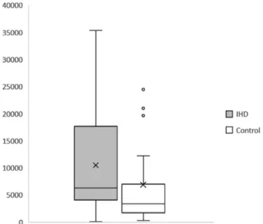
As shown inTable 3, we observed a significant increase in sLOX-1 levels in the pericardialfluid and urine of acute IHD cases compared to the control cases. The median levels of serum sLOX-1 in acute IHD and control cases were 18875.8 pg/ml (range: 181.3–49845.0 pg/ml) and 14213.3 pg/ml (range: 335.9–96697.6 pg/ml), respectively. The median levels of pericardialfluid sLOX-1 in acute IHD and control cases were 1085.5 pg/ml (range: 222.3–19274.6 pg/ml) and 487.1 pg/ml (range: 0–3720.7 pg/ml), respectively. The median values of urine sLOX-1 in acute IHD and control cases were 6415.7 pg/ml (range:
53.0–35397.6 pg/ml) and 3346.7 pg/ml (range: 323.7–62953.0 pg/ml), respectively.Fig. 1,Fig. 2, andFig. 3show the box charts generated for sLOX-1 level in the serum, pericardialfluid, and urine, respectively.
Although significant differences were observed in pericardialfluid and urine sLOX-1 levels, there was no correlation between the values for both the samples in the 54 cases from whom both the specimens were obtained (p = 0.35).
Fig. 4shows the ROC curve for pericardialfluid sLOX-1 levels for the postmortem diagnosis of acute IHD. The area under the curve was 0.70 (95% CI = 0.59–0.80). The pericardialfluid sLOX-1 cutoffvalue for diagnosing acute IHD was 551.7 pg/ml (sensitivity 91.7%, specifi- city 52.7%).
Fig. 5shows the ROC curve for urine sLOX-1 levels in the post- mortem diagnosis of acute IHD. Area under the curve was 0.66 (95%
CI = 0.54–0.78). The urine sLOX-1 cutoffvalue for diagnosing acute IHD was 4128.8 pg/ml (sensitivity 77.1%, specificity 55.3%).
Table 1
Number of samples.
IHD Control
Serum 70 79
Pericardialfluid 48 55
Urine 35 47
IHD, ischemic heart disease.
Table 2
Baseline characteristics (mean ± SD) in the IHD and control cases.
Baseline characteristics IHD (n = 70) control (n = 79) p value
Age (y) 61.9 ± 13.2 62.2 ± 18.7 0.9
BMI (kg/m2) 23.7 ± 5.2 21.8 ± 5.4 < 0.05
Heart weight (g) 457.7 ± 106.9 362.8 ± 94.3 < 0.05 BMI, body mass index; IHD, ischemic heart disease; SD, standard deviation.
3.3. Evaluation of sLOX-1 level and pathologicalfindings in the acute IHD cases
Among the acute IHD cases, coronary thrombosis/plaque rupture was observed in 23 cases (23 serum, 15 pericardialfluid, and 15 urine Table 3
Comparison of sLOX-1 level (mean ± SD) between the acute IHD and control cases.
Acute IHD Control p value
Serum (pg/ml) 20369.9 ± 11622.8 19423.4 ± 17367.1 0.07 Pericardialfluid (pg/ml) 1933.8 ± 2979.4 968.5 ± 1013.1 < 0.05 Urine (pg/ml) 11171.7 ± 10419.2 6904.2 ± 10285.7 < 0.05 sLOX-1, soluble lectin-like oxidized low-density lipoprotein receptor-1; SD, standard de- viation.
Fig. 1.Box chart of soluble lectin-like oxidized low-density lipoprotein receptor-1 (pg/
ml) in the serum of patients from the acute ischemic heart disease and control cases. × represents the mean value for each specimen.
Fig. 2.Box chart of soluble lectin-like oxidized low-density lipoprotein receptor-1 (pg/
ml) in the pericardialfluid of patients from the acute ischemic heart disease and control cases. × represents the mean value for each specimen.
Fig. 3.Box chart of soluble lectin-like oxidized low-density lipoprotein receptor-1 (pg/
ml) in the urine of patients from the acute ischemic heart disease and control cases. × represents the mean value for each specimen.
Fig. 4.ROC curve of pericardialfluid sLOX-1 for the postmortem diagnosis of acute IHD.
Fig. 5.ROC curve of urine sLOX-1 for the postmortem diagnosis of acute IHD.
samples). Pathological acute myocardial necrosis was observed in 28 cases (28 serum, 18 pericardialfluid, and 16 urine samples). The sLOX- 1 levels in acute IHD patients with and without acute coronaryfindings (thrombosis/plaque rupture) show no significant differences (Table 4).
The median level of serum sLOX-1 in acute IHD cases with and without acute coronary findings were 19901.1 pg/ml (range:
2665.2–49107.9 pg/ml) and 18224.2 pg/ml (range: 181.3–49845.0 pg/
ml), respectively. The median level of pericardialfluid sLOX-1 in acute IHD cases with and without acute coronaryfindings were 1090.9 pg/ml (range: 391.8–6674.4 pg/ml) and 1080.0 pg/ml (range:
222.3–19274.6 pg/ml), respectively. The median level of urine sLOX-1 in acute IHD cases with and without acute coronary findings were 13222.5 pg/ml (range: 728.4–35397.6 pg/ml) and 6095.1 pg/ml (range: 53.0–32405.8 pg/ml), respectively.
Although serum sLOX-1 was significantly higher in cases without pathological acute myocardial necrosis groups, there were no sig- nificant differences in pericardialfluid and urine sLOX-1 levels in pa- tients with IHD with and without pathological acute myocardial ne- crosis (Table 5). The median levels of serum sLOX-1 in acute IHD cases with and without acute myocardial necrosis were 21003.2 pg/ml (range: 10532.4–49107.9 pg/ml) and 16274.6 pg/ml (range:
181.3–49845.0 pg/ml), respectively. The median levels of pericardial fluid sLOX-1 in acute IHD cases with and without acute myocardial necrosis were 1104.3 pg/ml (range: 363.6–4430.8 pg/ml) and 1026.1 pg/ml (range: 222.3–19274.6 pg/ml), respectively. The median levels of urine sLOX-1 in acute IHD cases with and without acute myocardial necrosis were 7581.5 pg/ml (range: 728.4–34920.1 pg/ml) and 6135.4 pg/ml (range: 53.0–35397.6 pg/ml), respectively.
Among the acute IHD cases, both acute coronary findings and myocardial necrosis was absent in 33 cases.
3.4. Evaluation of sLOX-1 level and the degree of BMI in the control cases
Pericardialfluid and urine sLOX-1 levels were compared between the control cases with BMI above and below 25 (Table 6). No significant difference in sLOX-1 level was observed. The median levels of peri- cardialfluid sLOX-1 in BMI < 25 and 25 < BMI cases were 484.9 pg/
ml (range: 0–3699.4 pg/ml) and 727.2 pg/ml (range: 147.6–3720.7 pg/
ml), respectively. The median levels of urine sLOX-1 in BMI < 25 and 25 < BMI cases were 3182 pg/ml (range: 567.9–62953.0 pg/ml) and 3617.2 pg/ml (range: 323.7–21004.2 pg/ml), respectively.
3.5. Evaluation of sLOX-1 level and the degree of coronary atherosclerosis
In the control cases, the number of cases within non-coronary atherosclerosis, mild to moderate, and severe coronary athero- sclerosis groups was 46 (46 serum, 26 pericardialfluid, and 31 urine samples), 29 (29 serum, 25 pericardialfluid, and 12 urine samples), and 4 (4 serum, 4 pericardialfluid, and 4 urine samples) cases re- spectively. The median level of serum sLOX-1 in each degree was 16115.0 pg/ml (range: 335.9–83214.0 pg/ml), 13359.8 pg/ml (range: 1985.6–96697.6 pg/ml), and 28022.65 pg/ml (range:
7951.8–30186.4 pg/ml), respectively. The median level of peri- cardial fluid sLOX-1 in each degree was 421.1 pg/ml (range:
0–3720.7 pg/ml), 727.2 pg/ml (range: 0–3605.8 pg/ml), and 898.1 pg/ml (range: 257.6–1102.6 pg/ml), respectively. The median level of urine sLOX-1 in each degree was 3049.0 pg/ml (range:
323.7–24674.9 pg/ml), 5730.6 pg/ml (range: 737.0–62953.0 pg/
ml), and 3489.1 pg/ml (range: 2765.0–21004.2 pg/ml), respec- tively.
In the acute IHD cases, the number of single-vessel, double-vessel, and triple-vessel disease cases was 34 (34 serum, 19 pericardialfluid, and 18 urine samples), 23 (23 serum, 18 pericardialfluid, and 11 urine samples), and 13 (13 serum, 11 pericardial fluid, 6 urine samples) cases, respectively. The median level of serum sLOX-1 in each degree was 20530.5 pg/ml (range: 2665.2–48720.4 pg/ml), 16274.6 pg/ml (range: 181.3–49107.9 pg/ml), and 20996.7 pg/ml (range:
12279.8–49845.0 pg/ml), respectively. The median level of pericardial fluid sLOX-1 in each degree was 928.3 pg/ml (range: 222.3–1969.8 pg/
ml), 1081.5 pg/ml (range: 362.6–7919.6 pg/ml), and 1522.6 pg/ml (range: 369.1–19274.6 pg/ml), respectively. The median level of urine sLOX-1 in each degree was 7262.0 pg/ml (range: 53.0–29052.0 pg/ml), 6135.4 pg/ml (range: 3861.2–35397.6 pg/ml), and 11069.0 pg/ml (range: 728.4–34920.1 pg/ml), respectively.
No significant difference was observed in the sLOX-1 level between the varying degrees of coronary atherosclerosis (Tables 7 and 8).
3.6. Evaluation of sLOX-1 level and the degree of PMI in the control cases The cases with PMI under 24 h, 24–48 h, and 48–72 h were compared in the control cases (Table 9). The median levels of serum sLOX-1 in cases with PMI under 24 h, 24–48 h, and 48–72 h were 12981.5 pg/ml (range: 5083.5–52241.4 pg/ml), 16314.2 pg/ml (range: 3012.1–96697.6 pg/ml), and 10983.1 pg/ml (range:
335.9–41506.1 pg/ml), respectively. The median levels of peri- cardial fluid sLOX-1 in cases with PMI under 24 h, 24–48 h, and 48–72 h were 322.5 pg/ml (range: 47.4–2620.3 pg/ml), 726.8 pg/ml (range: 0–3720.7 pg/ml), and 975.2 pg/ml (range: 0–3699.4 pg/ml), respectively. The median levels of urine sLOX-1 in cases with PMI under 24 h, 24–48 h, and 48–72 h were 5694.3 pg/ml (range:
567.9–12223.3 pg/ml), 2934.1 pg/ml (range: 323.7–62953.0 pg/
ml), and 4134.9 pg/ml (range: 652.6–24674.9 pg/ml), respectively.
Table 4
Comparison of pericardialfluid and urine sLOX-1 levels (mean ± SD) between IHD cases with and without coronary thrombosis/plaque rupture.
With thrombosis/plaque rupture
Without thrombosis/
plaque rupture
p value
Serum (pg/ml) 22243.4 ± 11707.3 18816.9 ± 10863.3 0.23 Pericardial
fluid (pg/
ml)
1977.1 ± 1828.9 1914.2 ± 3401.9 0.33
Urine (pg/ml) 14681.4 ± 11528.7 8607.1 ± 8843.4 0.13 sLOX-1, soluble lectin-like oxidized low-density lipoprotein receptor-1; SD, standard de- viation; IHD, ischemic heart disease.
Table 5
Comparison of sLOX-1 (mean ± SD) between IHD cases with and without pathological acute myocardial necrosis.
With pathological acute myocardial necrosis
Without pathological acute myocardial necrosis
p value
Serum (pg/ml) 23152.1 ± 9764.9 17778.4 ± 11673.6 < 0.05 Pericardial
fluid (pg/
ml)
1444.6 ± 996.5 2227.4 ± 3683.3 0.65
Urine (pg/ml) 11640.5 ± 9633.2 10848.2 ± 11218.7 0.56 sLOX-1, soluble lectin-like oxidized low-density lipoprotein receptor-1; SD, standard de- viation; IHD, ischemic heart disease.
Table 6
Comparison of sLOX-1 levels (mean ± SD) between BMI in control cases.
BMI < 25 25 < BMI p value
Pericardialfluid (pg/ml) 888.1 ± 925.7 (n = 40)
1182.7 ± 1226.6 (n = 15)
0.50 Urine (pg/ml) 7378.8 ± 11460.2
(n = 35)
5520.2 ± 5801.9 (n = 12)
0.77
sLOX-1, soluble lectin-like oxidized low-density lipoprotein receptor-1; SD, standard de- viation; BMI, body mass index.
Although there was a time-depended tendency for elevation in pericardialfluid sLOX-1 level, we found no significant differences be- tween the groups.
3.7. Evaluation of sLOX-1 and the enforcement of cardiopulmonary resuscitation in control cases
There were 33 cases who underwent cardiopulmonary resuscitation in the control group. No significant difference in sLOX-1 level in all specimens were observed compared to the patient without cardio- pulmonary resuscitation.
4. Discussion
sLOX-1 is suggested to be associated with plaque vulnerability.38,39 Recent studies show serum sLOX-1 elevation in ACS.37–44 Hayashida et al.37discovered earlier peak value of serum sLOX-1 compared to TnT in ACS patients. Furthermore, the peak value of serum sLOX-1 in ACS patients were observed at the time of hospital arrival, suggesting that serum sLOX-1 levels may begin to rise before the onset of ACS.37Kume et al.40suggested that sLOX-1 elevation has a different etiology from myocardial injury.
In the present study, serum sLOX-1 levels were higher than clinical references in all cases. Postmortem increase in serum biochemical marker levels of myoglobin, CKMB, and cTnT have been previously reported.17–20Postmortem increases in NT-proBNP, hFABP, CKMB, and cTnT serum levels may be attributable to myocardium autolysis, whereas, the increase in serum myoglobin and cardiac myosin light chain I levels may be explained by myocardium and skeletal muscle autolysis.4–6,10,15,16,48–55 Although earlier studies have reported sig- nificant increases in serum sLOX-1 level in patients with ACS, no sig- nificant difference was observed between the acute IHD and control groups in the present study. Furthermore, the levels found in this study were remarkably higher than those found in living patients with ACS.37,38LOX-1 is reportedly expressedin vivoin the aortic intima and vascular-rich organs such as the placenta, lungs, brain, and liver.30,31 High serum sLOX-1 levels can be explained by the autolysis of these organs and postmortem redistribution. These results suggest that serum sLOX-1 levels cannot be used as a diagnostic marker in postmortem acute IHD diagnosis.
Pericardialfluid is an ultrafiltrate of blood plasma, and therefore, biomarker levels in the pericardialfluid are reflective of their levels in the plasma of living patients.56 Few studies have shown significant differences in the levels of brain natriuretic peptide, myoglobin, CKMB, and cTnT, between pericardial fluid samples obtained from normal patients and the deceased.17,20,24–29In the present study, the pericardial fluid level of sLOX-1 was significantly higher in the acute IHD group
compared to the control group (Table 3). Furthermore, measured values in the present study were comparable to the serum cutoff value (1000 pg/ml) in living patients as reported by Hayashida et al.37for diagnosing acute myocardial infarction (AMI).
Biochemical markers that exist in the myocardium may permeate through the epicardium because of postmortem myocardium autolysis.
Compared to some of the other biochemical markers that are used to diagnose IHD, such as NT-proBNP, hFABP, CKMB, myoglobin, cTnT, and cardiac myosin light chain I, sLOX-1 is absent in the myocardium.
Therefore, the pericardialfluid sLOX-1 level might be less affected by postmortem myocardium autolysis and is more likely to reflect the antemortem serum levels. However, the diffusion of sLOX-1 from the coronary arteries immediately beneath the epicardium must be con- sidered. LOX-1 is expressed on the atherosclerotic lesion of the arterial intima; thus, LOX-1 on the intima and sLOX-1 in the blood must permeate through the intima, tunica media, tunica adventitia, and epicardium to diffuse in to the pericardialfluid. The pericardialfluid sLOX-1 level in this study was comparable to the serum level in living individuals, and no significant time-dependent elevation was dis- covered (Tables 3 and 9). Therefore, we presumed the diffusion of sLOX-1 from the coronary artery to be negligible in cases with PMI shorter than 72 h.
Kobayashi et al.38compared serum sLOX-1 levels among patients with ACS having plaque rupture, patients with ACS without plaque rupture, and patients with stable angina pectoris. The sLOX-1 level was significantly higher in patients with ACS compared to patients with stable angina pectoris. Furthermore, sLOX-1 level was significantly higher in ACS patients with plaque rupture compared to those without plaque rupture. Therefore, sLOX-1 was suggested to be associated with plaque vulnerability.38,39Although significant differences in pericardial fluid sLOX-1 levels were observed between the acute IHD and control groups in the present study, we were unable to detect coronary thrombosis/plaque rupture (Table 4). Kume et al.40measured sLOX-1, hFABP, and cTnT levels in patients with ACS and showed that sLOX-1 had the highest sensitivity and specificity in diagnosing ACS. Further- more, no correlation was found between cTnT and sLOX-1 levels, sug- gesting that sLOX-1 elevation has a different etiology from myocardial injury. Consistent with the observations in previous studies,40 peri- cardialfluid sLOX-1 level was not affected by the degree of pathological acute myocardial necrosis in this study (Table 5).
In the present study, BMI and degree of coronary atherosclerosis were higher in the acute IHD group than in the control group, and might have contributed to the significant difference in sLOX-1 level.
However, because sLOX-1 level was unaffected by BMI and coronary atherosclerosis (Tables 6–8), it could be said that postmortem peri- cardialfluid sLOX-1 level does not strongly increase as a function of BMI and coronary atherosclerosis alone. Although we were unable to Table 7
Comparison of sLOX-1 (mean ± SD) between the degrees of coronary atherosclerosis in control cases.
Non Mild-moderate Severe p value
Serum (pg/ml) 19518.5 ± 16778.6 18704.0 ± 19308.9 23545.9 ± 10565.4 0.48
Pericardialfluid (pg/ml) 906.9 ± 1074.3 1061.3 ± 1034.4 789.1 ± 388.6 0.81
Urine (pg/ml) 5173.4 ± 5995.8 11114.7 ± 17169.9 7686.8 ± 8890.8 0.33
sLOX-1, soluble lectin-like oxidized low-density lipoprotein receptor-1; SD, standard deviation.
Table 8
Comparison of sLOX-1 (mean ± SD) between the degrees of coronary atherosclerosis in acute IHD cases.
Single-vessel Double-vessel Triple-vessel p value
Serum (pg/ml) 21230.4 ± 11012.6 16925.2 ± 10612.6 25904.0 ± 13324.5 0.18
Pericardialfluid (pg/ml) 1088.1 ± 529.3 1963.9 ± 2188.5 3419.1 ± 5401.2 0.24
Urine (pg/ml) 10193.8 ± 9314.5 11440.6 ± 11394.5 13752.5 ± 13741.8 0.93
sLOX-1, soluble lectin-like oxidized low-density lipoprotein receptor-1; SD, standard deviation; IHD, ischemic heart disease.
detect acute coronary thrombosis/plaque rupture in the acute IHD group, the usefulness of pericardialfluid sLOX-1 levels in postmortem acute IHD diagnosis in cases wherein PMI is 72 h or shorter is worth considering.
Urine levels of sLOX-1 were significantly higher in the acute IHD group than in the control group (Table 3). Several previous studies in living patients demonstrated an increase in urine hFABP in AMI pa- tients.7,8Nayashida et al.57demonstrated the importance of the renal clearance of hFABP compared to that of CKMB and TnT. Bjurman et al.58measured serum cTnT, cTnI, NT-proBNP, hFABP, and copeptin levels and showed that NT-proBNP, hFABP, and copeptin levels are elevated in patients with low glomerularfiltration rate. The authors suggested that compared to cTnT (37 kDa) and cTnI (24 kDa), low- molecular-weight molecules including NT-proBNP (8.5 kDa), hFABP (15 kDa), and copeptin (5 kDa) can easily permeate through the glo- merularfiltration membrane, and, the kidneys play a key role in their excretion.
sLOX-1 has a molecular weight of 35 kDa.34Therefore, we believe that sLOX-1 is not excreted in the urine of living patients. In the present study, urine sLOX-1 levels were relatively high, and no significant correlation was found between its urine and pericardial fluid levels, possibly owing to postmortem autolysis and LOX-1 expression in the bladder. Compared to the coronary artery directly beneath the epi- cardium, the number of vessels of the bladder wall is greater and the vessels are thinner. Therefore, the diffusion of sLOX-1 to the urine could have occurred earlier and more massively compared to pericardial fluid.
The limitations of the present study include the small sample size and lack of information regarding baseline characteristics. Levels of sLOX-1 are also known to increase in conditions such as diabetes, aortic dissection, and cerebral infarction. Information regarding antemortem diabetic conditions is often unknown in forensic autopsy cases.
Furthermore, we used preserved specimens in this study; thus, HbA1c could not be measured because of hemolysis. Fatal aortic dissection cases were often accompanied by hemopericardium. In such cases, pericardial fluid could not be obtained; thus, aortic dissection cases were excluded from this study. Cerebral infarction cases that fit the criteria of this study were not found during this study period.
In the present study, we measured sLOX-1 level in bodyfluids col- lected from postmortem specimens. Although further large-scale con- firmatory studies are required, the results suggest that pericardialfluid and urine sLOX-1 levels are useful in postmortem acute IHD diagnosis, in cases with a postmortem sampling interval of 72 h or shorter. In the field of forensic medicine, postmortem changes in biochemical markers are a major concern because of autolysis. sLOX-1 is a biochemical marker that is absent in the myocardium. Therefore, pericardialfluid sLOX-1 level was comparable to the serum cutofflevel in living pa- tients.
Coronary atherosclerosis without any demonstration of macroscopic or microscopic changes of myocardial necrosis is often the onlyfinding in acute IHD cases with a survival time under 6 h after onset. In such cases, the diagnosis is made by exclusion of other possible causes of death. In the present study, significant elevation of sLOX-1 level was observed not only in acute IHD cases with histopathologicalfindings,
but also in acute IHD cases without specific histopathologicalfindings.
Therefore, measuring sLOX-1 in pericardialfluid can be a useful addi- tional diagnostic tool for diagnosing acute IHD in cases where no his- topathologicalfindings are observed.
References
1. Lodge-Patch I. The ageing of cardiac infarcts, and its influence on cardiac rupture.Br Heart J.1951;13:37–42.
2. Staub D, Jonas N, Zellweger MJ, et al. Use of N-terminal pro-B-type natriuretic peptide to detect myocardial ischemia.Am J Med.2005;118:1287.
3. Haaf P, Balmelli C, Reichlin T, et al. N-terminal pro B-type natriuretic peptide in the early evaluation of suspected acute myocardial infarction.Am J Med.
2011;8:731–739.
4. Glatz JF, van der Vusse GJ, Simoons ML, Kragten JA, van Dieijen-Visser MP, Hermens WT. Fatty acid-binding protein and the early detection of acute myocardial infarc- tion.Clin Chim Acta Int J Clin Chem.1998;272:87–92.
5. Ishii J, Wang JH, Naruse H, et al. Serum concentrations of myoglobin vs human heart-type cytoplasmic fatty acid-binding protein in early detection of acute myo- cardial infarction.Clin Chem.1997;43:1372–1378.
6. Chan CP, Sanderson JE, Glatz JF, Cheng WS, Hempel A, Renneberg R. A superior early myocardial infarction marker. Human heart-type fatty acid-binding protein.
Zeitschrift fur Kardiologie.2004;93:388–397.
7. Tanaka T, Hirota Y, Sohmiya K, Nishimura S, Kawamura K. Serum and urinary human heart fatty acid-binding protein in acute myocardial infarction.Clin Biochem.
1991;24:195–201.
8. Tsuji R, Tanaka T, Sohmiya K, et al. Human heart-type cytoplasmic fatty acid-binding protein in serum and urine during hyperacute myocardial infarction.Int J Cardiol.
1993;41:209–217.
9. Kilpatrick WS, Wosornu D, McGuinness JB, Glen AC. Early diagnosis of acute myo- cardial infarction: CK-MB and myoglobin compared.Ann Clin Biochem.
1993;30:435–438.
10. Plebani M, Zaninotto M. Diagnostic strategies using myoglobin measurement in myocardial infarction.Clin Chim Acta Int J Clin Chem.1998;272:69–77.
11. Wagner GS, Roe CR, Limbird LE, Rosati RA, Wallace AG. The importance of identi- fication of the myocardial-specific isoenzyme of creatine phosphokinase (MB form) in the diagnosis of acute myocardial infarction.Circulation.1973;47:263–269.
12. Roberts R, Gowda KS, Ludbrook PA, Sobel BE. Specificity of elevated serum MB creatine phosphokinase activity in the diagnosis of acute myocardial infarction.Am J Cardiol.1975;36:433–437.
13. Mair J, Artner-Dworzak E, Lechleitner P, et al. Cardiac troponin T in diagnosis of acute myocardial infarction.Clin Chem.1991;37:845–852.
14. Katus HA, Remppis A, Neumann FJ, et al. Diagnostic efficiency of troponin T mea- surements in acute myocardial infarction.Circulation.1991;83:902–912.
15. Gere JB, Krauth GH, Trahern CA, Bigham DA. A radioimmunoassay for the mea- surement of human cardiac myosin light chains.Am J Clin Pathol.1979;71:309–318.
16. Katus HA, Yasuda T, Gold HK, et al. Diagnosis of acute myocardial infarction by detection of circulating cardiac myosin light chains.Am J Cardiol.1984;54:964–970.
17. Wang Q, Michiue T, Ishikawa T, Zhu BL, Maeda H. Combined analyses of creatine kinase MB, cardiac troponin I and myoglobin in pericardial and cerebrospinalfluids to investigate myocardial and skeletal muscle injury in medicolegal autopsy cases.
Leg Med.2011;13:226–232.
18. Zhu BL, Ishikawa T, Michiue T, et al. Postmortem pericardial natriuretic peptides as markers of cardiac function in medico-legal autopsies.Int J Leg Med.
2007;121:28–35.
19. Michaud K, Augsburger M, Donze N, et al. Evaluation of postmortem measurement of NT-proBNP as a marker for cardiac function.Int J Leg Med.2008;122:415–420.
20. Meng X, Ming M, Wang E. Heart fatty acid binding protein as a marker for post- mortem detection of early myocardial damage.Forensic Sci Int.2006;160:11–16.
21. Osuna E, Perez-Carceles MD, Vieira DN, Luna A. Distribution of biochemical markers in biologicfluids: application to the postmortem diagnosis of myocardial infarction.
Am J Forensic Med Pathol.1998;19:123–128.
22. Perez-Carceles MD, Noguera J, Jimenez JL, Martinez P, Luna A, Osuna E. Diagnostic efficacy of biochemical markers in diagnosis post-mortem of ischaemic heart disease.
Forensic Sci Int.2004;142:1–7.
23. Zhu BL, Ishikawa T, Michiue T, et al. Postmortem cardiac troponin I and creatine kinase MB levels in the blood and pericardialfluid as markers of myocardial damage in medicolegal autopsy.Leg Med.2007;9:241–250.
24. Batalis NI, Marcus BJ, Papadea CN, Collins KA. The role of postmortem cardiac Table 9
Comparison of sLOX-1 levels (mean ± SD) between PMI in control cases.
< 24 h 24–48 h 48–72 h p value
Serum (pg/ml) 16551.7 ± 13483.4 (n = 14) 21560.7 ± 19718.4 (n = 49) 15390.7 ± 11254.9 (n = 16) 0.61
Pericardialfluid (pg/ml) 514.6 ± 746.6 (n = 10)
989.4 ± 952.4 (n = 32)
1266.2 ± 1260.5 (n = 13)
0.09
Urine (pg/ml) 5790.1 ± 4083.0
(n = 9)
8115.4 ± 13549.5 (n = 24)
5544.2 ± 5806.9 (n = 14)
0.66
sLOX-1, soluble lectin-like oxidized low-density lipoprotein receptor-1; SD, standard deviation; PMI, postmortem interval.
markers in the diagnosis of acute myocardial infarction.J Forensic Sci.
2010;55:1088–1091.
25. Ghormade PS, Kumar NB, Tingne CV, Keoliya AN. Distribution & diagnostic efficacy of cardiac markers CK-MB & LDH in pericardialfluid for postmortem diagnosis of ischemic heart disease.J Forensic Leg Med.2014;28:42–46.
26. Ellingsen CL, Hetland O. Serum concentrations of cardiac troponin T in sudden death.Am J Forensic Med Pathol.2004;25:213–215.
27. Zhu BL, Ishikawa T, Michiue T, et al. Postmortem cardiac troponin T levels in the blood and pericardialfluid. Part 1. Analysis with special regard to traumatic causes of death.Leg Med.2006;8:86–93.
28. Zhu BL, Ishikawa T, Michiue T, et al. Postmortem cardiac troponin T levels in the blood and pericardialfluid. Part 2: analysis for application in the diagnosis of sudden cardiac death with regard to pathology.Leg Med.2006;8:94–101.
29. Gonzalez-Herrera L, Valenzuela A, Ramos V, Blazquez A, Villanueva E. Cardiac troponin T determination by a highly sensitive assay in postmortem serum and pericardialfluid.Forensic Sci Med Pathol.2016;12:181–188.
30. Sawamura T, Kume N, Aoyama T, et al. An endothelial receptor for oxidized low- density lipoprotein.Nature.1997;386:73–77.
31. Kataoka H, Kume N, Miyamoto S, et al. Expression of lectinlike oxidized low-density lipoprotein receptor-1 in human atherosclerotic lesions.Circulation.
1999;99:3110–3117.
32. Xu S, Ogura S, Chen J, Little PJ, Moss J, Liu P. LOX-1 in atherosclerosis: biological functions and pharmacological modifiers.Cell Mol Life Sci.2013;70:2859–2872.
33. Navarra T, Del Turco S, Berti S, Basta G. The lectin-like oxidized low-density lipo- protein receptor-1 and its soluble form: cardiovascular implications.J Atherosclerosis Thromb.2010;17:317–331.
34. Murase T, Kume N, Kataoka H, et al. Identification of soluble forms of lectin-like oxidized LDL receptor-1.Arterioscler Thromb Vasc Biol.2000;20:715–720.
35. Dreymueller D, Pruessmeyer J, Groth E, Ludwig A. The role of ADAM-mediated shedding in vascular biology.Eur J Cell Biol.2012;91:472–485.
36. Zhao XQ, Zhang MW, Wang F, et al. CRP enhances soluble LOX-1 release from macrophages by activating TNF-alpha converting enzyme.J Lipid Res.
2011;52:923–933.
37. Hayashida K, Kume N, Murase T, et al. Serum soluble lectin-like oxidized low-density lipoprotein receptor-1 levels are elevated in acute coronary syndrome: a novel marker for early diagnosis.Circulation.2005;112:812–818.
38. Pirillo A, Catapano AL. Soluble lectin-like oxidized low density lipoprotein receptor-1 as a biochemical marker for atherosclerosis-related diseases.Dis Markers.
2013;35:413–418.
39. Kobayashi N, Hata N, Kume N, et al. Soluble lectin-like oxidized LDL receptor-1 and high-sensitivity troponin T as diagnostic biomarkers for acute coronary syndrome.
Improved values with combination usage in emergency rooms.Circ J.
2011;75:2862–2871.
40. Kume N, Mitsuoka H, Hayashida K, Tanaka M, Kominami G, Kita T. Soluble lectin- like oxidized LDL receptor-1 (sLOX-1) as a sensitive and specific biomarker for acute coronary syndrome–comparison with other biomarkers.J Cardiol.2010;56:159–165.
41. Mitsuoka H, Kume N, Hayashida K, et al. Interleukin 18 stimulates release of soluble lectin-like oxidized LDL receptor-1 (sLOX-1).Atherosclerosis.2009;202:176–182.
42. Kobayashi N, Takano M, Hata N, et al. Soluble lectin-like oxidized LDL receptor-1 (sLOX-1) as a valuable diagnostic marker for rupture of thin-capfibroatheroma:
verification by optical coherence tomography.Int J Cardiol.2013;168:3217–3223.
43. Kobayashi N, Hata N, Kume N, et al. Soluble lectin-like oxidized low-density lipo- protein receptor-1 as an early biomarker for ST elevation myocardial infarction:
time-dependent comparison with other biomarkers.Circ J.2011;75:1433–1439.
44. Kume N, Mitsuoka H, Hayashida K, Tanaka M, Kita T. Soluble lectin-like oxidized low-density lipoprotein receptor-1 predicts prognosis after acute coronary syn- drome–a pilot study.Circ J.2010;74:1399–1404.
45. Kobayashi N, Hata N, Kume N, et al. Detection of acute aortic dissection by extremely high soluble lectin-like oxidized LDL receptor-1 (sLOX-1) and low troponin T levels in blood.Int J Cardiol.2013;165:557–559.
46. Tan KC, Shiu SW, Wong Y, Leng L, Bucala R. Soluble lectin-like oxidized low density lipoprotein receptor-1 in type 2 diabetes mellitus.J Lipid Res.2008;49:1438–1444.
47. Nomata Y, Kume N, Sasai H, et al. Weight reduction can decrease circulating soluble lectin-like oxidized low-density lipoprotein receptor-1 levels in overweight middle- aged men.Metab, Clin Exp.2009;58:1209–1214.
48. Mukoyama M, Nakao K, Hosoda K, et al. Brain natriuretic peptide as a novel cardiac hormone in humans. Evidence for an exquisite dual natriuretic peptide system, atrial natriuretic peptide and brain natriuretic peptide.J Clin Invest.1991;87:1402–1412.
49. Dagnino L, Drouin J, Nemer M. Differential expression of natriuretic peptide genes in cardiac and extracardiac tissues.Mol Endocrinol.1991;5:1292–1300.
50. Tanaka T, Hasegawa K, Fujita M, et al. Marked elevation of brain natriuretic peptide levels in pericardialfluid is closely associated with left ventricular dysfunction.J Am Coll Cardiol.1998;31:399–403.
51. Hunt PJ, Richards AM, Nicholls MG, Yandle TG, Doughty RN, Espiner EA.
Immunoreactive amino-terminal pro-brain natriuretic peptide (NT-PROBNP): a new marker of cardiac impairment.Clin Endocrinol.1997;47:287–296.
52. Offner GD, Brecher P, Sawlivich WB, Costello CE, Troxler RF. Characterization and amino acid sequence of a fatty acid-binding protein from human heart.Biochem J.
1988;252:191–198.
53. McMahon CG, Lamont JV, Curtin E, et al. Diagnostic accuracy of heart-type fatty acid-binding protein for the early diagnosis of acute myocardial infarction.Am J Emerg Med.2012;30:267–274.
54. Van Nieuwenhoven FA, Kleine AH, Wodzig WH, et al. Discrimination between myocardial and skeletal muscle injury by assessment of the plasma ratio of myo- globin over fatty acid-binding protein.Circulation.1995;92:2848–2854.
55. Frearson N, Perry SV. Phosphorylation of the light-chain components of myosin from cardiac and red skeletal muscles.Biochem J.1975;151:99–107.
56. Ben-Horin S, Shinfeld A, Kachel E, Chetrit A, Livneh A. The composition of normal pericardialfluid and its implications for diagnosing pericardial effusions.Am J Med.
2005;118:636–640.
57. Nayashida N, Chihara S, Tayama E, et al. Influence of renal function on serum and urinary heart fatty acid-binding protein levels.J Cardiovasc Surg.2001;42:735–740.
58. Bjurman C, Petzold M, Venge P, Farbemo J, Fu ML, Hammarsten O. High-sensitive cardiac troponin, NT-proBNP, hFABP and copeptin levels in relation to glomerular filtration rates and a medical record of cardiovascular disease.Clin Biochem.
2015;48:302–307.