Acta Med, Nagasaki 28: 129-135
Experimental Assessment of Restoration of the Bronchial Artery in Sleeve Lobectomy
Combined with Pulmonary Angioplasty
Katsunobu KAWAHARA, Masao TOMITA,
Yuzuru NAKAMURA, Hiroyoshi AYABE,
Toshiyasu KUGIMIYA
The First Department of Surgery, Nagasaki University School of Medicine
Received for publication, May 20, 1983
The restoration of the bronchial artery after bronchoplasty combined with pulmona- ry angioplasty was studied in relation to the degree of stenosis of the pulmonary artery by means of the microangiographic technique. Thirty-three dogs were used in this study.
They were divided into Group A (15 dogs) with sleeve lobectomy alone, Group B (10 dogs) with sleeve lobectomy and a 50% stenosis of the left main pulmonary artery and Group C (8 dogs) with sleeve lobectomy and a 75% stenosis of the left main pulmonary artery.
The restoration of the bronchial artery was assessed by microangiography.
1) The restoration of th bronchial artery after bronchoplasty was completed on day 14.
The trend toward a ready regeneration of the bronchial artery becomes apparent if
some degree of stenosis of the pulmonary artery exists after a duration of seven days.
2) The interrupted bronchial artery starts to restore at the adventitial face and extends throughout the whole wall of the bronchus after the 7th day.
3) From these results, an operative procedure of sleeve lobectomy with pulmonary an- gioplasty is applicable even though stenosis of the pulmonary artery remains to some
degree.
INTRODUCTION
With advances in surgery, it has become popular to use an operative procedure of bronchoplasty in the surgical treatment of hilar type of lung cancer.
Furthermore, the bronchoplastic procedures and their combinations with pulmonary
川 原 克 信 ・富 田 正 雄 ・中村 譲 ・綾部 公 盤 ・釘 宮 敏 定
angioplastic procedures are not infrequently indicated for those who have limited cancer invasion in the wall of the pulmonary artery.
The outcome of using this operative technique was satisfactory in the avoidance of postoperative cardiopulmonary crisis and subsequent cor pulmonale. The use of this operative technique offers the advantage of either preserving the postoperative cardiopul- monary function in older patients or enhancing the surgical curability.
Increasing attention has been given to the nutritional blood flow of the lung, which is interrupted by the bronchoplastic procedure. It is clear that the blood supply of the pulmonary artery plays an important role in maintaining the nutritional blood flow through the bronchial artery which is interrupted by the bronchoplastic procedure itself. On the basis of clinical results, the bronchoplastic procedure gives better pulmonary function fol- lowing surgery without any nutritional deficency of the lung.
A stenosis of pulmonary artery to some extent following angioplasty inevitably en- sue due to technical errors, distortion in the anastomotic site and compression by scar for- mation around the tissue and so on.
The aim of this study is to clarify the role of pulmonary arterial blood flow with respect to the nutritional aspect in the lung undergoing bronchoplasty combined with an- gioplasty.
METHOD
Mongrel dogs weighing from 7 to 15 Kg were anesthetized with 25 mg/Kg of so- dium pentobarbital, intubated with a cuffed endotracheal tube and ventilated with room air using a Havard volume respirator with 200-300 ml of tidal volume.
Thirty-three dogs were divided into three grougs.
Group A consisted of 15 dogs with sleeve anastomosis between the left main bron- chus and the left lower bronchus after a left upper lobectomy. Group B consisted of 10 dogs with a sleeve lobectomy and snaring of the left main pulmonary artery with the artificial dura mater (Silastic sheeling 501-1, Dow Corning . Co.) 0.5-0.7 cm wide, to make a 50% stenosis which corresponds to two-third of the outside diameter of the pul- monary artery. Group C consisted of 8 dogs with a sleeve lobectomy and a 75% stenosis of the left main pulmonary artery.
These dogs underwent the pulmonary angiography by means of the right cardiac catherization using the Cournand 7F catheter to evaluate the degree of stenosis in the pulmonary artery and were sacrificed during a period of the first day to 7 months. At autopsy, the postmortem bronchial arteriography was performed to evaluate the vascular anastomosis between the bronchial artery and the pulmonary artery as shown in the fol-
lowing manner.
Left thoracotomy was made at the fifth intercosal space. The descending aorta
was separated at both levels just distal to the origin of the left subclavian artery and
proximal to the diaphragm. The No. 7 Nelaton catheter was introduced into the sepa-
rated descending aorta, 500 ml of saline with heparin of 10 mg was infused and blood
in the aorta was washed out.
The cut-down tube (c-o type) was also inserted into the left main pulmonary artery and 500 ml of saline was infused with gravity drip of 60 cm high, keeping the wall of the left artium opened. Thirty to sixty ml of the radiopaque medium of 30%
barium sulfate heated to 40°C was given through the No. 7 Nelaton catheter and intro- duced into the descending aorta. During this procedure the left thoracic cavity was cooled with ice slush filled in place.
It took about 30 minutes until the infused contrast medium became stiff.
Thus, the trachea and the bilateral lung were removed together. In the resected specimens, the trachea and bronchus, including the anastomotic site, were longitudinally opened and fixed in 10% formalin during a period of 7 days. A roentogenological examination with soft ray was conducted using Softex Type EMB (Softex Co.).
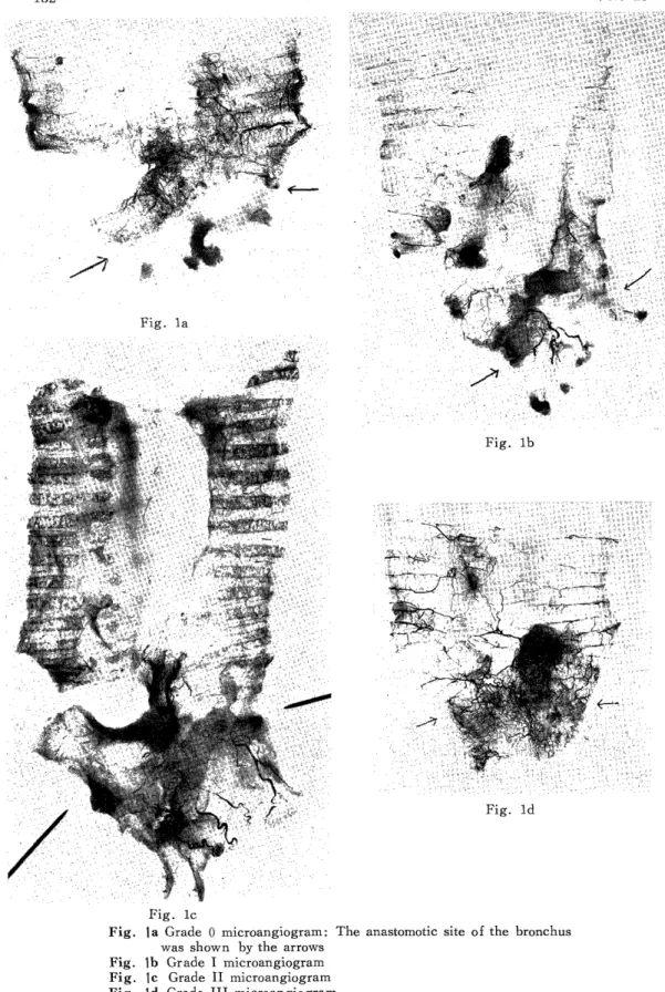
The regeneration of the bronchial artery was evaluated on the basis of findings or the microangiography by the following grading. The microangiographic findings of Grade 0 corresponds to unregenerated bronchial artery extending across the anastomotic site which is shown by the arrows.
Grade I indicates a few regenerated bronchial artery in the adventitia extending across the anastomotic site.
Grade II reveals a moderately developed bronchial artery in both the adventitia and the mucosa. Grade III demonstrates a markedly developed bronchial artery throughout the bronchial wall. These findings were shown in Fig. la to Id.
Dogs with evidence of atelectasis, pneumonia, intraparenchymal bleeding and ab- scess formation at autopsy were excluded from this study.
RESULTS
The degree of stenosis on the pulmonay artery was adjusted by snaring it with ar- tificial dura mater into 50% and 75% in diameter. Postmortem examination in the site of created stenosis on the pulmonary artery revealed that stenosis resulted from develop- ment of fibrogranulomatous tissue around the artificial dura mater had increased with time. In group B, complete obstruction was achieved on day of 13, whereas in Group C, it was within 7 days' duration.
On the basis of these results, it was determined that the degree of stenosis of the pulmonary artery became gradually manifest and exceeded the predicted degrees with the elapse of time and occlusion of the pulmonary artery finally developed. The restoration of the bronchial artery was evaluated according to grading on the microangi- ographic findings.
Fig. 2 showed the rate of regeneration of the bronchial artery extending beyond the
anastomotic site with time in relation to the stenosis of the pulmonary artery. The restora-
tion of the bronchial artery is initiated in the adventitial face on Day 7 as shown in
grade I, and it is completed on Day 14 or after as shown in grade II or III.
Fig. la
Fig. lb
Fig. ld
Fig. lc
Fig. la Grade 0 microangiogram: The anastomotic site of the bronchus was shown by the arrows
Fig. lb Grade I microangiogram Fig. lc Grade II microangiogram Fig. Id Grade III microangiogram
Group B revealed the tendency for the restoration of the bronchial artery to be enhanced compared to those in Group A, whereas in Group C, the development of the bronchial artery was significantly depressed.
The existence of stenosis in the pulmonary artery seems to contribute to early restoration of the interrupted bronchial blood flow. The ready regeneration of the bronchial artery may as well require the stenosis of the pulmonary artery, not exceeding a 50% stenosis.
Fig. 2 The degree of the regenerated bronchial artery was shown with grading classified by microangisgraphic
findings according to a classification of group A, B and C in relation to the elapsed time.