Introduction
Hepatocellular carcinoma (HCC) is one of the common causes of cancer death in Asia. Hepatic resection has been established as a curative treatment for hepatocellular carcinoma. Nevertheless, the prognosis remains poor because postoperative recurrences fre- quently occur (50 - 60%).
1-3Such recurrences could originate from the intrahepatic metastases of the primary HCC
4,5and the multicentric occurrence of new tumors in the postoperative liver remnant.
6-8These unique features might be due to underlying liver diseases such as chronic active hepatitis with hepatitis B and C viral infection. There were several reports that HCC development
was significantly linked to underlying liver diseases.
6,9,10Shuto et al.
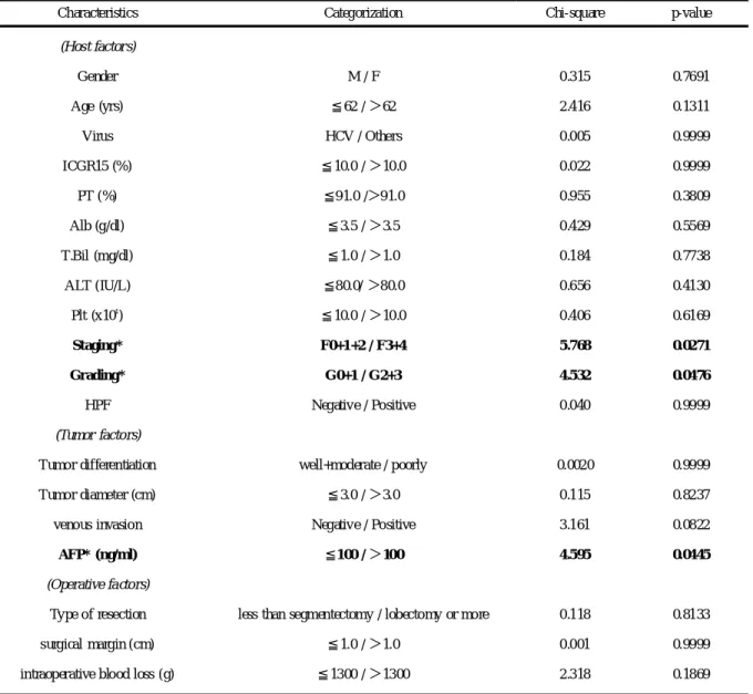
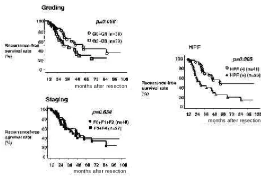
11suggested that hyperplastic foci (HPF), which are defined as a focal hepatic parenchymal lesion where the hepatocytes have dense and small nuclei as well as eosinophilic cytoplasm, was an impor- tant predictor of recurrence of HCC after hepatic resection.
Therefore, not only HCC tumor factors but also the underlying liver status should be carefully examined in order to select the op- timal treatments and also better predict tumor recurrence after curative resection.
The present study was conducted to clarify the risk factors asso- ciated with intrahepatic recurrences in HCC patients who under- went curative hepatic resection by investigating tumor factors, op- Address correspondence: Hikaru Fujioka, M.D., Ph.D. National Hospital Organization Nagasaki Medical Center, Department of Surgery & Clinical Research Center, 2-1001-1 Kubara, Omura, Nagasaki 856-8562 JAPAN
TEL: +81-(0)957-52-3121, FAX: +81-(0)957-53-6675, E-mail: [email protected] Received January 14, 2009; Accepted February 6, 2009
MS#AMN 07041
Underlying histological activity of hepatitis Plays an Important Role for Tumor Recurrence After Curative Resection of Hepatocellular Carcinoma
Kazuma K OBAYAS HI ,
1Hikaru F UJIOKA ,
2Yukio K AMOHARA ,
3Sadayuki O KUDAIR A ,
1Katsuhiko Y ANAGA ,
3Junichiro F URUI ,
1Takashi K ANEMATSU
11
De partment of Tr ansplant and Digestive Surgery, Nagasaki University Gra duate School of Biom edical Sciences, Nagasa ki, Japan
2
Na tional Hospital Or ganization Nagasaki M edical Center, Omura, Ja pan
3