Flavonoids from Iris sanguinea var. tobataensis and
Chemotaxonomic and Molecular Phylogenetic Comparisons with Iris sanguinea var. sanguinea
Takayuki Mizuno * , Yudai Okuyama and Tsukasa Iwashina
Department of Botany, National Museum of Nature and Science, Amakubo 4–1–1, Tsukuba, Ibaraki 305–0005, Japan
* E-mail address: [email protected] (Received 8 May 2018; accepted 28 June 2018)
Abstract Iris sanguinea var. tobataensis (Japanese name, “Tobata-ayame”) is endemic to Tobata, Kita-Kyushu City, Fukuoka Pref., Japan, and has been extinct in the wild. To revalidate the taxo- nomic placement of this Iris plant, we performed chemotaxonomic and molecular phylogenetic comparisons of this variety with I. sanguinea var. sanguinea. Two anthocyanins, delphinidin 3-O-[(4‴-p-coumaroylrhamnosyl)-(1→6)-glucoside]-5-O-glucoside (An1) and petunidin 3-O-[(4‴- p-coumaroylrhamnosyl)-(1→6)-glucoside]-5-O-glucoside (An2), and twelve C-glycosylflavones, vicenin-2 (CF1), swertiajaponin X″-O-hexoside (CF2), isoorientin (CF3), swertiajaponin (CF4), isovitexin 2″-O-glucoside (CF5), swertisin 2″-O-glucoside (CF6), isovitexin (CF7), swertisin (CF8), isovitexin 2″-O-glucoside (CF9), acetylated flavoayamenins (CF10 and CF11), and acety- lated swertisin (CF12), and two flavonols, myricetin 3-O-rhamnoside (FL1), and quercetin 3-O-rhamnoside (FL2), were isolated and characterized from the flowers and leaves of two I. san- guinea varieties. It was shown that flavonoid composition of their varieties is essentially the same except for CF11, which was detected only in the leaves of I. sanguinea var. tobataensis. Further- more, a phylogenetic analysis using chloroplast matK gene and trnL-F intron DNA sequence data of Iris species including these two Iris sanguinea varieties was performed. The resultant tree indi- cated that two I. sanguinea varieties were closely related. These results supported that the taxo- nomic position of var. tobataensis as a variety of I. sanguinea is appropriate.
Key words : acetylated C-glycosylflavone, anthocyanins, Iridaceae, Iris sanguinea var. tobataensis, Iris sanguinea var. sanguinea, phylogeny.
Introduction
Iris sanguinea Hornem. var. tobataensis S.
Akiyama & Iwashina has been recorded as a new variety of I. sanguinea by Akiyama and Iwashina (2009). The variety is known as “Tobata-Ayame”
in Tobata, Kita-kyushu City, Fukuoka Pref., Japan. In this area, although the variety is culti- vated as an ornamental, the wild population has already been extinct (Akiyama and Iwashina, 2009). I. sanguinea var. tobataensis can be dis- tinguished with other Iris species by the morpho-
logical characters. Its shorter stature, 10–15 cm tall, is easily distinguishable with I. sanguinea var. sanguinea. Furthermore, its bigger flower size, 5–7 cm across, is differ from the Japanese dwarf stature iris, Iris rossii Baker (Akiyama and Iwashina, 2009). However, chemotaxonomic and molecular phylogenetic features of I. sanguinea var. tobataensis have not been reported.
Flavonoid distribution including anthocyanins,
flavones, flavonols, and isoflavones etc. in the
genus Iris has been reviewed as an important
resources and chemotaxonomic markers (Iwa-
shina and Ootani, 1998; Wang et al,. 2010). In this study, flavonoids were isolated and identified from the flowers and leaves of I. sanguinea var.
sanguinea and var. tobataensis and compared between their varieties. Furthermore, we obtained the chloroplast matK gene and trnL-F intron DNA sequence data from the two I. san- guinea varieties to understand their phylogenetic relationship within the genus Iris. These data were added in the dataset for the previously reported phylogenetic tree of 43 species and vari- eties of section Limniris of the genus Iris (Wil- son, 2009; Mizuno et al., 2012a).
Materials and Methods Plant materials
Iris sanguinea var. tobataensis and var. san- guinea were each collected from Tobata, Kita- kyushu City, Fukuoka Pref., Japan, and Omachi City, Nagano Pref., Japan, respectively, and then cultivated in Tsukuba Botanical Garden, National Museum of Nature and Science, Tsukuba, Japan.
The voucher specimens of I. sanguinea var. toba- taensis, and var. sanguinea were deposited in the herbarium of National Museum of Nature and Science (TNS), Japan.
Extraction and isolation
Fresh flowers (4.5 g and 96.0 g) and leaves (19.0 g and 696.4 g) of I. sanguinea var. toba- taensis and var. sanguinea were extracted with HCOOH/MeOH (8 : 92) for flowers and MeOH for leaves. After filtration and concentration, the extracts were applied to preparative paper chro- matography (PC) using solvent systems, BAW (n-BuOH/HOAc/H
2O=4 : 1 : 5, upper phase), 15% HOAc and BEW (n-BuOH/EtOH/H
2O=
4 : 1 : 2.2). After PC separation, each fraction was purified by Sephadex LH-20 column chromatog- raphy using MAW (MeOH/HOAc/H
2O=
70 : 5 : 25 for anthocyanins) or 70% MeOH for other flavonoids. Moreover, preparative high per- formance liquid chromatography (HPLC) were performed with Tosoh HPLC systems using Inertsil ODS-4 (I.D. 10×250 mm, GL Science
Inc., Japan) or L-column2 ODS (I.D.
10×250 mm, Chemicals Evaluation and Research Institute (CERI), Japan), at a flow-rate of 3.0 ml/min; injection of 300–350 μl; detection wavelength of 530 nm for anthocyanins and 340 nm for other flavonoids, and eluent of HCOOH/MeCN/H
2O (5 : 15 : 80) or HCOOH/
MeCN/H
2O (1 : 18 : 81).
Analytical HPLC
HPLC analysis of anthocyanins and other fla- vonoids were performed using Shimadzu HPLC system with Inertsil ODS-4 column (I.D.
6.0×150 mm, GL Science Inc.), at flow-rate:
1.0 ml/min, detection: 530 nm and eluents:
MeCN/HOAc/H
2O/H
3PO
4(10 : 8 : 79 : 3) for anthocyanins (Method 1), and L-column2 ODS column (I.D. 6.0×150 mm, Chemical Evaluation and Research Institute), at flow-rate: 1.0 ml/min, detection: 340 nm and eluents: MeCN/H
2O/
H
3PO
4(15 : 85 : 0.2) for other flavonoids (Method 2).
Identification of flavonoids
Isolated flavonoids were identified by UV-Vis spectral survey (anthocyanins: 700–220 nm, fla- vones and flavonols: 500–220 nm) according to Mabry et al. (1970), LC-MS, alkaline and acid hydrolysis, and direct TLC (BAW, 15%HOAc and BEW) and HPLC comparisons with authen- tic samples.
Liquid chromatograph-mass spectra (LC-MS) was performed with Shimadzu LC-MS systems using Inertsil ODS-4 (I.D. 2.1×100 mm, GL Science Inc.) or L-column2 ODS (I.D.
2.1×100 mm, CERI), at a flow-rate of 0.2 ml/
min, detection wavelength of 500 and 530 nm for anthocyanins or 350 and 300 nm for flavones and flavonols, and the eluent, HCOOH/MeCN/H
2O (5 : 12 : 83) or HCOOH/MeCN/H
2O (1 : 12 : 87), ESI
+4.5 kV and ESI
−3.5 kV, 250°C.
Alkaline hydrolysis was performed in water,
and a few drops of xylene. After neutralization
and removal of xylene, diethyl ether was added
to mother liquor. Organic acid and deacylated
compounds were surveyed by HPLC. Acid
hydrolysis was performed in 12% aq. HCl for 30 min at 100°C. After cooling, the solution was shaken with diethyl ether. The hydrolysates, fla- vones and flavonol aglycones, were identified by HPLC. Glycosidic sugars were identified by PC with authentic sugars using solvent systems:
BBPW (n-BuOH/benzene/pyridine/H
2O=
5 : 1 : 3 : 3) and BTPW (n-BuOH/toluene/pyridine/
H
2O=5 : 1 : 3 : 3). TLC, HPLC, UV, and LC-MS data of the isolated flavonoids were as followed.
Delphinidin 3-O-[(4‴-p-coumaroylrhamnosyl)- (1→6)-glucoside]-5-O-glucoside (delphanin, An1, Fig. 1). UV–Vis:
λmax (nm) 0.1% HCl-MeOH 282, 310, 542; E
440/E
max10%; E
acid/E
max77%;
+AlCl3bathochromic shift; LC-MS: m/z 919 [M]
+; HPLC: tR (min) 17.2 (Method 1).
Petunidin 3-O-[(4‴-p-coumaroylrhamnosyl)- (1→6)-glucoside]-5-O-glucoside (petanin, An2, Fig. 2). UV–Vis:
λmax (nm) 0.1% HCl-MeOH281, 314, 543 nm; E
440/E
max11%; E
acid/E
max159%; +AlCl
3bathochromic shift; LC-MS: m/z 933 [M]
+; HPLC: tR (min) 29.4 (Method 1).
Vicenin-2 (CF1, Fig. 3). TLC: Rf 0.13 (BAW), 0.38 (15%HOAc), 0.33 (BEW); color UV (365 nm): deep purple, UV/NH
3: dark yellow.
HPLC: tR (min) 5.7 (Method 2). UV: λmax (nm) MeOH 273, 332; +NaOMe 282, 334, 399 (inc.);
+AlCl3
260sh, 280, 305, 350, 385; +AlCl
3/HCl 260sh, 279, 304, 346, 380; +NaOAc 282, 387;
+NaOAc/H3
BO
3286, 321, 355, 410sh; LC-MS:
m/z 595 [M+H]
+, 593 [M−H]
−.
Swertiajaponin hexoside (CF2). TLC: Rf 0.28 (BAW), 0.76 (15%HOAc), 0.42 (BEW); color UV (365 nm): deep purple, UV/NH
3: yellow.
HPLC: tR (min) 8.4 (Method 2). UV: λmax (nm) MeOH 243, 255sh, 272, 347; +NaOMe 276, 310sh, 394 (inc.); +AlCl
3279, 300sh, 325, 422;
+AlCl3
/HCl 265sh, 280, 300sh, 366, 390;
+NaOAc 270, 310sh, 416; +NaOAc/H3
BO
3270, 381, 425sh; LC-MS: m/z 625 [M+H]
+, 623 [M−H]
−.
Isoorientin (CF3, Fig. 4). TLC: Rf 0.35 (BAW), 0.25 (15%HOAc), 0.44 (BEW); color UV (365 nm): deep purple, UV/NH
3: yellow.
HPLC: tR (min) 8.9 (Method 2). UV: λmax (nm) MeOH 255, 271, 351; +NaOMe 277, 325sh, 415 (inc.); +AlCl
3275, 300sh, 330, 421;
Fig. 2. Petunidin 3-O-[(4‴-p-coumaroylrham- nosyl)-(1→6)-glucoside]-5-O-glucoside (pet- anin, An2) .
Fig. 1. Delphinidin 3-O-[(4‴-p-coumaroylrham- nosyl)-(1→6)-glucoside]-5-O-glucoside (del-
phanin, An1). Fig. 3. Vicenin-2 (CF1).
+AlCl3
/HCl 265sh, 277, 295sh, 362, 385;
+NaOAc 272, 320sh, 405; +NaOAc/H3
BO
3269, 382, 420sh; LC-MS: m/z 449 [M+H]
+, 447 [M−H]
−.
Swertiajaponin (CF4, Fig. 5). TLC: Rf 0.39 (BAW), 0.33 (15%HOAc), 0.46 (BEW); color UV (365 nm): deep purple, UV/NH
3: yellow.
HPLC: tR (min) 10.1 (Method 2). UV:
λmax(nm) MeOH 244, 258sh, 270, 348; +NaOMe 270, 300sh, 401 (inc.); +AlCl
3277, 300sh, 328, 422;
+AlCl3/HCl 260sh, 277, 295sh, 363, 378;
+NaOAc 268, 300sh, 405; +NaOAc/H3
BO
3265, 377, 420sh; LC-MS: m/z 463 [M+H]
+, 461 [M−H]
−.
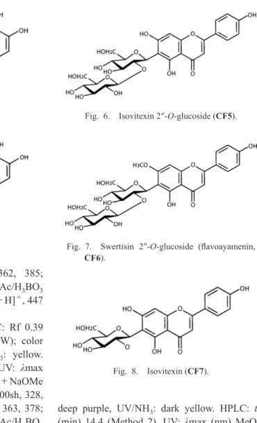
Isovitexin 2″-O-glucoside (CF5, Fig. 6). TLC:
Rf 0.32 (BAW), 0.72 (15%HOAc), 0.46 (BEW);
color UV (365 nm): deep purple, UV/NH
3: dark yellow. HPLC: tR (min) 11.4 (Method 2). UV:
λmax (nm) MeOH 272, 335; +NaOMe 280,
330, 399 (inc.); +AlCl
3279, 304, 350, 385;
+AlCl3
/HCl 280, 304, 346, 380; +NaOAc 279, 309, 333, 395; +NaOAc/H
3BO
3274, 327sh, 347, 395sh; LC-MS: m/z 595 [M+H]
+, 593 [M−H]
−.
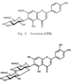
Swertisin 2″-O-glucoside (flavoayamenin, CF6, Fig. 7). TLC: Rf 0.36 (BAW), 0.78 (15%HOAc), 0.48 (BEW); color UV (365 nm):
deep purple, UV/NH
3: dark yellow. HPLC: tR (min) 14.4 (Method 2). UV:
λmax (nm) MeOH273, 332; +NaOMe 274, 307, 385 (inc.);
+AlCl3
260sh, 280, 302, 351, 380; +AlCl
3/HCl 260sh, 281, 302, 349, 380; +NaOAc 271, 330sh, 389; +NaOAc/H
3BO
3278, 337; LC-MS:
m/z 609 [M+H]
+, 607 [M−H]
−.
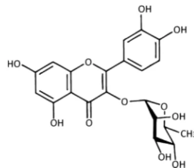
Isovitexin (CF7, Fig. 8). TLC: Rf 0.61 (BAW), 0.33 (15%HOAc), 0.59 (BEW); color UV (365 nm): deep purple, UV/NH
3: dark yel- low. HPLC: tR (min) 15.3 (Method 2). UV: λmax (nm) MeOH 271, 335; +NaOMe 273, 330, 398 (inc.); +AlCl
3279, 300, 348, 385; +AlCl
3/HCl 280, 302, 348, 380; +NaOAc 278, 301, 391;
+NaOAc/H3
BO
3272, 310sh, 345, 400sh;
LC-MS: m/z 433 [M+H]
+, 431 [M−H]
−. Swertisin (CF8, Fig. 9). TLC: Rf 0.64 (BAW),
Fig. 5. Swertiajaponin (CF4).
Fig. 6. Isovitexin 2″-O-glucoside (CF5).
Fig. 7. Swertisin 2″-O-glucoside (flavoayamenin, CF6).
Fig. 8. Isovitexin (CF7).
Fig. 4. Isoorientin (CF3).
0.49 (15%HOAc), 0.67 (BEW); color UV (365 nm): deep purple, UV/NH
3: dark yellow.
HPLC: tR (min) 18.3 (Method 2). UV:
λmax(nm) MeOH 272, 333; +NaOMe 272, 308, 360sh, 386 (inc.); +AlCl
3260sh, 279, 301, 353, 370;
+AlCl3/HCl 260sh, 279, 301, 349, 380;
+NaOAc 270, 335sh, 388; +NaOAc/H3
BO
3271, 339; LC-MS: m/z 447 [M+H]
+, 445 [M−H]
−.
Isoorientin 2″-O-glucoside (CF9, Fig. 10).
TLC: Rf 0.26 (BAW), 0.65 (15%HOAc), 0.38 (BEW); color UV (365 nm): deep purple, UV/
NH
3: yellow. HPLC: tR (min) 8.3 (Method 2).
UV:
λmax (nm) MeOH 255sh, 271, 352;+NaOMe 279, 330sh, 412 (inc.); +AlCl3
271, 305sh, 325, 421; +AlCl
3/HCl 262sh, 276, 295sh, 360, 390; +NaOAc 273, 325sh, 404;
+NaOAc/H3
BO
3269, 385, 425sh; LC-MS: m/z 611 [M+H]
+, 609 [M−H]
−.
Acetylated swertisin 2″-O-glucoside (acety- lated flavoayamenin, CF10). TLC: Rf 0.58 (BAW), 0.90 (15%HOAc), 0.64 (BEW); color UV (365 nm): deep purple, UV/NH
3: dark yel- low. HPLC: tR (min) 19.2 (Method 2). UV: λmax (nm) MeOH 272, 334; +NaOMe 277, 310, 354sh, 383 (inc.); +AlCl
3260sh, 281, 302, 354,
385;
+AlCl3/HCl 260sh, 281, 303, 350, 380;
+NaOAc 271, 295sh, 391; +NaOAc/H3
BO
3272, 340; LC-MS: m/z 651 [M+H]
+, 649 [M−H]
−.
Acetylated swertisin 2″-O-glucoside (acety- lated flavoayamenin, CF11). TLC: Rf 0.49 (BAW), 0.83 (15%HOAc), 0.53 (BEW); color UV (365 nm): deep purple, UV/NH
3: dark yel- low. HPLC: tR (min) 23.0 (Method 2). UV: λmax (nm) MeOH 272, 334; +NaOMe 278, 309, 355sh, 384 (inc.); +AlCl
3260sh, 281, 303, 354, 385;
+AlCl3/HCl 265sh, 280, 303, 350, 380;
+NaOAc 272, 340sh, 391; +NaOAc/H3
BO
3272, 340; LC-MS: m/z 651 [M+H]
+, 649 [M−H]
−.
Acetylated swertisin (CF12). TLC: Rf 0.91 (BAW), 0.65 (15%HOAc), 0.93 (BEW); color UV (365 nm): deep purple, UV/NH
3: dark yel- low. HPLC: tR (min) 44.0 (Method 2). UV: λmax (nm) MeOH 271, 333; +NaOMe 277, 305, 360sh, 384 (inc.); +AlCl
3260sh, 280, 302, 354, 380;
+AlCl3/HCl 260sh, 280, 301, 349, 380;
+NaOAc 270, 325sh, 389; +NaOAc/H3
BO
3271, 340; LC-MS: m/z 489 [M+H]
+, 487 [M−H]
−.
Myricetin 3-O-rhamnoside (myricitrin, FL1, Fig. 11). TLC: Rf 0.57 (BAW), 0.39 (15%HOAc), 0.62 (BEW); color UV (365 nm):
dark yellow, UV/NH
3: orange. HPLC: tR (min) 15.1 (Method 2). UV:
λmax (nm) MeOH 257,265sh, 300sh, 354; +NaOMe decomposition;
+AlCl3
270, 314, 421; +AlCl
3/HCl 271, 310, 365sh, 398; +NaOAc 271, 323, 383; +NaOAc/
Fig. 9. Swertisin (CF8).
Fig. 10. Isoorientin 2″-O-glucoside (CF9). Fig. 11. Myricetin 3-O-rhamnoside (myricitrin, FL1).
H
3BO
3259, 300sh, 374; LC-MS: m/z 465 [M+H]
+, 463 [M−H]
−, and 319 [M−146
+H]+
.
Quercetin 3-O-rhamnoside (quercitrin, FL2, Fig. 12). TLC: Rf 0.78 (BAW), 0.46 (15%HOAc), 0.77 (BEW); color UV (365 nm):
deep purple, UV/NH
3: yellow. HPLC: tR (min) 30.0 (Method 2). UV:
λmax (nm) MeOH 257,265sh, 352; +NaOMe 271, 326sh, 400; +AlCl
3274, 300sh, 360sh, 431; +AlCl
3/HCl 270, 300sh, 360, 395sh; +NaOAc 271, 320sh, 384;
+NaOAc/H3
BO
3260, 300sh, 370; LC-MS: m/z 447 [M−H]
−, 303 [M−146+H]
+Authentic samples
Origins of the authentic samples were as fol- lows: delphinidin 3-O-[(4‴-p-coumaroylrham- nosyl)-(1→6)-glucoside]-5-O-glucoside, and petunidin 3-O-[(4‴-p-coumaroylrhamnosyl)- (1→6)-glucoside]-5-O-glucoside from the flow- ers of Iris ensata Thunb. (Hayashi, 1940), vicenin-2 from the fronds of Asplenium normale D.Don (Aspleniaceae) (Iwashina et al., 1990), isoorientin, swertiajaponin, isovitexin and swerti- sin from the flowers and leaves of Iris rossii (Mizuno et al., 2012a), isovitexin 2″-O- glucoside and isoorientin 2″-O-glucoside from the fronds of Cyrtomium falcatum (L.f.) C.Presl (Dryopteridaceae) (Iwashina et al., 2006), swerti- sin 2″-O-glucoside from the flowers of bearded iris (Iris germanica L.) cultivar
ʻVictoria Fallsʼ(Mizuno et al., 2012b), and myricetin 3-O-rham- noside from the bark of Myrica rubra Sieb. et
Zucc. (Myricaceae) (Hattori and Hayashi, 1931).
Quercetin 3-O-rhamnoside from Extrasynthese (Genay, France).
DNA extraction, PCR and sequencing
Total genomic DNAs of two Iris sanguinea varieties were extracted from the fresh leaves using CTAB procedure (Doyle and Doyle, 1987).
The protein-coding matK gene plus the flanking trnK introns, and trnL-F intergenic spacer region in the chloroplast DNA were sequenced. Specifi- cally, the matK+trnK region was separated in two sections using the primer pairs 3914mF/1235R and 1176-iF/trnk2R (Wilson, 2004). The PCR amplifications were performed in a predenaturation step at 94°C for 4 min, fol- lowed by 30 cycles of denaturation at 94°C for 30 sec., annealing at 52‒56°C for 30 sec and extension at 72°C for 2.5 min, and a final exten- sion at 72°C for 7 min. 2.5 μl of amplified PCR products were treated with 0.5 μl of ExoSAP-IT (USB corporation, USA) for 15 min at 37°C, fol- lowed by the enzyme inactivation at 80°C for 15 min. The amplified DNA was then sequenced with BigDye Terminator v3.0 (Applied Biosys- tems, California) according to the manufacturerʼs protocol on a 3130xl Genetic Analyzer (Applied Biosystems).
Molecular phylogenetic analysis
In addition to sequenced DNA of two Iris sanguinea varieties, 90 (45+45) nucleotide sequences of corresponding chloroplast DNA regions of 43 species of the genus Iris subgenus Limniris, and other two species, Dietes robinso- niana (C.Moore & F.Muell.) Klatt, and Moraea pritzeliana Diels. were obtained from Genbank (Wilson, 2009, 2011; Reeves et al., 2001). Align- ment was conducted using ClustalW imple- mented in Seaview (Galtier et al., 1996) and obvious errors were corrected manually. A maxi- mum likelihood (ML) tree search was performed with PAUP*4.0a (build159) (Swofford, 2002) using a heuristic search with tree bisection–
reconnection (TBR) branch swapping, 10 ran- dom-addition sequence replicates, and Multrees
Fig. 12. Quercetin 3-O-rhamnoside (quercitrin, FL2).
option in effect. Topological uncertainty was assessed using non-parametric bootstrapping with 1000 replicates under the GTRCAT model as implemented in RaxML ver. 8.2.0 (Stamata- kis, 2014).
Result and Discussion
It was shown by HPLC survey of flower extracts that anthocyanin composition between two Iris sanguinea varieties is the same. These anthocyanins were identified as delphinidin 3-O-[(4‴-p-coumaroylrhamnosyl)-(1→6)- glucoside]-5-O-glucoside (A1), and petunidin 3-O-[(4‴-p-coumaroylrhamnosyl)-(1→6)- glucoside]-5-O-glucoside (A2) by LC-MS, UV- Vis spectra, and direct HPLC comparisons with authentic samples. Ishikura and Yamamoto (1978) isolated their anthocyanins from the flow- ers of I. sanguinea var. sanguinea and identified by TLC. We reconfirmed anthocyanin composi- tion of I. sanguinea var. sanguinea flowers by HPLC comparisons with authentic samples. On the other hand, anthocyanins in var. tobataensis
flowers were reported for the first time in this paper. These anthocyanins were widely present in the flowers of many Japanese Iris species, e.g.
I. japonica Thumb., I. laevigata Fisch., I. ensata Thumb., and three I. setosa varieties (Ishikura and Yamamoto, 1978; Yabuya, 1991; Hayashi et al., 1989).
HPLC chromatograms of the flowers and leaves of two I. sanguinea varieties were shown in Figs. 13 and 14. Fourteen compounds (CF1–
12, and FL1, 2) were found and characterized.
Of their compounds, three ones (CF4, 6 and 8) were detected in the both flowers and leaves of two varieties. Other eleven compounds were detected in either flower or leaves. However, CF11 was found in the leaves only of I. san- guinea var. tobataensis.
UV spectral properties showed that CF1, 5 and 7 are flavones having free 5-, 7- and 4′-hydroxyl groups (Mabry et al., 1970). By LC-MS survey, molecular ion peaks, m/z 595 [M+H]
+and 593 [M–H]
–(CF1 and 5) and m/z 433 [M+H]
+and 431 [M−H]
−(CF7) appeared, showing that they are apigenin which attached 2
Fig. 13. HPLC comparisons of C-glycosylflavones from the flowers of Iris sanguinea var. sanguinea and var.
tobataensis. CF1=vicenin-2, CF2=swertiajaponin X″-O-hexoside, CF3=isoorientin, CF4=swertiajaponin, CF5=isovitexin 2″-O-glucoside, CF6=swertisin 2″-O-glucoside (flavoayamenin), CF7=isovitexin, and CF8=swertisin. Wavelength is 340 nm.
and 1 mol hexose, respectively. Since CF1 and CF7 were unhydrolyzable by hot acid treatment, they are C-glycosylflavones. Retention time of hydrolysed CF5 was the same with that of CF7.
Finally, CF1, 5 and 7 were identified as vicenin-2, isovitexin 2″-O-glucoside and isovi- texin by direct HPLC and TLC comparisons with authentic samples, respectively.
Compounds CF2 and CF4 were characterized as the flavones having free 5-, 3′- and 4′- hydroxyl and a substituted 7-hydroxyl groups by UV spectral survey. Finally, CF4 was identified as swertiajaponin by HPLC and TLC comparison with authentic sample. CF2 was partially charac- terized as swertiajaponin which attached 1 mol hexose by LC-MS.
UV spectral properties showed that CF3 and CF9 are 5,7,3′,4′-tetrahydroxylflavones (luteolin type). Molecular ion peaks, m/z 449 [M+H]
+and 447 [M−H]
−(CF3), and m/z 611 [M+H]
+and 609 [M−H]
−(CF9) appeared on LC-MS, showing that they are luteolin monohexoside and dihexoside, respectively. Compound CF3 was unhydrolyzable. On the other hand, retention
time of hydrolysed CF9 is the same with that of CF3. Finally, CF3 and CF9 were identified as isoorientin and isoorientin 2″-O-glucoside by TLC and HPLC comparisons with authentic sam- ples.
Compounds CF6, 8, 10–12 had essentially the same UV spectral properties, i.e. flavones having free 5- and 4′-hydroxyl, and a substituted 7-hydroxyl groups. Compound CF6 revealed molecular ion peaks, m/z 609 [M+H]
+and 607 [M−H]
−(dihydroxy-monomethoxyflavone which attached 2 mol hexose) and was identified as swertisin 2″-O-glucoside (flavoayamenin) by direct HPLC and TLC comparison with authentic sample. Compounds CF10–12 were deacylated by alkaline saponification. LC-MS survey of deacylated compounds presented molecular ion peaks, m/z 609 [M+H]
+and 607 [M−H]
−(deacylated CF10 and 11), and m/z 447 [M+H]
+and 445 [M−H]
−(deacylated CF12), meaning that CF10–12 are flavones which attached 1 mol acetic acid. Finally, CF10 and 11 were partially identified as acetylated swertisin 2″-O-glucosides. On the other hand, CF12 was
Fig. 14. HPLC comparisons of C-glycosylflavones and flavonols from the leaves of Iris sanguinea var.
sanguinea and var. tobataensis. CF4=swertiajaponin, CF6=swertisin 2”-O-glucoside (flavoayamenin), CF8=swertisin, CF9=isoorientin 2″-O-glucoside, CF10, 11=acetylated swertisin 2″-O-glucosides, CF12=acetylated swertisin, FL1=myricetin 3-O-rhamnoside, and FL2=quercetin 3-O-rhamnoside. Wave- length is 340 nm.
partially identified as acetylated swertisin.
UV spectral patterns of FL1 and FL2 were those of flavonols having a substituted 3-hydroxyl group. Their structures were deter- mined as myricetin 3-O-rhamnoside (myricitrin) and quercetin 3-O-rhamnoside (quercitrin) by LC-MS, acid hydrolysis, TLC and HPLC com- parisons with authentic samples.
In HPLC comparison of the flavonoids from two I. sanguinea varieties, they showed the dif- ference of the foliar flavonoid composition, i.e.
presence or absence of CF10 (acetylated swerti- sin 2″-O-glucoside). We presumed that CF10 and CF11 are isomers by their chemical properties.
Iris rossii, which is occasionally misidentified
as I. sanguinea var. tobataensis because of its dwarf statue, has been reported that their floral and foliar flavonoids are anthocyanin, delphanin, and seven C-glycosylflavones, isoorientin, swertiajaponin, isovitexin, swertisin, schaftoside, isoschaftoside and apigenin 6,8-di-C-arabino- side, and C-glycosylxanthone, mangiferin (Mizuno et al., 2012a). By contrast, in two I.
sanguinea varieties, C-arabinosylflavones and xanthone were not found. As the results, it was confirmed that I. sanguinea var. tobataensis and I. rossii are chemotaxonomically different.
In this study, three acetylated C-glycosylfla- vones were isolated. There are some reports of acetylated C-glycosylflavones from Iris species.
Fig. 15. A maximum likelihood (ML) tree based on chloroplast DNA sequences of matK and trnL-F regions. The black arrows are the samples newly added in this study. Others are already published by Wilson (2009) and Mizuno et al. (2012a). Numbers above branches are bootstrap supports.
Embigenin 2″-O-(2″′-acetylrhamnoside) has been isolated from the leaves of I. lactea Pallas (Pryakhina et al., 1984). Irislactin A (acacetin 6-C-[(6′″-acetylglucosyl)-(1→2)-glucoside]-7-O- (2″″,4″″-diacetylrhamnoside) and irislactin B (apigenin 7,4′-dimethyl ether 6-C-[(2′″,3′″- diacetylrhamnosyl)-(1→2)-glucoside] have been isolated from the leaves of I. lactea var. chinensis (Fisch.) Koidz, (Shen et al., 2008). Apigenin 7,4′-dimethyl ether 6-C-[(3′″-acetylrhamnosyl)- (1→2)-glucoside] has been isolated from the leaves of I. tectorum Maxim. (Ma et al., 2012).
Isovitexin and swertisin 2″-O-(2′″-acetylrham- nosides) and their 4′-O-glucosides have been iso- lated from the flowers of Dutch Iris cultivars (Mizuno et al., 2013). Apigenin 7,4′-dimethyl ether 6-C-[(4′″-acetylrhamnosyl-(1→2)-xyloside) has been isolated from the leaves of I. gracilipes A.Gray (Mizuno et al., 2015). Acetylated swerti- sin 2″-O-glucosides (CF10 and 11) have not been reported from Iris species until now. These results suggested that I. sanguinea var. sanguinea and var. tobataensis are closely related. How- ever, they are chemotaxonomically different by the presence of CF11 in the leaves of I. san- guinea var. tobataensis. Thus, it was presumed that acetylated swertisin 2″-O-glucoside (CF11) is chemotaxonomically useful to distinguish two I. sanguinea varieties.
In the present study, we also performed a molecular phylogenetic analysis, and four ML trees (-logL=8959.6) were obtained using chlo- roplast DNA (matK gene and trnL-F region com- bined), of which 23 out of 44 internal branches were supported with >70% bootstrap replicates (Fig. 15). The resultant phylogenetic trees indi- cated that two I. sanguinea varieties were closely related, and it is concordant with the present tax- onomic treatment. However, two I. sanguinea varieties analyzed in the present study were also shown to be closely related to I. typhifolia Kitag and I. sanguinea var. sanguinea collected from South Korea (Wilson, 2009).
Taken together, the evidences obtained from the present chemotaxonomic and molecular phy- logenetic studies have verified that the taxonomic
placement of I. sanguinea var. tobataensis as a variety of I. sanguinea would be appropriate, although some uncertainty remained concerning the taxonomic relationships of I. sanguinea with
I. typhifolia. Further chemotaxonomic and phylo-genetic evidences for I. typhifolia and other I.
sanguinea varieties and populations would be necessary to solve this problem.
Acknowledgments
The authors are grateful to late Mr. Kazuaki Tsunemori and Tobata Ward Office, Kita-Kyushu City, Fukuoka-Pref., Japan for the opportunity to this study and the offer of plant materials.
References
Akiyama, S. and Iwashina, T. 2009. Tobata-ayame: A new variety of Iris sanguinea Hornem. from northern Kyusyu, Japan. Bulletin of the National Museum of Nature and Science, Series B 35: 205–209.
Doyle, J. and Doyle, J. L. 1987. Genomic plant DNA preparation from fresh tissue-CTAB method. Phyto- chemical Bulletin 19: 11–15.
Galtier, N., Gouy, M. and Gautier, C. 1996. SEAVIEW and PHYLO_WIN: Two graphic tools for sequence alignment and molecular phylogeny. Bioinformatics 12: 543–548.
Hattori, S. and Hayashi, K. 1931. Konstitution des Myric- itrins. Acta Phytochimica 5: 213–218.
Hayashi, K. 1940. Isolation of ensatin, an acylated antho- cyanin from the flowers of Japanese Iris. Proceedings of the Imperial Academy, Tokyo 16: 478–481.
Hayashi, K., Ootani, S. and Iwashina, T. 1989. Compara- tive analysis of the flavonoid and related compounds occurring in three varieties of Iris setosa in the flora of Japan. Science Report of the Research Institute of Evo- lutionary Biology 6: 30–60.
Ishikura, N. and Yamamoto, E. 1978. Anthocyanins in the flowers of Japanese garden iris belonging to ʻHigoʼ line.
Kumamoto Journal of Science, Series B, Biology 14:
9–15.
Iwashina, T., Matsumoto, S., Ozawa, K. and Akuzawa, K.
1990. Flavone glycosides from Asplenium normale.
Phytochemistry 29: 3543–3546.
Iwashina, T. and Ootani, S. 1998. Flavonoids of the genus Iris: Structures, distribution and function (review).
Annals of the Tsukuba Botanical Garden 17: 147–183.
Iwashina, T., Kitajima, J. and Matsumoto, S. 2006. Flavo- noids in the species of Cyrtomium (Dryopteridaceae)
and related genera. Biochemical Systematics and Ecol- ogy 34: 14–24.
Ma, Y., Li, H., Lin, B., Wang, G. and Qin, M. 2012.
C-Glycosylflavones from the leaves of Iris tectorum Maxim. Acta Pharmaceutica Sinica B 2: 598–601.
Mabry, T. J., Markham, K. R. and Thomas, M. B. 1970.
The Systematic Identification of Flavonoids. Springer, New York.
Mizuno, T., Okuyama, Y. and Iwashina, T. 2012a. Pheno- lic compounds from Iris rossii, and their chemotaxo- nomic and systematic significance. Biochemical Sys- tematics and Ecology 44: 157–160.
Mizuno, T., Yabuya, T., Kitajima, J. and Iwashina, T.
2013. Identification of novel C-glycosylflavones and their contribution to flower colour of the Dutch iris cul- tivars. Plant Physiology and Biochemistry 72: 116–124.
Mizuno, T., Yabuya, T., Sasaki, N. and Iwashina, T.
2012b. Phenolic compounds, including novel C-glyco- sylflavone, from the flowers of the tall bearded iris cul- tivar ʻVictoria Fallsʼ. Natural Product Communications 7: 1591–1594.
Mizuno, T., Kamo, T., Sasaki, N., Yada, H., Murai, Y. and Iwashina, T. 2015. Novel C-xylosylflavones from the leaves and flowers of Iris gracilipes. Natural Product Communications 10: 441–444.
Pryakhina, N. I., Sheichenko, V. I. and Blinova, K. F.
1984. Acylated C-glycosides of Iris lactea. Chemistry of Natural Compounds 20: 554–559.
Reeves, G., Chase, M. W., Goldblatt, P., Rudall, P., Fay,
M. F., Cox, A. V., Lejeune, B. and Souza-Chies, T.
2001. Molecular systematics of Iridaceae: Evidence from four plastid DNA regions. American Journal of Botany 88: 2074–2087.
Shen, W. J., Qin, M. J., Shu, P. and Zhang, C. F. 2008.
Two new C-glycosylflavones from the leaves of Iris lactea var. chinensis. Chinese Chemical Letters 19:
821–824.
Stamatakis, A. 2014. RAxML version 8: A tool for phylo- genetic analysis and post-analysis of large phylogenies.
Bioinformatics 30: 1312–1313.
Swofford, D. L. 2002. Phylogenetic Analysis Using Parsi- mony. Sunderland, MA: Sinauer Associates Inc, soft- ware.
Wang, H., Cui, Y. and Zhao, C. 2010. Flavonoids of genus Iris (Iridaceae). Mini-Reviews in Medicinal Chemistry 10: 643–661.
Wilson, C. A. 2004. Phylogeny of Iris based on chloro- plast matK gene and trnK intron sequence data. Molec- ular Phylogenetics and Evolution 33: 402–412.
Wilson, C. A. 2009. Phylogenetic relationships among the recognized series in Iris section Limniris. Systematic Botany 34: 277–284.
Wilson, C. A. 2011. Subgeneric classification in Iris re- examined using chloroplast sequence data. Taxon 60:
27–35.
Yabuya, T. 1991. High-performance liquid chromato- graphic analysis of anthocyanins in Japanese garden Iris and its wild forms. Euphytica 52: 215–219.