九州大学学術情報リポジトリ
Kyushu University Institutional Repository
Association of preoperative serum CRP with PD- L1 expression in 508 patients with non-small cell lung cancer: A comprehensive analysis of systemic inflammatory markers
赤嶺, 貴紀
http://hdl.handle.net/2324/2236085
出版情報:九州大学, 2018, 博士(医学), 課程博士 バージョン:
権利関係:
Association of preoperative serum CRP with PD-L1 expression in 508 patients with non-small cell lung cancer: A comprehensive analysis of systemic in fl ammatory markers
Takaki Akamine
a, Kazuki Takada
a,*, Gouji Toyokawa
a, Fumihiko Kinoshita
a, Taichi Matsubara
a, Yuka Kozuma
a, Naoki Haratake
a, Shinkichi Takamori
a, Fumihiko Hirai
a, Tetsuzo Tagawa
a, Tatsuro Okamoto
a, Yasuto Yoneshima
b, Isamu Okamoto
b, Mototsugu Shimokawa
c, Yoshinao Oda
d, Yoichi Nakanishi
b, Yoshihiko Maehara
aaDepartment of Surgery and Science, Graduate School of Medical Sciences, Kyushu University, 3-1-1 Maidashi, Higashi-ku, Fukuoka-shi, Fukuoka 812-8582, Japan
bResearch Institute for Disease of the Chest, Graduate School of Medical Sciences, Kyushu University, 3-1-1 Maidashi, Higashi-ku, Fukuoka-shi, Fukuoka 812-8582, Japan
cClinical Research Institute, National Kyushu Cancer Center, 3-1-1 Notame, Minami-ku, Fukuoka-shi, Fukuoka 811-1395, Japan
dDepartment of Anatomic Pathology, Graduate School of Medical Sciences, Kyushu University, 3-1-1 Maidashi, Higashi-ku, Fukuoka-shi, Fukuoka 812-8582, Japan
a r t i c l e i n f o
Article history:
Received 10 December 2017 Received in revised form 30 December 2017 Accepted 8 January 2018
Keywords:
Programmed death-ligand 1 C-reactive protein Non-small cell lung cancer
a b s t r a c t
Objectives: Programmed death-1 (PD-1) and programmed death-ligand 1 (PD-L1) inhibitors have been approved as a standard therapy for metastatic non-small cell lung cancer (NSCLC). Although PD-L1 expression serves as a predictive biomarker for the efficacy of immunotherapy, there are no estab- lished biomarkers to predict the expression of PD-L1. The inflammatory markers C-reactive protein (CRP) and neutrophil-lymphocyte ratio (NLR) were recently shown to predict the efficacy of nivolumab for NSCLC patients. Therefore, here we investigated the potential association of PD-L1 expression with systemic inflammatory markers, including CRP, NLR, lymphocyte-monocyte ratio and platelet- lymphocyte ratio.
Methods:We retrospectively examined tumor PD-L1 expression in 508 surgically resected primary NSCLC cases by immunohistochemical analysis (cut-off value: 1%). The association of PD-L1 expression with preoperative systemic inflammatory markers was assessed by univariate and multivariate analyses.
We generated a PD-L1 association score (A-score) from serum CRP level (cut-off value: 0.3 mg/dl) and smoking status to predict PD-L1 expression.
Results:Among the total 508 patients, 188 (37.0%) patients were positive for PD-L1 expression at the 1%
cut-off value and 90 (17.5%) had elevated serum CRP level. Multivariate logistic regression revealed that PD-L1 positivity was significantly associated with advanced stage, the presence of vascular invasion and high serum CRP level (P¼.0336, .0106 and 0.0018, respectively). Though not significant, smoking history tended to be associated with PD-L1 protein expression (P¼.0717). There was no correlation with other inflammatory markers. Smoking history with elevated CRP level (A-score: 2) was strongly associated with PD-L1 protein expression (odds ratio: 5.18,P<.0001), while it was inversely associated withEGFR mutation (odds ratio: 0.11,P<.0001).
Conclusions: Our results indicate that among all systemic inflammatory markers examined, serum CRP seems to predict PD-L1 expression in patients with NSCLC however the clinical applicability is limited given the obtained area under the receiver operating characteristic curve values.
©2018 Elsevier Ltd. All rights reserved.
*Corresponding author.
E-mail address:[email protected](K. Takada).
Contents lists available atScienceDirect
Surgical Oncology
j o u r n a l h o m e p a g e :w w w . e l s e v i e r . c o m/ l o ca t e / s u r o n c
https://doi.org/10.1016/j.suronc.2018.01.002 0960-7404/©2018 Elsevier Ltd. All rights reserved.
1. Introduction
The therapeutic options for lung cancer, especially non-small cell lung cancer (NSCLC), have expanded over the last two de- cades. Patients with NSCLC harboringepidermal growth factor re- ceptor (EGFR) mutation or anaplastic lymphoma kinase (ALK) rearrangement can be successfully treated with specific tyrosine kinase inhibitors (TKIs), such as gefitinib and crizotinib [1]. How- ever, resistance to these inhibitors is a major clinical problem.
Although the mechanism of acquired resistance to these inhibitors has been determined and second- and third-generation TKIs have been developed, the progression-free survival (PFS) for NSCLC pa- tients still remains poor, even in patients treated with TKIs [2].
Therefore, novel and more effective therapies are required to improve prognosis.
Immune checkpoint inhibitors, such as nivolumab, pem- brolizumab and atezolizumab, have attracted much attention because of their superiority to conventional cytotoxic chemo- therapy [3e6]. A line of reports revealed that the expression of programmed death-ligand 1 (PD-L1) is significantly associated with the anti-tumor efficacy of programmed death-1 (PD-1) and PD-L1 inhibitors [7,8]. In a phase II/III study (KEYNOTE-010), pem- brolizumab significantly improved PFS in the group with PD-L1 positive expression in50% of tumor cells, and pembrolizumab is therefore now used as thefirst-line treatment of advanced NSCLC with high PD-L1 expression (tumor proportion score50%) [6,8].
Despite the predictive role of PD-L1 expression for the efficacy of immunotherapy, there are no established biomarkers to predict its expression in tumors prior to such therapy. Furthermore, it is difficult to biopsy the tissue to evaluate PD-L1 expression in some cases, such as brain metastasis. Therefore, the discovery of novel, inexpensive, convenient and less invasive markers for PD-L1 expression would help physicians select patients who would benefit from immune checkpoint inhibitor treatments.
Inflammatory markers, such as C-reactive protein (CRP), neutrophil-lymphocyte ratio (NLR), lymphocyte-monocyte ratio (LMR) and platelet-lymphocyte ratio (PLR), serve as indexes for the immune status of the host and the degree of tumor progression [9e12]. Among these markers, NLR and serum CRP level were shown to be predictive of the therapeutic efficacy of nivolumab [13]. In a cohort of NSCLC patients treated with nivolumab, NLR5 prior to therapy was associated with inferior outcome compared with patients with NLR<5 [13]. In addition, patients with lower serum CRP level showed substantially longer median time to treatment failure than those with higher serum CRP level [14].
However, no reports have examined the potential association be- tween these markers and PD-L1 expression.
Herein, we examined the relationships of inflammatory markers, including NLR, CRP, PLR, and LMR, with PD-L1 expression in NSCLC.
2. Materials and methods 2.1. Patients and samples
We retrospectively examined patients with primary NSCLC including adenocarcinoma and squamous cell carcinoma who un- derwent complete surgical resection between January 2003 and December 2013 at the Department of Surgery and Science, Grad- uate School of Medical Sciences, Kyushu University. We selected 508 patients in which PD-L1 had been previously determined [15,16]. Patients who received neoadjuvant therapy were excluded because a previous report showed inconsistency in the expression of PD-L1 on tumor cells before and after neoadjuvant chemo- therapy [17]. We also excluded patients with inflammatory diseases
such as rheumatic disease and patients who were treated for obstructive pneumonia or cardiovascular events before surgery.
Finally, 439 patients with adenocarcinoma and 69 patients with squamous cell carcinoma were included in this study. A total of 508 paraffin-embedded specimens were retrieved from the registry of the Department of Anatomic Pathology, Graduate School of Medical Sciences, Kyushu University.
The clinicopathological features, including age at surgery, sex, smoking status, pathologic tumor-node-metastasis (TNM) stage (7th edition of the Lung Cancer Staging System), pleural or lym- phovascular invasion, carcinoembryonic antigen (CEA), CRP, NLR, LMR and PLR were examined.EGFRstatus had been determined in tumor tissue using the peptide nucleic acid-locked nucleic acid (PNA-LNA) polymerase chain reaction clamp method (Mitsubishi Chemical Medicine, Tokyo, Japan) in 265 specimens [18]. The clin- ical information and follow-up data were obtained from the pa- tients' medical records. This study was approved by our institutional review board (Kyushu University, IRB No. 28e100).
2.2. Serum CRP measurement and inflammatory markers
The preoperative blood samples were obtained routinely before surgery. Serum CRP level was measured by a Hitachi H-7600s (from 2003 to 2007) and Hitachi H-7600S transmission electron micro- scope (from 2007 to 2013) (Hitachi, Tokyo, Japan). The cut-off value of serum CRP level was set at 0.3 mg/dl, which is widely used as a normal baseline and was used in a previous study [19]. The defi- nition and cut-off values of NLR, LMR and PLR were based on pre- vious reports [10e12].
2.3. Association score of PD-L1 expression (A-score)
The A-score is a combination score of the serum CRP level and smoking history to predict PD-L1 expression (Table 1). Patients who had neither an elevated CRP level (>0.3 mg/dl) nor smoking history were allocated an A-score of 0. Patients with an elevated serum CRP level or smoking history were allocated an A-score of 1, and pa- tients who had both were allocated an A-score of 2.
2.4. Immunohistochemical analysis of resected primary NSCLC
Immunohistochemistry was performed in 508 surgically resec- ted primary NSCLC cases using formalin-fixed and paraffin- embedded tumor tissue sections according to the previously described PD-L1 immunohistochemistry protocol [16]. The primary antibody was an anti-human PD-L1 rabbit monoclonal antibody (clone SP142, dilution 1:100; Spring Bioscience, Ventana, Tucson, AZ). Carcinoma cells showing membranous staining for PD-L1 were considered PD-L1-positive cells. The proportion of PD-L1-positive cells was independently estimated as the percentage of total car- cinoma cells in whole sections by three investigators (K.T., M.K., and G.T.). If the independent assessments did not agree, the slides were
Table 1
Association score of PD-L1 expression.
Score Factors
0 CRP<0.3 mg/dl
and never smoker 1 CRP0.3 mg/dl
or smoker 2 CRP0.3 mg/dl
and smoker PD-L1: programmed death-ligand 1, CRP: C-reactive protein.
T. Akamine et al. / Surgical Oncology 27 (2018) 88e94 89
reviewed by all three investigators together to achieve consensus.
The consensus judgments were adopted as thefinal results. Cases with <1% tumor membrane staining were considered PD-L1- negative. In the analysis of association between A-score and PD- L1 expression, we used 1% and 5% cut-off values. Sections from human placentas were used as positive controls.
2.5. Statistical analysis
The associations between PD-L1 expression and patient char- acteristics were analyzed using Fisher's exact test, and univariate and multivariate analyses of the relationships between PD-L1 expression and other patient characteristics were performed by logistic regression analysis. All statistical analyses were performed by JMP Statistical Discovery Software (v11.0; SAS Institute, Cary, NC, USA).Pvalues<0.05 were considered statistically significant.
3. Results
3.1. Association between PD-L1 expression and clinicopathological characteristics in patients with primary NSCLC
The clinicopathological characteristics of the 508 patients with primary NSCLC (439 with adenocarcinoma and 69 with squamous cell carcinoma) who underwent surgical resection are listed in Supplementary Table 1. Two hundred seventy-six (54.3%) patients were male, and 236 (46.5%) had never smoked; the median age of all patients was 69 years (range, 29e87 years).EGFR status was available for 265 patients, and 142 (53.5%) and 123 (46.5%) had wild-type and mutantEGFR, respectively.
The associations between PD-L1 expression and clinicopatho- logical characteristics of patients are listed inTable 2. One hundred eighty-eight (27.0%) patients were positive for PD-L1 at the 1% cut- off value. PD-L1 expression was significantly higher in men than in women, in smokers than in never smokers, and in patients with wild-typeEGFRthan in those withEGFRmutation. In addition, PD- L1 expression was significantly associated with more advanced stage cancer (including T and N factors), squamous cell carcinoma histology, and pleural or vascular invasion. Elevated serum CRP level and CEA values were more frequently observed in patients with PD-L1-positive expression compared with those with PD-L1- negative expression.
3.2. Univariate and multivariate analyses of the association between PD-L1 expression and clinicopathological factors in primary NSCLC
We examined the association between PD-L1 expression and other patient characteristics, including serum CRP level and other inflammatory markers. Multivariate analysis revealed that only serum CRP level, and no other inflammatory marker examined, was an independent predictor of PD-L1 expression in NSCLC patients (Table 3). In the subset analysis of adenocarcinoma and squamous cell carcinoma, serum CRP level also independently predicted PD- L1 expression in patients with both adenocarcinoma and squa- mous cell carcinoma (Table 4). When confined to patients with pathological stage I, similar results were obtained (data not shown).
We also examined the potential associations between PD-L1 expression and clinical factors that were preoperatively obtained to predict PD-L1 expression. These results also showed that only serum CRP level was an independent predictor of PD-L1 expression (Supplementary Table 2).
3.3. Predictive score of PD-L1 expression
To preoperatively predict PD-L1 expression, we generated an A- score from serum CRP level and smoking status, which were associated with PD-L1 expression in clinical factors (Table 3). Pa- tients were divided into three groups according to A-score (0, 1, and 2). The corresponding odds ratio (OR) for PD-L1 expression for the 1 and 2 score groups compared with the 0 score group was 1.90 [95%
confidence interval (CI), 1.27e2.86, P¼.0018] and 5.18 (95% CI, 2.91e9.42, P<.0001), respectively (Fig. 1 and Supplementary Table 3). We also examined associations between A-score and PD-L1 expression at 5% cut-off value andEGFRmutation. As the A- score increased, the OR for positive PD-L1 expression significantly increased, while the OR for mutant EGFRmutation significantly Table 2
Association between PD-L1 protein expression and clinicopathological factors in all non-small cell lung cancer patients.
Factors N PD-L1,N(%) Pvalue
Negative Positive
Age (years) <70 264 165 (51.6) 99 (52.7) .8542
70 244 155 (48.4) 89 (47.3)
Sex Female 232 168 (52.5) 64 (34.0) <.0001
Male 276 152 (47.5) 124 (66.0) Smoking status Never-smoker 236 171 (53.4) 65 (34.6) <.0001
Smoker 272 149 (46.6) 123 (65.4) Tumor size (cm) 3.0 325 222 (69.4) 103 (54.8) .0011
>3.0 183 98 (30.6) 85 (45.2)
Pathological T1 271 189 (59.1) 82 (43.6) .0009
T status T2 237 131 (40.9) 106 (56.4)
Pathological N0 406 270 (84.4) 136 (72.3) .0013
N status N1 102 50 (15.5) 52 (27.7)
Pathological M0 501 315 (98.4) 186 (98.9) 1.0000
M status M1 7 5 (1.6) 2 (1.1)
Pathological stage Ⅰ 359 247 (77.2) 112 (59.6) <.0001
II 149 73 (22.8) 76 (40.4)
pl Absent 380 255 (79.7) 125 (66.5) .0014
Present 128 65 (20.3) 63 (33.5)
ly Absent 433 271 (84.7) 162 (86.2) .6988
Present 75 49 (15.3) 26 (13.8)
v Absent 352 246 (76.6) 106 (56.5) <.0001
Present 156 74 (23.4) 82 (43.5) Histological type Ad 439 289 (90.3) 159 (79.8) .0012
Sq 69 31 (9.7) 38 (20.2)
CEAa(ng/ml) <3.2 231 157 (56.8) 74 (44.6) .0014
3.2 211 119 (43.1) 92 (55.4)
CRP (mg/dl) <0.3 419 282 (88.4) 135 (71.8) <.0001
0.3 90 37 (11.6) 53 (28.2)
NLR 2.48 324 210 (65.6) 114 (60.6) .2930
>2.48 184 110 (34.4) 74 (39.4)
PLR 150 335 215 (67.2) 120 (63.8) .4401
>150 173 105 (32.8) 68 (36.2)
LMR 3.68 77 42 (13.1) 35 (18.6) .0978
>3.68 431 278 (86.9) 153 (81.4)
EGFRa Wild-type 142 87 (46.5) 55 (70.5) .0004
Mutant 123 100 (53.5) 23 (29.5)
PD-L1: programmed death-ligand 1, pl: pleural invasion, ly: lymphatic invasion, v:
vascular invasion, Ad: adenocarcinoma, Sq: squamous cell carcinoma, CEA: carci- noembryonic antigen, CRP: C-reactive protein, NLR: neutrophil-lymphocyte ratio, PLR: platelet-lymphocyte ratio, LMR: lymphocyte-monocyte ratio,EGFR:epidermal growth factor receptor.
aCases for which data were available.
decreased (Supplementary Table 3). Additionally, receiver oper- ating characteristic (ROC) curve analysis was applied to detect the efficacy of the A-score. The area under the ROC curve values (AUC) for PD-L1 expression at 1% and 5% cut-off values were 0.633 and 0.691, respectively (Supplementary Fig. 1). In the subset analysis of adenocarcinoma and squamous cell carcinoma, the OR for PD-L1 expression also increased as the A-score increased (Supplementary Tables 4 and 5).
4. Discussion
In the present study, we investigated the relationship between PD-L1 expression and inflammatory markers, such as CRP, NLR, PLR and LMR. Among the inflammatory markers examined in this study, only serum CRP level was significantly associated with PD-L1 expression. Additionally, multivariate analysis revealed that elevated serum CRP level was a predictor of PD-L1 expression in patients with adenocarcinoma and squamous cell carcinoma.
In previous reports, we evaluated the association between PD- L1 expression with computed tomography (CT) characteristics and18F-fluorodeoxyglucose positron emission tomography/CT (18F- FDG PET/CT) [20]. We showed that PD-L1-positive adenocarcinoma cases showed convergence and cavitation on CT more frequently than did PD-L1-negative cases. We also found that the maximum standardized uptake value (SUVmax) in preoperative18F-FDG PET/
CT was a predictor of PD-L1 protein expression in NSCLC patients [21].
Besides these imaging modalities, clinical factors, such as smoking history, were reported to be associated with the PD-L1 expression [16]. In this study, we generated the A-score from CRP
and smoking status, and our results suggest that the A-score may preoperatively serve as a noninvasive and convenient method of predicting PD-L1 expression. Ourfindings revealed that as the A- score increased, the OR for positive PD-L1 expression significantly increased. Similar results were obtained in advanced NSCLC as well.
Therefore, this approach may be useful to determine whether to perform biopsy or surgical resection aggressively to evaluate PD-L1 expression.
Based on thefindings obtained in this study, we speculated that higher serum CRP level could be a predictive biomarker for efficacy of immunotherapy. However, two retrospective studies reported results different from ours. According to a study conducted on 22 consecutively nivolumab-treated patients with squamous cell car- cinoma, patients with serum CRP level below medium (6.4 mg/dl) had substantially longer median time to treatment failure than patients with serum CRP level above medium [14]. Moreover, in another study examining correlations between peripheral blood tests, including counts of lymphocytes, neutrophils, and CRP, and the efficacy of nivolumab monotherapy, there were no correlations in pretreatment absolute counts, but changes in the ratios pre- and post-treatment were observed [22]. Specifically, the authors found that elevation of CRP and neutrophils and depression of lympho- cytes after treatment were significantly observed in patients with progressive disease. Thus, our assumption differs from these re- sults. The main reason for this discrepancy may be the difference in the population enrolled in each study and the cut-off value of serum CRP. Because nivolumab or other immunotherapies are used for patients with advanced NSCLC, the serum CRP levels of these patients are often higher compared to our cut-off value (0.3 mg/dL).
Another reason is that the previous studies focused on a small Table 3
Univariate and multivariate analyses of PD-L1 protein expression and clinicopathological factors in all non-small cell lung cancer patients.
Factors Univariate analysis Multivariate analysis
OR (95%CI) OR (95%CI)
P P
Age (years) 70 .95 (.67e1.37)
<70 .8111
Sex Male 2.14 (1.48e3.12) 1.20 (.73e1.97)
Female <.0001 .4754
Smoking status Smoker 2.17 (1.50e3.16) 1.56 (.96e2.54)
Never-smoker <.0001 .0717
Pathological Stage II 2.30 (1.55e3.40) 1.63 (1.04e2.55)
I <.0001 .0336
pl Present 1.98 (1.32e2.97) 1.03 (.62e1.70)
Absent .0012 .8961
ly Present .89 (.52e1.47)
Absent .6480
v Present 2.57 (1.75e3.80) 1.86 (1.16e2.99)
Absent <.0001 .0011
Histological subtype Sq 2.36 (1.42e3.97) 1.27 (.71e2.78)
Ad .0010 .4175
CRP (mg/dl) 0.3 3.09 (1.94e4.98) 2.27 (1.35e3.84)
<0.3 <.0001 .0018
NLR >2.48 1.24 (.85e1.80)
2.48 .2599
PLR >150 1.16 (.79e1.69)
150 .4415
LMR >3.68 .66 (.40e1.08)
3.68 .0991
PD-L1: programmed death-ligand 1, OR: odds ratio, CI: confidence interval, pl: pleural invasion, ly: lymphatic invasion, v: vascular invasion, Ad: adenocarcinoma, Sq:
squamous cell carcinoma, CRP: C-reactive protein, NLR: neutrophil-lymphocyte ratio, PLR: platelet-lymphocyte ratio, LMR: lymphocyte-monocyte ratio.
T. Akamine et al. / Surgical Oncology 27 (2018) 88e94 91
number of the patients and also did not exclude patients with in- fectious events, such as pneumonia. Therefore, a prospective study should be performed to confirm whether inflammatory markers, such as CRP, could be a predictive biomarker of PD-L1 expression and the efficacy of immunotherapy.
Ourfindings demonstrated that serum CRP levels were associ- ated with PD-L1 expression, and two speculations can be made with regard to this association from the biological viewpoint. As inflammation is well known to be both a cause and a consequence of tumor development and growth, the elevation of serum CRP levels may represent the host's chronic inflammatory status and host immune response to tumor (Fig. 2). The elevation of serum CRP levels may represent the host's chronic inflammatory status, which may itself cause induced PD-L1 expression (Fig. 2A). CRP is syn- thesized by hepatocytes mainly in response to interleukin-6 (IL-6) or other proinflammatory cytokines, such as IL-1, tumor necrosis factor alpha and transforming growth factorb[23]. Several of these factors that induce CRP were also reported to upregulate PD-L1 expression [3,24,25]. In fact, Chen et al. reported that PD-L1 expression is associated with IL-6 activation [26], and moreover, inhibition of IL-6 was reported to enhance the efficacy of anti-PD- L1 in a mouse model [27]. Together this suggests that elevation of inflammatory cytokines, such as IL-6, which is seen in chronic inflammation, may lead to elevated serum CRP levels and increased expression of PD-L1 (Fig. 2A). The elevation of serum CRP levels may also reflect the result of the host immune response to tumors, which may induce PD-L1 expression (Fig. 2B). Lee et al. reported that preoperative serum CRP level was associated with tumor size and lymphovascular invasion in resected NSCLC [28]. A positive correlation between CRP level and pathologic tumor size has been Table 4
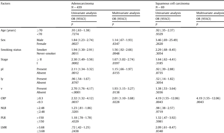
Univariate and multivariate subgroup analyses of PD-L1 protein expression and clinicopathological factors in patients with adenocarcinoma and squamous cell carcinoma.
Factors Adenocarcinoma
N¼439
Squamous cell carcinoma N¼69
Univariate analysis Multivariate analysis Univariate analysis Multivariate analysis
OR (95%CI) OR (95%CI) OR (95%CI) OR (95%CI)
P P P P
Age (years) 70 .93 (.63e1.38) .92 (.35e2.37)
<70 .7274 .9329
Sex Male 1.84 (1.23e2.74) 1.14 (.67e1.93) 3.46 (.69e25.49)
Female .0027 .6347 .2620
Smoking status Smoker 1.94 (1.30e2.91) 1.56 (.92e2.66) 2.29 (.68e8.45)
Never-smoker .0011 .0948 .3054
Stage II 2.30 (1.49e3.56) 1.67 (1.02e2.74) 1.64 (.62e4.41)
I .0002 .0397 .3185
pl Present 2.11 (1.34e3.32) 1.15 (.66e1.97) .92 (.39e2.88)
Absent .0012 .6155 .8735
ly Present .96 (.54e1.67) .52 (.14e1.82)
Absent .8797 .3054
v Present 2.70 (1.76e4.17) 1.93 (1.15e3.27) 1.38 (.53e3.64)
Absent <.0001 .0138 .5036
CRP 0.3 2.32 (1.32e4.12) 2.01 (1.10e3.68) 4.19 (1.55e12.06) 4.19 (1.55e12.06)
<0.3 .0037 .0228 .0043 .0043
NLR >2.48 1.23 (.81e1.86) .98 (.38e2.57)
2.48 .3201 .9719
PLR >150 1.18 (.78e1.78) 1.32 (.47e3.92)
150 .4329 .5981
LMR >3.68 .72 (.42e1.25) 2.09 (.61e8.47)
3.68 .2438 .8340
PD-L1: programmed death-ligand 1, OR: odds ratio, CI: confidence interval, pl: pleural invasion, ly: lymphatic invasion, v: vascular invasion, CRP: C-reactive protein, NLR:
neutrophil-lymphocyte ratio, PLR: platelet-lymphocyte ratio, LMR: lymphocyte-monocyte ratio.
Fig. 1.Association score of programmed death-ligand 1 (PD-L1) expression (A-score) and odds ratio for PD-L1 expression (1% and 5% cut-off values) andEGFRmutation.
proposed, because a large tumor cell burden is likely to increase inflammatory cytokine levels, stimulating CRP production [29,30].
Concerning PD-L1 expression, our previous data also showed that PD-L1 was associated with advanced stage cancer and lympho- vascular invasion [15,16]. Thus, a consequence of the host immune response to tumors, which is seen with aggressive tumors, may lead to both elevated CRP levels and PD-L1 expression (Fig. 2B). Thus, we speculate that tumors expressing PD-L1 correspond to those with elevated CRP levels (Fig. 2B).
Our study has several limitations. First, serum CRP level is easily influenced by various physiological and pathological factors like acute and chronic infection and use of anti-inflammatory drugs.
Even though we excluded patients with inflammatory diseases or in which infectious events occurred before surgery, we might not have been able to exclude all patients whose serum CRP level was elevated by these factors. Moreover, because offluctuating serum CRP levels, at least two measurements of serum CRP level before surgical operation might be needed for more accurate data. Second, the current study included patients with only operable NSCLC.
Further studies focusing on a larger cohort of advanced NSCLC including inoperable cases should be performed to confirm the findings obtained in the current study. Third, the PD-L1 analysis for surgically resected NSCLC was conducted using a specific antibody against PD-L1 (SP142). According to the report by the Blueprint Working Group, the positive rate in detecting PD-L1 expression was lower using SP142 in comparison to other antibodies, such as 28e8, 22C3, and SP263 [31]. Thus, the association between serum CRP level and PD-L1 expression should be evaluated using other anti- bodies or cut-off values in future studies.
In conclusion, this is thefirst report to show a statistically sig- nificant association between serum CRP level and PD-L1 expression in surgically resected NSCLC. Thesefindings suggest that serum CRP seems to predict PD-L1 expression in patients with NSCLC however the clinical applicability is limited given the obtained AUC values.
Acknowledgements
We are grateful to Dr. Kenichi Kohashi for the helpful comments about pathologicalfindings.
Appendix A. Supplementary data
Supplementary data related to this article can be found at
https://doi.org/10.1016/j.suronc.2018.01.002.
References
[1] D. Morgensztern, M.J. Campo, S.E. Dahlberg, R.C. Doebele, E. Garon, D.E. Gerber, S.B. Goldberg, P.S. Hammerman, R.S. Heist, T. Hensing, L. Horn, S.S. Ramalingam, C.M. Rudin, R. Salgia, L.V. Sequist, A.T. Shaw, G.R. Simon, N. Somaiah, D.R. Spigel, J. Wrangle, D. Johnson, R.S. Herbst, P. Bunn, R. Govindan, Molecularly targeted therapies in non-small-cell lung cancer annual update 2014, J. Thorac. Oncol. 10 (1 Suppl 1) (2015) S1eS63.
[2] G.R. Oxnard, A. Binder, P.A. Janne, New targetable oncogenes in non-small-cell lung cancer, J. Clin. Oncol. 31 (8) (2013) 1097e1104.
[3] D.M. Pardoll, The blockade of immune checkpoints in cancer immunotherapy, Nat. Rev. Canc. 12 (4) (2012) 252e264.
[4] J. Brahmer, K.L. Reckamp, P. Baas, L. Crino, W.E. Eberhardt, E. Poddubskaya, S. Antonia, A. Pluzanski, E.E. Vokes, E. Holgado, D. Waterhouse, N. Ready, J. Gainor, O. Aren Frontera, L. Havel, M. Steins, M.C. Garassino, J.G. Aerts, M. Domine, L. Paz-Ares, M. Reck, C. Baudelet, C.T. Harbison, B. Lestini, D.R. Spigel, Nivolumab versus docetaxel in advanced squamous-cell non- small-cell lung cancer, N. Engl. J. Med. 373 (2) (2015) 123e135.
[5] L. Fehrenbacher, A. Spira, M. Ballinger, M. Kowanetz, J. Vansteenkiste, J. Mazieres, K. Park, D. Smith, A. Artal-Cortes, C. Lewanski, F. Braiteh, D. Waterkamp, P. He, W. Zou, D.S. Chen, J. Yi, A. Sandler, A. Rittmeyer, P.S. Group, Atezolizumab versus docetaxel for patients with previously treated non-small-cell lung cancer (POPLAR): a multicentre, open-label, phase 2 randomised controlled trial, Lancet 387 (10030) (2016) 1837e1846.
[6] R.S. Herbst, P. Baas, D.W. Kim, E. Felip, J.L. Perez-Gracia, J.Y. Han, J. Molina, J.H. Kim, C.D. Arvis, M.J. Ahn, M. Majem, M.J. Fidler, G. de Castro Jr., M. Garrido, G.M. Lubiniecki, Y. Shentu, E. Im, M. Dolled-Filhart, E.B. Garon, Pembrolizumab versus docetaxel for previously treated, PD-L1-positive, advanced non-small- cell lung cancer (KEYNOTE-010): a randomised controlled trial, Lancet 387 (10027) (2016) 1540e1550.
[7] H. Borghaei, L. Paz-Ares, L. Horn, D.R. Spigel, M. Steins, N.E. Ready, L.Q. Chow, E.E. Vokes, E. Felip, E. Holgado, F. Barlesi, M. Kohlhaufl, O. Arrieta, M.A. Burgio, J. Fayette, H. Lena, E. Poddubskaya, D.E. Gerber, S.N. Gettinger, C.M. Rudin, N. Rizvi, L. Crino, G.R. Blumenschein Jr., S.J. Antonia, C. Dorange, C.T. Harbison, F. Graf Finckenstein, J.R. Brahmer, Nivolumab versus docetaxel in advanced nonsquamous non-small-cell lung cancer, N. Engl. J. Med. 373 (17) (2015) 1627e1639.
[8] M. Reck, D. Rodriguez-Abreu, A.G. Robinson, R. Hui, T. Csoszi, A. Fulop, M. Gottfried, N. Peled, A. Tafreshi, S. Cuffe, M. O'Brien, S. Rao, K. Hotta, M.A. Leiby, G.M. Lubiniecki, Y. Shentu, R. Rangwala, J.R. Brahmer, K. Investigators, Pembrolizumab versus chemotherapy for PD-L1-positive non-small-cell lung cancer, N. Engl. J. Med. 375 (19) (2016) 1823e1833.
[9] X.Y. Jing, C.B. Huang, H.Y. Zhou, C.P. Li, L.L. Fan, J.G. Chen, G. Zhang, Y.Y. Liu, Z. Cui, D.L. Qi, J. Ma, Association between serum C-reactive protein value and prognosis of patients with non-small cell lung cancer: a meta-analysis, Int. J.
Clin. Exp. Med. 8 (7) (2015) 10633e10639.
[10] J. Wang, N. Kalhor, J. Hu, B. Wang, H. Chu, B. Zhang, Y. Guan, Y. Wu, Pre- treatment neutrophil to lymphocyte ratio is associated with poor survival in patients with stage I-III non-small cell lung cancer, PLoS One 11 (10) (2016), e0163397.
[11] P. Hu, H. Shen, G. Wang, P. Zhang, Q. Liu, J. Du, Prognostic significance of systemic inflammation-based lymphocyte- monocyte ratio in patients with lung cancer: based on a large cohort study, PLoS One 9 (9) (2014), e108062.
Fig. 2.Association of C-reactive protein and programmed death-ligand 1 from the aspect of host's chronic inflammatory status (A) and host immune response to tumor (B).
T. Akamine et al. / Surgical Oncology 27 (2018) 88e94 93
[12] N. Ding, Z. Pang, H. Shen, Y. Ni, J. Du, Q. Liu, The prognostic value of PLR in lung cancer, a meta-analysis based on results from a large consecutive cohort, Sci.
Rep. 6 (2016) 34823.
[13] S.J. Bagley, S. Kothari, C. Aggarwal, J.M. Bauml, E.W. Alley, T.L. Evans, J.A. Kosteva, C.A. Ciunci, P.E. Gabriel, J.C. Thompson, S. Stonehouse-Lee, V.E. Sherry, E. Gilbert, B. Eaby-Sandy, F. Mutale, G. DiLullo, R.B. Cohen, A. Vachani, C.J. Langer, Pretreatment neutrophil-to-lymphocyte ratio as a marker of outcomes in nivolumab-treated patients with advanced non-small- cell lung cancer, Lung Canc. 106 (2017) 1e7.
[14] O.T. Brustugun, M. Sprauten, A. Helland, C-reactive protein (CRP) as a pre- dictive marker for immunotherapy in lung cancer, J. Clin. Oncol. 34 (15_suppl) (2016), e20623.
[15] K. Takada, T. Okamoto, G. Toyokawa, Y. Kozuma, T. Matsubara, N. Haratake, T. Akamine, S. Takamori, M. Katsura, F. Shoji, Y. Oda, Y. Maehara, The expression of PD-L1 protein as a prognostic factor in lung squamous cell carcinoma, Lung Canc. 104 (2017) 7e15.
[16] K. Takada, T. Okamoto, F. Shoji, M. Shimokawa, T. Akamine, S. Takamori, M. Katsura, Y. Suzuki, T. Fujishita, G. Toyokawa, Y. Morodomi, S. Okano, Y. Oda, Y. Maehara, Clinical significance of PD-L1 protein expression in surgically resected primary lung adenocarcinoma, J. Thorac. Oncol. 11 (11) (2016) 1879e1890.
[17] J. Sheng, W. Fang, J. Yu, N. Chen, J. Zhan, Y. Ma, Y. Yang, Y. Huang, H. Zhao, L. Zhang, Expression of programmed death ligand-1 on tumor cells varies pre and post chemotherapy in non-small cell lung cancer, Sci. Rep. 6 (2016) 20090.
[18] M. Kohno, T. Okamoto, K. Suda, M. Shimokawa, H. Kitahara, S. Shimamatsu, H. Konishi, T. Yoshida, M. Takenoyama, T. Yano, Y. Maehara, Prognostic and therapeutic implications of aromatase expression in lung adenocarcinomas with EGFR mutations, Clin. Canc. Res. 20 (13) (2014) 3613e3622.
[19] M. Yotsukura, T. Ohtsuka, K. Kaseda, I. Kamiyama, Y. Hayashi, H. Asamura, Value of the glasgow prognostic score as a prognostic factor in resectable non- small cell lung cancer, J. Thorac. Oncol. 11 (8) (2016) 1311e1318.
[20] G. Toyokawa, K. Takada, T. Okamoto, M. Shimokawa, Y. Kozuma, T. Matsubara, N. Haratake, S. Takamori, T. Akamine, M. Katsura, F. Shoji, Y. Oda, Y. Maehara, Computed tomography features of lung adenocarcinomas with programmed death ligand 1 expression, Clin. Lung Canc. 18 (6) (2017) e375ee383.
[21] K. Takada, G. Toyokawa, T. Okamoto, S. Baba, Y. Kozuma, T. Matsubara, N. Haratake, T. Akamine, S. Takamori, M. Katsura, F. Shoji, H. Honda, Y. Oda, Y. Maehara, Metabolic characteristics of programmed cell death-ligand 1- expressing lung cancer on (18) F-fluorodeoxyglucose positron emission to- mography/computed tomography, Cancer Med 6 (11) (2017) 2552e2561.
[22] Y. Kawashima, S. Nishikawa, R. Ariyasu, J. Koyama, M. Saiki, T. Sonoda,
N. Takano, T. Oguri, S. Kitazono, Y. Tambo, N. Yanagitani, F. Ohyanagi, A. Horiike, T. Horai, M. Nishio, Monitoring of peripheral lymphocyte and neutrophil counts to predict efficacy of nivolumab (nivo), J. Clin. Oncol. 35 (15_suppl) (2017), e20583.
[23] M.I. Guillen, M.J. Gomez-Lechon, T. Nakamura, J.V. Castell, The hepatocyte growth factor regulates the synthesis of acute-phase proteins in human he- patocytes: divergent effect on interleukin-6-stimulated genes, Hepatology 23 (6) (1996) 1345e1352.
[24] A.T. Parsa, J.S. Waldron, A. Panner, C.A. Crane, I.F. Parney, J.J. Barry, K.E. Cachola, J.C. Murray, T. Tihan, M.C. Jensen, P.S. Mischel, D. Stokoe, R.O. Pieper, Loss of tumor suppressor PTEN function increases B7-H1 expression and immunoresistance in glioma, Nat. Med. 13 (1) (2007) 84e88.
[25] M. Marzec, Q. Zhang, A. Goradia, P.N. Raghunath, X. Liu, M. Paessler, H.Y. Wang, M. Wysocka, M. Cheng, B.A. Ruggeri, M.A. Wasik, Oncogenic kinase NPM/ALK induces through STAT3 expression of immunosuppressive protein CD274 (PD-L1, B7-H1), Proc Natl Acad Sci U S A 105 (52) (2008) 20852e20857.
[26] M.F. Chen, P.T. Chen, W.C. Chen, M.S. Lu, P.Y. Lin, K.D. Lee, The role of PD-L1 in the radiation response and prognosis for esophageal squamous cell carcinoma related to IL-6 and T-cell immunosuppression, Oncotarget 7 (7) (2016) 7913e7924.
[27] H. Liu, J. Shen, K. Lu, IL-6 and PD-L1 blockade combination inhibits hepato- cellular carcinoma cancer development in mouse model, Biochem. Biophys.
Res. Commun. 486 (2) (2017) 239e244.
[28] J.G. Lee, B.C. Cho, M.K. Bae, C.Y. Lee, I.K. Park, D.J. Kim, S.V. Ahn, K.Y. Chung, Preoperative C-reactive protein levels are associated with tumor size and lymphovascular invasion in resected non-small cell lung cancer, Lung Canc. 63 (1) (2009) 106e110.
[29] J.M. Jones, N.C. McGonigle, M. McAnespie, G.W. Cran, A.N. Graham, Plasma fibrinogen and serum C-reactive protein are associated with non-small cell lung cancer, Lung Canc. 53 (1) (2006) 97e101.
[30] M. Hara, Y. Matsuzaki, T. Shimuzu, M. Tomita, T. Ayabe, Y. Enomoto, T. Onitsuka, Preoperative serum C-reactive protein level in non-small cell lung cancer, Anticancer Res. 27 (4C) (2007) 3001e3004.
[31] F.R. Hirsch, A. McElhinny, D. Stanforth, J. Ranger-Moore, M. Jansson, K. Kulangara, W. Richardson, P. Towne, D. Hanks, B. Vennapusa, A. Mistry, R. Kalamegham, S. Averbuch, J. Novotny, E. Rubin, K. Emancipator, I. McCaffery, J.A. Williams, J. Walker, J. Longshore, M.S. Tsao, K.M. Kerr, PD-L1 immunohistochemistry assays for lung cancer: results from phase 1 of the Blueprint PD-L1 IHC assay comparison project, J. Thorac. Oncol. 12 (2) (2017) 208e222.