Biology
Biology fields
Okayama University Year 2007
C. elegans model for studying
tropomyosin and troponin regulations of
muscle contraction and animal behavior
Hiroaki Kagawa∗ Tomohide Takaya† Razia Ruksana‡
Frederick Anokye-Danso∗∗ Md. Ziaul Amin†† Hiromi Terami‡‡
∗Okayama University, [email protected] †Okayama University
‡Okayama University ∗∗Okayama University ††Okayama University ‡‡Okayama University
This paper is posted at eScholarship@OUDIR : Okayama University Digital Information Repository.
1
C. ELEGANS MODEL FOR STUDYING TROPOMYOSIN
AND TROPONIN REGURATIONS OF MUSCLE
CONTRACTION AND ANIMAL BEHAVIOR
Kagawa, H., Takaya, T., Ruksana, R., Anokye-Danso, F., Amin, Z., and Terami, H. Div. of Bioscience, Grad. Sch. of Sci. and Tech., Okayama University, JAPAN
1. BACKGROUND
There are two muscle tissues in the nematode Caenorhabditis elegans: the pharynx for feeding and the body wall for locomotion. These correspond to cardiac and skeletal muscles in vertebrates, respectively. Study of the muscle genes of C. elegans can be classified into three stages; first, mutant isolation and gene mapping, second, cloning and sequencing of the gene, and third, complete sequences of all genes. Many uncoordinated mutant animals have been isolated (Brenner 1974, Waterston, 1988, Moerman and Fire 1997) and the complete amino acid sequence of myosin heavy chain, twitchin, and paramyosin, (invertebrate specific core protein of thick filament), and were the first determined in any animals by analyzing the unc-54, unc-22, and unc-15 mutants, respectively (Karn et al., 1983, Benian
et al., 1989, Kagawa et al., 1989). Tropomyosin and troponin components are also present but as with
actin and myosin heavy chain in the worm, there are some differences in gene structure and sequence compared to those in other animals (Kagawa et al., 1995, Myers et al., 1996, Moerman and Fire 1997). Deficiencies of body wall troponin C or tropomyosin in C. elegans cause the Pat (paralyzed arrest at embryonic two-fold stage) phenotype (Williams and Waterston 1994, Terami et al., 1999) and those of troponin T cause Mup (muscle position abnormal) phenotype (Myers et al., 1996). After determining the complete genome sequences of the nematode (The C. elegans Sequence Consortium, 1998), we can find out how isoforms are related to each other. Only one troponin C gene, pat-10/tnc-1, is expressed in the body wall muscles and the gene defect causes a developmental arrest of the animals (Terami et al., 1999). This is the first report analyzing a troponin C mutant. Recently we analyzed the tissue expression patterns of the four troponin I genes and their interaction with two troponin C isoforms (Ruksana et al., 2005). It now possible to compare how the troponin complex works in muscle contraction in invertebrates since Ebashi, Ohtsuki and their school had described these systems in vertebrates (Ohtsuki et al., 1986). In this paper we review how the mutant genes for tropomyosin and the troponin components of C. elegans affect animal behavior. A summary of expression, mutation and RNA interference results on these isoforms are provided in Table 1.
2.1. Tropomyosin mutant shows lethal or Ca2+ signaling defect
In contrast to one gene encoding one protein in most other muscle genes in C. elegans the tropomyosin gene, tmy-1/lev-11, encodes four isoforms, which are two each for the pharynx and
2 VOLUME TITLE body wall muscles produced under the control of distal and internal promoters and splicing choice, respectively (Kagawa et al., 1995, Anyanful et al., 2001). The lev-11(st557) mutant animal, which was isolated as a Pat animal (Williams and Waterston 1994) produces no tropomyosin due to a mutation at the splice donor site of exon 1 (Kagawa et al., 1997). Another mutant lev-11(x12) animal, which was isolated as a levamisol-resistant animal (Lewis et al., 1980) has an amino acid substitution of glutamic acid to lysine at position 234 of tropomyosin (Kagawa et al., 1997).
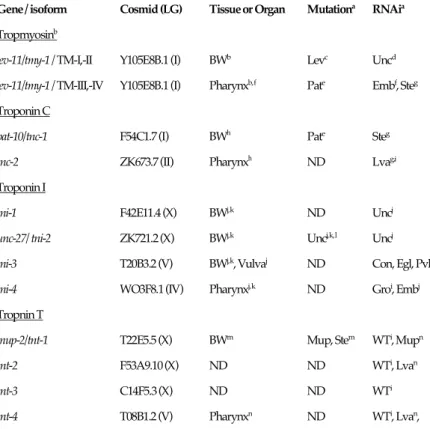
Table 1 Summary of gene, tissue expression and phenotype of tropomyosin and troponin isoforms
in C. elegans
Gene / isoform Cosmid (LG) Tissue or Organ Mutationa RNAia Tropmyosinb
lev-11/tmy-1 / TM-I,-II Y105E8B.1 (I) BWb Levc Uncd
lev-11/tmy-1 / TM-III,-IV Y105E8B.1 (I) Pharynxb, f Pate Embf, Steg
Troponin C
pat-10/tnc-1 F54C1.7 (I) BWh Pate Steg
tnc-2 ZK673.7 (II) Pharynxh ND Lvag,i
Troponin I
tni-1 F42E11.4 (X) BWj, k ND Uncj
unc-27/ tni-2 ZK721.2 (X) BWj, k Uncj, k, l Uncj
tni-3 T20B3.2 (V) BWj, k, Vulvaj ND Con, Egl, Pvlj
tni-4 WO3F8.1 (IV) Pharynxj, k ND Groi, Embj
Tropnin T
mup-2/tnt-1 T22E5.5 (X) BWm Mup, Stem WTi, Mupn
tnt-2 F53A9.10 (X) ND ND WTi, Lvan
tnt-3 C14F5.3 (X) ND ND WTi
tnt-4 T08B1.2 (V) Pharynxn ND WTi, Lvan,
aDesignations for phenotypes: Lev, levamisole-resistant; Unc, uncoordinated; Pat, paralyzed arrest at embryonic two-fold stage;
Emb, embryonic lethal; Ste, sterile; Con, constipated; Egl, egg laying-defective; Pvl, protruding vulva; Mup, muscle positioning; WT, wild type; ND, not determined. bThe tropomyosin gene, lev-11/tmy-1 encodes four isoforms (Kagawa et al., 1995). bKagawa et al., 1995; cLewis et al., 1980; dOno and Ono, 2002; eWilliams and Waterston, 1994; fAnyanful et al., 2001; gOno and
Ono, 2004; hTerami et al., 1999; iKamath et al., 2003; jRuksana et al., 2005; kBurkeen et al., 2004; lBrenner, 1974; mMyers et al.,
1996; nWormbase (on TnT by Allen et al.,) .
Exon 1 encodes the N-terminal part of CeTMI and CeTMII isoforms that are only expressed in body wall muscles. Mutant animals lacking CeTMI and CeTMII show a Pat phenotype suggesting that tropomyosin in body wall muscle is essential for the late development of C. elegans. As levamisole is a potent agonist of acetylcholine lev-11(x12) mutant animals cannot transmit the Ca2+
signal from the post-synaptic membrane to cause actin-myosin sliding step due to the position of the mutant tropomyosin. The mutation site of lev-11(x12) is in exon 7 a constitutive exon for all isoforms and causes the Glu234Lys substitution in tropomyosin (Kagawa et al., 1997). How this mutation affects interactions between troponin and tropomyosin or tropomyosin and actin is not
currently known. This mutant will be useful in solving how tropomyosin functions in Ca2+ signal
transduction.
2.2. A Pat animal has missed Ca2+ binding to site II and troponin I binding to the H-helix of the body wall troponin C
Two different genes; pat-10/tnc-1 and tnc-2, encode the two troponin C isoforms that are expressed in body wall and pharyngeal muscles, respectively (Terami et al., 1999). Comparisons to other troponin C sequences show that body wall troponin C, TnC-1, has the potential for Ca2+
binding at sites II and IV. Using TnC-1 produced in bacteria, the apparent Ca2+ binding constants,
Kapp, of both sites have been confirmed by fluorescence titration method to be 7.9 x1 05 M-1 and 1.2
x 106 M-1, respectively (Ueda et al., 2001). Study of the Mg2+-dependence of the Kapp showed that
both Ca2+-binding sites II and IV do not bind Mg2+ competitively. This means that the Ca2+ binding
of nematode TnC-1 is low affinity, fast dissociation and Ca2+-specific.
A troponin C mutant animal was isolated as one of the Pat mutants. These show paralyzed arrest phenotype at embryonic two-fold stage (Williams and Waterston 1994). We report that the
pat-10(st575) animal has two mutations in the body wall troponin C; an Asp64Asn (D64N)
substitution at site II and a deletion of the H-helix of the C-terminus (Terami et al., 1999). Recombinant mutant troponin C proteins produced in bacteria were assayed by SDS-Page to determine the molecular size and by Western blot analysis to assay their interactions with troponin I.
Figure 1 Experimental strategy of transgenic worms by injecting mutant gene of the troponin C. How designed mutant gene (top) affects on the animal is analyzed the phenotypic ratio by counting F2 animals (bottom). (Terami, PhD thesis, 2002)
4 VOLUME TITLE TnC-1m1: D64N showed no change in mobility with or without Ca2+ suggesting that site II has lost
Ca2+ binding. TnC-1m2: W153stop exhibited a mobility shift dependent on the presence or absence
of Ca2+, but did not bind troponin I due to missing H-helix.
A series of amino acid substitution experiments destroying and shortening the H-helix in the C-terminal region of TnC-1 suggest that the length and direction of the H-helix are important for troponin I binding. Especially two residues F152 and W153 were found to be essential for Ca2+
binding and troponin I binding (Takaya and Kagawa, unpublished). These results confirm that the H-helix of TnC-1 is essential for Ca2+ binding at site IV and troponin I binding. The topographical
position of the C-terminal of troponin C close to troponin I is consistent with the crystal structure of the troponin complex (Vassylyev et al., 1998).
We have also analyzed transgenic animals to study how troponin C mutants affect the animal phenotype by injecting recombinant mutant genes with the marker plasmid, pTG96 (sur-5::gfp) (Fig. 1) and found that development of mutant animals of m1: D64N (Ca2+ binding site II of TnC-1)
was almost to the same as wild type, but that adult transgenic animals dramatically lose their motility and show an egg-laying defect phenotype (Takaya, Terami and Kagawa, unpublished). This mutation is the first example of the miss-sense mutant worm that is homozygous for a mutant troponin C gene; the pat-10(st575) animal has two mutations in TnC-1 and is only kept in the heterozygous state (Williams and Waterston 1994, Terami et al., 1999). On the other hand the mutation of m2: W153stop in TnC-1 caused the Pat phenotype in transgenic animals. Both in vitro and in vivo experiments indicate that the Pat phenotype of the worm comes from a loss of Ca2+
binding to site IV or of troponin I binding to the H-helix.
2.3. Pharynx and body wall muscles express one- and three-troponin I isoforms, respectively
Four troponin I isoforms of the worm were characterized and compared to those of other animals (Fig. 2). It is interesting that the three body wall troponin I isoforms have unique glutamate- rich C-terminal extensions. The C-termini of troponin T in Drosophila and crayfish Astacus also show conserved glutamate-rich extensions (Fyrberg et al., 1990; Benoist et al., 1998; Domingo et al., 1998). It has been proposed that the C-terminal extension might enhance cooperation of troponin-tropomyosin complexes within thin filaments (White et al., 1987). The C. elegans troponin I extensions may have a homologous function. Glutamate has a negative charge, which may contribute to protein-protein interactions under different Ca2+ concentrations. The C-terminal
hydrophilic regions of the three body wall type troponin I isoforms could be important for interactions with TnC-1 or TnT in the worm body wall muscles.
Tissue expression patterns were determined using lacZ/gfp/rfp reporter gene assays. The tni-1,
tni-2/unc-27 and tni-3 genes, each encoding one troponin I isoform, are expressed in body wall,
vulval and anal muscles at different intensities, but tni-4 was expressed solely in the pharynx. Knock down of tni-1 or tni-2 gene by RNA interference caused motility defects similar to the unc-27 (e155) mutant, which is a tni-2/unc-27 null allele. An RNAi experiment for tni-3 produced egg-laying defects, while similar experiment with tni-4 RNAi caused arrest at gastrulation (Table 1). These show that body wall troponin I isoforms are important for animal motility and egg-laying and that the pharynx troponin I is essential for animal development (Ruksana et al., 2005).
2.4. Body wall troponin I isoforms interact with only that of troponin C
In vitro overlay assays have been used to analyse interactions between the four troponin I isoforms to the two troponin C isoforms. The three body wall troponin I isoforms interacts with the body wall and pharyngeal troponin C isoforms, reciprocally, but the pharyngeal TnI-4 interacts only with the pharyngeal TnC-2. Our results suggest that body wall TnI genes have evolved following duplication of the pharynx gene and provide important data about gene duplication and functional differentiation of nematode troponin I isoforms (Ruksana et al., 2005). Recently we confirmed that the N-terminal part of troponin I interacts with troponin C of the worm (Amin and Kagawa, unpublished). This molecular nature is similar to the molluscan troponin I that interacts with the
troponin C of the Akazara scallop (Tanaka et al., 2005). The importance of the N-terminal function of troponin I could be common throughout the invertebrates.
Figure 2. Structural homology of troponin I isoforms of invertebrates and vertebrates
(A) The two regulatory regions, TnC-binding and actin/TnC-binding sites showed conserved homology among invertebrates. Numbers represent percentage homology. TnI-1, TnI-2 and TnI-3 of C. elegans had unique C-terminal extensions. (B) Phylogenic alignment of four TNI isoforms of C. elegans; CeTNI-1, CeTNI-2, CeTNI-3 and CeTNI-4 and TNIs of Drosophila, crayfish, Ciona body wall, Ciona heart, rabbit cardiac (Ra cardiac), rabbit slow (Ra slow) and rabbit fast (Ra fast). The tree was derived using CLUSTALW and TreeView software. (from Ruksana et al., 2005)
6 VOLUME TITLE Mutants for a troponin T heat-sensitive allele, mup-2(e2346ts) and for a putative null,
mup-2(up1) are defective in embryonic body wall muscle cell contraction and sarcomere
organization (Myers et al., 1996). The troponin T gene abundantly expresses in body-wall muscles. The mup-2(up1) mutation causes a termination codon near the NH2 terminus (Glu94) and
mup-2(e2346ts) is a termination codon in the C-terminal invertebrate specific tail (Trp342) (Myers et al., 1996). These results indicate that the body-wall troponin T is also essential for muscle
formation. At least three troponin T genes are expressed in the body wall and one is in the pharynx of the worm (WormBase, from a report of Allen et al.). This expression profile is similar to that of the troponin I isoforms. How isoforms function in muscle cells and interact with other troponin components will be known in future.
3. DISCUSSION
Mutants of tropomyosin and three troponin components of C. elegans have been isolated and characterized (Table1). The isolated mutants can be divided into two groups; one group are missense alleles having single amino acid substitutions in the protein, the other groups are nulls producing no product. The null mutant phenotypes are consistent with the results obtained in gene knock down experiments using RNAi. Recently we have been able to use the experimental data of the total genome sequence together with the mutant genes, and phenotypes of RNAi animals (WormBase). In combination with biochemical and biophysical approaches on tropomyosin and troponin mutants, we have been able to determine the relationships between the molecular nature of the proteins and the behavior of the animals. Interestingly, the mutants isolated map to one of the genes expressed in body wall muscles. Body wall troponin I and troponin T of the worm are encoded by three different genes, but by only one troponin C gene. This is the reason that troponin C mutant animal is isolated under control of genetic skills, analyzing the F2 ratio of the mutant animals that are kept in the heterozygous state (Williams and Waterston, 1994, Terami et al., 1999). This is also applicable to isolating null mutants of the pharynx type isoform because only one gene encodes one isoform. There are three muscle tissues of the body wall type; body wall for locomotion, vulva for egg-laying and anus for defecation. Isolated mutants of troponin I and troponin T are found only in the abundant body wall isoform genes of tni-2/unc-27 and mup-2/tnt-1, which are expressed in body wall muscles, respectively (Table 1). Some differences of expression control and tissue localization of the three troponin I genes are detected between these three muscle groups, but functional differences have not been found in comparisons of the amino acid sequences (Ruksana et
al., 2005). It is of interest to know how three isoforms of troponin I and troponin T interact each
other and are utilized in different tissues. How minor isoforms can function and interact to other components in different tissues is also of interest in understanding the molecular interactions and evolutionary relationships.
Sequence comparisons between the different isoforms and with other animals indicate that tropomyosin and troponin components have common interaction sites to other proteins and additional unique sequences in their molecules. The binding sites for troponin C and actin/troponin C are common for all isoforms of troponin I (Figure 2). The N-terminal extension is found in vertebrate cardiac and all invertebrate troponin I isoforms. There is a C-terminal hydrophilic regions in the three troponin I isoforms of C. elegans (Ruksana et al., 2005). This may be important for interactions with other proteins. Recently the crystal structure of the core troponin complex has been determined. The authors stated that the interaction sites between troponin and tropomyosin/actin might be unique to each molecule of the complex in different tissues and animals (Takeda et al., 2003). Ohtsuki and Morimoto and their colleagues have established an in vitro physiological testing system for studying interactions between exchanged troponin components in myofibrils (Shiraishi et
al., 1992). Using our system, a reverse genetic approach in which a designed gene can be introduced
into the animal is possible (Figure 2). With this we can determine which part of the molecule functions in muscle and subsequently contributes to animal behavior. These experiments offer two approaches to troponin function; one is to compare proteins from different species and another is related to mutant proteins. The former is useful in studying how molecules are changed during evolution. The later is applicable to understanding how mutations disturb tissue functions of animals
or even humans. As we have mentioned, troponin mutants that cause functional defects of these proteins in the nematode will only be apparent in animals homozygous for the mutants. Even in some human disorders in human symptoms will be detected by a combination of genome or gene sequencing approaches of the gene and phenotypic analysis of the transgenic model animal.
4. ACKNOWLEDGEMENT
This article will dedicate to Profs Setsuro Ebashi, Iwao Ohtsuki and their school for the fortieth anniversary of their seminal discoveries relating to the troponin system. This study originated the time when one of the authors, H. K. had learned biophysics from Profs. Fumio Oosawa and Sho Asakura, and molecular biology of the worm from Profs. Sydney Brenner and Jon Karn. Some information on the troponin C and tropomyosin mutants was owed from Profs. Robert Waterston and Benjamin Williams. We acknowledge Prof. Takayoshi Iio for analyzing Ca2+ binding
constant of the troponin C and Prof. John C. Sparrow for his generous help in critically reading the manuscript.
5. REFERENCES
Anyanful, A., Sakube, Y., Takuwa, K. and Kagawa, H., 2001, The third and fourth tropomyosin isoforms of Caenorhabditis
elegans are expressed in the pharynx and intestines and are essential for development and morphology. J. Mol. Biol.
313:525-537.
Benian, G.M., Kiff, J.E., Neckelmann, N., Moerman, D.G., and Waterston, W.H., 1989, Sequence of unusually large protein implicated in regulation of myosin activity in C. elegans. Nature 342:45-50.
Benoist, P., Mas, J.A., Marco, R., and Cervera, M., 1998, Different muscle-type expression of the Drosophila troponin T gene. J.
Biol. Chem. 273:7538-7546.
Brenner, S., 1974, The genetics of Caenorhabditis elegans. Genetics. 77: 71-94.
Burkeen, A.K., Maday, S.L., Rybicka, K.K., et al., 2004, Disruption of Caenorhabditis elegans muscle structure and function caused by mutation troponin I. Biophys. J. 86:991-1001.
Domingo, A., Gonzalez-Jurado, J., Maroto, M., et al., 1998, Troponin T is a calcium-binding protein in insect muscle in vivo phosphorylation, muscle-specific isoforms and developmental profile in Drosophila melanogaster. J. Muscle Res. Cell Motil.
19:393-403.
Fyrberg, E., Fyrberg, C.C., Beall, C. and Saville, D.L., 1990, Drosophila melanogaster troponin T mutations engender three distinct syndromes of myofibrillar abnormalities. J. Mol. Biol. 216: 657-675.
Kagawa H., Gengyo, K, McLachlan, A.D., Brenner, S. and Karn, J., 1989, Paramyosin gene (unc-15) of Caenorhabditis elegans- Molecular cloning, nucleotide sequence and models for thick filament structure. J. Mol. Biol. 207:311-333.
Kagawa, H., Sugimoto, K., Matsumoto, S., Inoue, T., Imadzu, H., Takuwa, K., and Sakube, Y., 1995, Genome structure, mapping and expression of the tropomyosin gene tmy-1 of Caenorhabditis elegans. J. Mol. Biol. 251:603-613.
Kagawa, H., Takuwa, K. and Sakube, Y., 1997, Mutations and expressions of the tropomyosin gene and the troponin C gene of
Caenorhabditis elegans. Cell Str. Funct. 22:213-218.
Kamath, R.S., Fraser, A.G., Dong, Y., et al., 2003 Systematical functional analysis of the Caenorhabditis elegans genome using RNAi. Nature. 421:231-237.
Karn J., Brenner, S. and Barnett, L., 1983, Protein structural domains in the Caenorhabditis elegans unc-54 myosin heavy chain gene are not separated by introns. Proc. Nat. Acad. Sci., U. S. A. 80:4253-4257.
8 VOLUME TITLE
Lewis, J.A., Wu, C-H., Berg, H., and Levin, J. H., 1980, The genetics study levamisole resistance in the nematode Caenorhabditis elegans. Genetics. 95:905-928.
Moerman, D.G. and Fire, A., 1997, Muscle: Structure, function and development. In C. elegans II (eds Riddle, D.L., Blumenthal, T., Meyer, B.J., and Priess, J.R.), pp 417–470, Cold Spring Harbor Laboratory Press, New York
Myers, C.D., Goh, P.Y., Allen, T.S., Bucher, E.A. and Bogaert, T., 1996, Developmental genetics analysis of troponin T mutations in striated and nonstriated muscle cells of Caenorhabditis elegans. J. Cell Biol. 132:1061-1077.
Ohtsuki, I., Maruyama, K. and Ebashi, S., 1986, Regulatory and cytoskeletal proteins of vertebrate skeletal muscle. Advan. Protein
Chem. pp38-67.
Ono, S. and Ono, K., 2002, Tropomyosin inhibits ADF/cofilin-dependent actin filament dynamics. J. Cell Biol. 156:1065-1076. Ono, K. and Ono, S., 2004, Tropomyosin and troponit are required for ovarian contraction in the Caenorhabditis elegans
reproductive system. Mol. Biol. Cell, 15:2782-2793.
Ruksana, R., Kuroda, K., Terami, H., Bando, T., Kitaoka, S., Takaya, T., Sakube, Y., and Kagawa, H., 2005, Tissue expression of four troponin I genes and their molecular interactions with two troponin C isoforms in Caenorhabditis elegans, Genes to Cells,
10:261-276.
Shiraishi, F., Kambara, M., and Ohtsuki, I., 1992, Replacement of troponin components in myofibrils.J. Biochem. (Tokyo)
111:61-65.
Takeda, S., Yamashita, A., Maeda, K. and Maeda, Y., 2003, Structure of the core domain of human cardiac troponin in the Ca2+-saturated form. Nature, 424:35-41.
Tanaka, H., Takeya, Y., Doi, T., Yumoto, F., Tanokura, M., Ohtsuki, I., Nishita, K., and Ojima, T., 2005, Comparative studies on the functional roles of N- and C-terminal regions of molluskan and vertebrate troponin I, FEBS J. 272:4475-4486.
Terami, H., Williams, B.D., Kitamura, S., Sakube, Y., Matsumoto, S., Doi, S., Obinata, T., and Kagawa, H., 1999, Genomic organization, expression and analysis of the Troponin C gene pat-10 of Caenorhabditis elegans, J. Cell Biol. 146:93-202. The C. elegans Sequence Consortium, 1998, Genome sequence of the nematode C. elegans: A platform for investigating biology.
Science, 282:2012-2018.
Ueda,T, Katsuzaki, H., Terami, H., Ohtsuka, H., Kagawa, H., Murase, T., Kajiwara, Y., Yoshioka, O., and Iio, T., 2001, Calcium-binding of wild type and mutant troponin Cs of Caenorhabditis elegans. Biochim. Biophys. Acta 1548:220-228. Vassylyev, D.G., Takeda, S., Wakatsuki, S., Maeda, K. and Maeda, Y., 1998, Crystal structure of troponin C in complex with
troponin I fragment at 2.3-A° resolution. Proc. Natl. Acad. Sci. USA. 95:4847-4852.
Waterston, R. H., 1988, Muscle. In The nematode Caenorhabditis elegans (ed Wood, W. B.), pp. 281–335Cold Spring Harbor Laboratory Press, New York
White, S.P., Cohen, C. and Phillips, G.N.,Jr., 1987, Structure of co-crystals of tropomyosin and troponin. Nature. 325:826-828. Williams, B.D. and Waterston, R.H., 1994, Genes critical for muscle development and function in Caenorhabditis elegans identified
through lethal mutations. J. Cell Biol. 124:475-490. WormBase; http://www.wormbase.org/