INTRODUCTION
Crohn’s disease was first reported as regional ileitis in 1932 (1). It is an intractable disease mainly noted in young adults:ulcers are formed in regions
ORIGINAL
The use of electrogastrography and external ultrasonography
to evaluate gastric motility in Crohn’s disease
Nao Kohno, Masahiro Nomura
*, Hiroshi Okamoto, Masako Kaji, and Susumu Ito
Department of Digestive and Cardiovascular Medicine, Institute of Health Biosciences, The University of Tokushima Graduate School, and *
Faculty of Integrated Art and Sciences, Depart-ment of Human and Social Sciences, The University of Tokushima, Tokushima, Japan
Abstract : Although Crohn’s disease is associated with various digestive symptoms, there have been few reports on gastric motility. In this study, we conducted a study of gastric motility in Crohn’s disease using 20 healthy subjects (N group) and 15 patients with Crohn’s disease (C group) by electrogastrography (EGG) using a Nipro electrogastrog-raph. An EGG was recorded for 30 minutes in a fasting state and after ingestion of 300 ml of a liquid meal. As an index of gastric emptying, the rate of change in the cross-sectional area of the gastric antrum was measured 1 and 15 minutes after ingestion of the liquid meal by external ultrasonography. In an EGG frequency analysis, waveforms with a peak of 3 cycles/minute (cpm) were noted in the N group, and the peak amplitude in-creased significantly after the ingestion of food. In the C group, division of the normal-gastria component was noted after the ingestion of food in 5 patients (33.3%%). In a com-parison of the peak amplitudes of fasting brady-gastria, normal-gastria, and tachy-gastria between the N and C groups, the peak amplitude was significantly increased in normal-gastria in the N group, and in brady-normal-gastria and tachy-normal-gastria in the C group. In a com-parison of the rates of food ingestion-induced changes in the peak amplitudes for brady-gastria, normal-brady-gastria, and tachy-gastria between the N and C groups, the peak ampli-tudes were significantly increased in normal-gastria in the N group, but not in the C group. In the case of gastric emptying investigated by external ultrasonography, the rate of food ingestion-induced change in the cross-sectional antrum area was signifi-cantly lower in the C group (50.5±9.2%%) than in the N group (65.0±8.5%%). For gastrointes-tinal motility, a 3 cpm normal-gastria represents efficient gastric motility. In the C group, the peak amplitudes of brady-gastria and tachy-gastria were significantly in-creased, but were low in normal-gastria in the fasting EGG, postprandial division of the normal-gastria component was noted, and the rate of food ingestion-induced increase in the normal-gastria peak amplitude was significantly lower than that in the N group, sug-gesting that patients with Crohn’s disease have a functional abnormality in, not only the small and large intestine, but also the stomach. J. Med. Invest. 53 : 277-284, August, 2006
Keywords : Crohn’s disease, gastric motility, electrogastrography, gastric emptying
Received for publication May 1, 2006 ; accepted June 12, 2006. Address correspondence and reprint requests to Nao Kohno, M.D., Department of Digestive and Cardiovascular Medicine, Institute of Health Biosciences, The University of Tokushima Graduate School, Kuramoto-cho, Tokushima 770-8503, Japan and Fax : +81-88-633-9235.
The Journal of Medical Investigation Vol. 53 2006
of the digestive tract from the oral cavity to the anus, accompanied by abdominal pain, diarrhea, and bloody stool. The ileum is affected (ileal type) in about 35% of diagnosed patients, and the ileum and colon (ileocolic type) in 45%, the right colon in many cases. Only the colon is affected in about 20% (granulomatous colitis), but the entire small intestine (jejunoileitis), stomach/duodenum, or esophagus are affected in rare cases. The perianal region is also affected in 1/4-1/3 of diagnosed patients.
Crohn’s disease frequently develops at the ter-minal of the small intestine, as described above, but it may cause an abnormality in gastric motility, even though the organic lesion does not extend to the stomach (2-6). In the present study, we inves-tigated gastric motility in Crohn’s disease by electrogastrography (EGG) and evaluating gastric emptying using external ultrasonography.
SUBJECTS AND METHODS
1. Subjects
The subjects were 20 healthy individuals (N group, mean age : 33.2 ± 8.4 years, 12 males and 8 females) and 15 patients with Crohn’s disease with lesions in the small and large intestine (C group, mean age: 35 ± 7.2 years, 10 males and 5 females). The subjects in the N group had no prior medical history of heart, lung, or digestive dis-eases and were clinically diagnosed as healthy based on normal physical findings and the ab-sence of any abnormality on standard lead electro-cardiography, a chest X-ray examination, urinaly-sis, or blood chemistry.
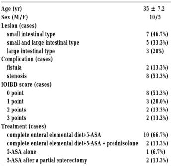
Table 1 shows the characteristics of the patients in the C group. Of the 15 patients, 7, 5, and 3 pa-tients, respectively, had small intestinal, small and large intestinal, and large intestinal types. Two and 8 patients had complications of an anal fistula and stenosis, respectively. Upper gastrointestinal endoscopy was performed on all patients, and no lesion was noted in the stomach, nor were any complications associated with gastrointestinal ob-struction detected. The International Organization of Inflammatory Bowel Disease (IOIBD) score (7) was 0 point in 8 patients, 1 point in 3, 2 points in 2, and 3 points in 2.
Before this study, 10 patients had been treated with a complete enteral elemental diet pulse of 5-aminosalicylic acid (5-ASA), 2 with a complete
enteral elemental diet pulse of 5-ASA, a pulse of prednisolone, 1 with 5-ASA alone, and 2 with 5-ASA afer a partial enterectomy. Crohn’s disease had been definitely diagnosed pathologically in all pa-tients.
The subjects refrained from eating and drinking from 21:00 on the day before the study, and the test was initiated at 9 : 00 at which time, percutaneous EGG and external ultrasonography were performed. Written informed consent was obtained before the test from all subjects.
2. Electrogastrography
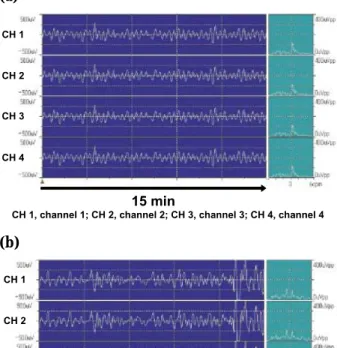
Percutaneous EGG was continuously recorded for 30 minutes in a fasting state and after ingestion of 300 ml of Elental!, using a portable electrogas-trograph (Nipro, EGG, A&D, Tokyo Japan). As shown in Figure 1, surface electrodes were at-tached to 4 sites (CH1-CH4) so as to surround the stomach, and the central electrode (C) was placed at the mid point between the navel and the xiphoid process. EGG was recorded by bipolar derivation between the central and 4 surface electrodes. The sampling period was 1 second, and the frequency of measurement was set to 2.1-6.0 cpm. Data were recorded at 13 bits. The influence of breathing was completely eliminated via the use of a 10th-degree filter, and body movement was minimized using a linear-phase filter to reduce the distortion of EGG signals.
The data were recorded using a portable elec-Table 1. Patient characteristics in Crohn’s disease
Age (yr) 35 ± 7.2
Sex (M/F) 10/5
Lesion (cases)
small intestinal type 7 (46.7%)
small and large intestinal type 5 (33.3%)
large intestinal type 3 (20%)
Complication (cases)
fistula 2 (13.3%)
stenosis 8 (53.3%)
IOIBD score (cases)
0 point 8 (53.3%)
1 point 3 (20.0%)
2 points 2 (13.3%)
3 points 2 (13.3%)
Treatment (cases)
complete enteral elemental diet+5-ASA 10 (66.7%) complete enteral elemental diet+5-ASA + prednisolone 2 (13.3%)
5-ASA alone 1 (6.7%)
5-ASA after a partial enterectomy 2 (13.3%) IOIBD, International Organization of Inflammatory Bowel Disease; 5-ASA, 5-aminosalicylic acid
N. Kohno, et al. Gastric motility in Crohn’s disease
trogastrograph (weighing 300 g), which was then transferred to a personal computer (Windows XP) through RC232C, and a Fast Fouriertransform (FFT) analysis was applied to 512 points using the EG-exclusive software program (NIPRO ESCI, A& D, Tokyo) (8-10). In the EGG analysis, the domi-nant frequencies at the 4 channels and their peak amplitudes were obtained. The peak amplitudes of brady-gastria (<2.4 cpm), normal-gastria (2.4-3.6 cpm), and tachy-gastria (>3.6 cpm) in the EGG spectrum analysis, and rates of change in peak amplitude after the ingestion of food were com-pared.
3. Gastric emptying examinations
Based on the method reported by Haruma et al., the gastric antrum area was determined 1 and 15 minutes after ingestion of the test meal using an external ultrasonic diagnosis system (central fre-quency of the probe : 3.75 MHz, Toshiba, Tokyo, Japan), and the rate of change (%) was evaluated (11-13). In a morning fasting state, the ultrasonic probe was attached to a site at which the abdomi-nal aorta, superior mesenteric artery, and the cross-section of the gastric vestibule could be imaged in the same field, and the cross-sectional area of the antrum was measured using a caliper built into the system. For the liquid test meal, 300 ml of Elental! (room temperature, 300 kcal, Ajinomoto Pharma,
Tokyo, Japan) was used.
4. Statistical analysis
All values are given as the mean ± standard deviation. Statistical analyses were performed using a personal computer. The data for the three groups were compared using one-way factorial analysis of variance (ANOVA) and two-way re-peated measures ANOVA (two-tail) and p<0.05 was considered to be statistically significant. StatView 5.0 software (SAS Institute Inc., Cary, North Caroline, USA) was used for the statistical analysis.
RESULTS
1. EGG waveform and frequency analysis in the N group
Figure 2 shows the fasting (panel a) and postpran-dial (panel b) waveforms at 4 EGG channels and their frequency analyses. Both the fasting and postprandial EGG waveforms recorded for 15 minutes at the 4 channels and the waveforms ap-peared at a frequency of approximately 3 times per minute (3 cpm). A frequency spectral analysis of Figure. 1 Recording positions of electrogastrogram.
CH 1, channel 1 ; CH 2, channel 2 ; CH 3, channel 3 ; CH 4, channel 4 ; C, central ; N, navel ; RMCL, right mid-clavicular line ; LMCL, left mid-clavicular line.
Figure. 2 Fasting (panel a) and postprandial (panel b) EGG waveforms at 4 channels and their frequency analyses in a reprensentative subject from the N group (35-year-old male) (a)
(b)
the EGG waveforms indicated that the dominant frequency was 2.9 cpm before and after ingestion of the test meal, suggesting that no change was induced by the ingestion of food. The mean peak amplitude of the waveforms at the 4 channels was 90µV in the fasting state, which increased to 230µV after ingestion.
2. EGG waveform and frequency analyses in the C group
1) Case 1
Figure 3 shows the fasting (panel a) and postpran-dial (panel b) waveforms at 4 EGG channels and their frequency analyses in the case of a 32-year-old male with large intestinal type Crohn’s disease. In a frequency spectral analysis, the dominant fre-quency was found to be 3.2 cpm in the fasting state. In the postprandial period, a bimodal divi-sion to 2.6 and 3.4 cpm waveforms was noted. The mean fasting and postprandial peak amplitudes for the 4 channels were 108µV and 98µV, respectively, suggesting no food ingestion-induced changes. A similar postprandial division of the normal-gastria component was noted in 5 of the 15 patients (33.3%) in the C group.
2) Case 2
Figure 4 shows the fasting (panel a) and postpran-dial (panel b) waveforms at 4 EGG channels and their frequency analyses in the case of a 43-year-old male with small/large intestinal type Crohn’s disease. No 3 cpm waveforms were noted at any of the 4 EGG channels in either the fasting or postpran-dial period, and relatively low-frequency wave-forms were noted. In a frequency spectral analy-sis, the dominant frequency was found to be 1.0 cpm, showing brady-gastria, and the peak amplitude was 53µV in the fasting state. A second small peak with an amplitude of 25µV was noted near 3.6 cpm. The postprandial dominant frequency was 1.2 cpm, showing brady-gastria, and a slight 2.9 cpm normal-gastria component appeared. The peak amplitudes of brady-gastria and normal-gastria were 35 and 48µV, respectively.
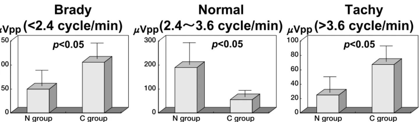
3) Comparison of the peak amplitudes of fasting brady-gastria, normal-gastria, and tachy-gastria be-tween the N and C groups
Figure 5 shows a comparison of the peak ampli-Figure. 3 Fasting (panel a) and postprandial (panel b) EGG
waveforms at 4 channels and their frequency analyses in a patient with large intestinal type Crohn’s disease (32-year-old male)
Figure. 4 Fasting (panel a) and postprandial (panel b) EGG waveforms at 4 channels and their frequency analyses in a pa-tient with small and large intestinal type Crohn’s disease (43-year-old male)
(a)
(a)
(b)
(b)
N. Kohno, et al. Gastric motility in Crohn’s disease
tudes of fasting brady-gastria, normal-gastria, and tachy-gastria between the N and C groups. The peak amplitude of normal-gastria was significantly larger in the N group than in the C group (p<0.05). In contrast, the peak amplitudes of brady-gastria and tachy-gastria were significantly larger in the C group than in the N group (p<0.05).
4) Comparison of the rates of food ingestion-induced changes in the peak amplitudes of brady-gastria, normal-gastria, and tachy-gastria between the N and C groups Figure 6 shows a comparison of the rates of food ingestion-induced changes in the peak
ampli-tudes of brady-gastria, normal-gastria, and tachy-gastria between the N and C groups. The normal-gastria peak amplitude increased 3.4 ± 1.1 and 1.6 ± 0.7 times in the N and C groups, respectively, showing a significantly higher rate of change in the N group (p<0.05). In contrast, no significant differences were noted in the rate of change of the brady-gastria or tachy-gastria components between the 2 groups.
3. Comparison of gastric emptying between the N and C groups
Figure 7 shows some typical gastric emptying
Figure.7 Examples of the gastric emptying test in a healthy subject (panel a) and a patient with Crohn’s disease (panel b) Figure. 6 Comparison of the rates of food ingestion-induced changes in the peak amplitudes of brady-gastria, normal-gastria, and tachy-gastria between the N and C groups
Figure. 5 Comparison of the peak amplitudes of fasting brady-gastria, normal-gastria, and tachy-gastria between the N and C groups
(a) (b)
test results after the oral ingestion of 300 ml of Elental!for a healthy subject (panel a) and a pa-tient with Crohn’s disease (panel b) using exter-nal ultrasonography. Gastric emptying in the healthy subject and the patient with Crohn’s disease were 72.0% and 46.3%, respectively, showing a decreased gastric emptying ability in Crohn’s disease.
Figure 8 shows a comparison of the rates of food ingestion-induced changes in the cross-sectional gastric antrum area measured by exter-nal ultrasonography between the N and C groups. The rates of change were 65.0 ± 8.5% and 50.5 ± 9.2% in the N and C groups, respectively, showing a significant decrease in the C group (p<0.05). However, there was no relationship between gas-tric motility and IOIBD score.
DISCUSSION
Only a few studies of gastrointestinal motility in Crohn’s disease have been reported, in which gas-trointestinal motility was investigated by manome-try and cinematography. The results generally indicated that gastrointestinal motility is impaired in Crohn’s disease (3,14). Delayed gastric empty-ing in Crohn’s disease patients with large intesti-nal lesions, as evidenced by scintigraphic gastric emptying tests have been reported (2). However, there has been only a single study, in which EGG was used to investigate gastric motility in 8 patients with Crohn’s disease. The results indicated that the 3 cpm waves or the peak of dominant frequency did not significantly increase after the ingestion of food compared to healthy subjects (15), but no further investigation was performed. In our study, EGG 3 cpm waves, which may represent effective
motility with food ingestion, were divided, the brady-gastria frequency increased, and gastric emptying decreased in Crohn’s disease, as evi-denced by external ultrasonography, suggesting that not only the small and large intestine but also gastric electrical activity and gastric emptying are impaired in Crohn’s disease.
In 1932, Crohn et al.(1) initially reported Crohn’s disease as an idiopathic intestinal disease that fre-quently occurs in the ileal terminal. Impairment of the entire digestive tract from the oral cavity to anus was subsequently clarified. The etiology re-mains unknown, and it progresses with repeating episodes of exacerbation and remission. The main lesion is present in the small or large intestine, or both in many cases, and less frequently in the stomach, but symptoms related to gastric motility are frequently noted. The close involvement of im-paired intestinal motility in the development of digestive symptoms has been noted, and changes in gastric motility may be a particularly important factor of digestive symptoms. Since the detection of changes in gastric emptying as changes in gastric motility has been reported (16,17), we investi-gated this issue using simple and non-invasive EGG and external abdominal ultrasonography.
Regarding studies of EGG, Alvarez first re-ported the presence of periodic changes in electri-cal potential in the stomach and small intestine in 1922 (18,19). Later studies confirmed that gastric electrical activity is an electrical phenomenon transmitted from the pacemaker located in the upper cardiac region of the stomach body toward the pyloric side (20, 21). The spontaneous action potential transmitted from the pacemaker is de-fined as electrical control activity, and is transmit-ted to the pyloric side at a frequency of 3 cpm and controls gastric motility (18-22). Electrical control activity is not derived from gastric motility, and electrical response activity is derived from con-tractile motility of the stomach. EGG is currently used to measure gastric electrical phenomena. Experimentally, EGG waveforms are similar to electrical activity and gastric motility is directly recorded from the gastric mucosa and serous membrane (23, 24). EGG is a simple non-invasive percutaneous method (21, 25, 26). Stable EGG re-cordings, have recently become possible, and clinical applications are now attracting attention.
Regarding Crohn’s disease, abnormal gastroin-testinal motilities have been detected by various methods (2-6). Bracci et al . reported that the 3 Figure. 8 Comparison of the rates of food ingestion-induced
changes in the cross-sectional gastric antrum area measured by external ultrasonography between the N and C groups
N. Kohno, et al. Gastric motility in Crohn’s disease
cpm waves did not significantly increase in pa-tients with Crohn’s disease compared to healthy subjects, and that the peak of the dominant fre-quency did not increase after food ingestion (15). Knoblauch et al .(14) reported that an impairment in gastrointestinal motiliy should be considered when Crohn’s disease is complicated by disph-agia. Furthermore, Annese et al .(3) reported on the Crohn’s disease-induced impairment of gas-trointestinal motility using conventional manome-try. Our study detected abnormal gastric motility in Crohn’s disease, consistent with previous reports. In an EGG frequency analysis, brady-gastria and tachy-gastria significantly increased and normal-gastria decreased in the fasting state, while the division of the normal-gastria component and a significantly lower rate in the food ingestion-induced increase in the normal-gastria peak ampli-tude were noted in the patients with Crohn’s disease compared to the healthy subjects, and these factors may be the cause of the abnormal gastric motility.
There were no relation between severity of Crohn’s disease and gastric motility using EGG and external ultrasonography in the present study. IOIBD score is an index to evaluate severity of Crohn disease. Crohn’s patients with remission (IOIBD score ; 0-1 point) were 11 cases (73.3%) in this study, and it was suggested that these patients influenced the correlation between gastric motility and IOIBD score.
It has been reported that 3 cpm normal-gastria represents the most efficient gastrointestinal motility (18-22), suggesting that the functional abnormal-ity is present in not only the small and large intestine but also the stomach in causes of Crohn’s disease. The rate of food ingestion-induced change in the cross-sectional gastric antrum area simultane-ously measured by external ultrasonography is considered to be a direct measure of gastric emp-tying (11-13, 27). Compared to the healthy subjects, gastric emptying was decreased in patients with Crohn’s disease, thus confirming the EGG find-ings.
REFERENCES
1. Crohn BB, Ginzburg L, Oppenheimer GD : Regional ileitis : a pathologic and clinical entity. JAMA 99 : 1323-1329, 1932
2. Annese V, Bassotti G, Napolitano G, Frusciante V,
Bruno M, Conoscitore P, Germani U, Morelli A, Andriulli A : Gastric emptying of solids in patients with nonobstructive Crohn’s disease is some-times delayed. J Clin Gastroenterol 21 : 279-282, 1995
3. Annese V, Bassotti G, Napolitano G, Usai P, Andriulli A, Vantrappen G : Gastrointestinal motility disorders in patients with inactive Crohn’s disease. Scand J Gastroenterol 32 : 1107-1117, 1997
4. Fielding JF, Toye DK, Beton DC, Cooke WT. Crohn’s disease of the stomach and duode-num. Gut 11 : 1001-1006, 1970
5. Grill BB, Lange R, Markowitz R, Hillemeier AC, McCallum RW, Gryboski JD : Delayed gastric emptying in children with Crohn’s dis-ease. J Clin Gastroenterol 7 : 216-226, 1985 6. Gryboski JD, Burger J, McCallum R, Lange R :
Gastric emptying in childhood inflammatory bowel disease : nutritional and pathologic cor-relates. Am J Gastroenterol 87:1148-1153, 1992 7. de Dombal FT, Softley A : IOIBD report no 1. Observer variation in calculating indices of severity and activity in Crohn’s disease. Inter-national Organisation for the Study of Inflam-matory Bowel Disease. Gut 28 : 474-481, 1987 8. Smout AJ, van der Schee EJ, Grashuis JL :
What is measured in electrogastrography? Dig Dis Sci 25 : 179-187, 1980
9. Pezzolla F, Riezzo G, Maselli MA, Giorgio I : Electrical activity recorded from abdominal surface after gastrectomy or colectomy in hu-mans. Gastroenterology 97 : 313-320, 1989 10. Stern RM, Koch KL, Stewart WR, Lindblad
IM : Spectral analysis of tachygastria recorded during motion sickness. Gastroenterology 92 : 92-97,1987
11. Kusunoki H, Haruma K, Hata J, Tani H, Okamoto E, Sumii K, Kajiyama G : Real-time ultrasonographic assessment of antroduode-nal motility after ingestion of solid and liquid meals by patients with functional dyspepsia. J Gastroenterol Hepatol 15 : 1022-1027, 2000 12. Fujimura J, Haruma K, Hata J, Yamanaka H,
Sumii K, Kajiyama G : Quantitation of duode-nogastric reflux and antral motility by color Doppler ultrasonography. Study in healthy volunteers and patients with gastric ulcer. Scand J Gastroenterol 29 : 897-902, 1994 13. Okamoto E, Haruma K, Hata J, Tani H, Sumii K,
Kajiyama G : Effects of octreotide, a somato-statin analogue, on gastric function evaluated
by real-time ultrasonography. Aliment Phar-macol Ther 11 : 177-184, 1997
14. Knoblauch C, Netzer P, Scheurer U, Seibold F: Dysphagia in Crohn’s disease : a diagnostic challenge. Dig Liver Dis 34 : 660-664, 2002 15. Bracci F, Iacobelli BD, Papadatou B, Ferretti
F, Lucchetti MC, Cianchi D, Francalanci P, Ponticelli A : Role of electrogastrography in detecting motility disorders in children affected by chronic intestinal pseudo-obstruction and Crohn’s disease. Eur J Pediatr Surg 13 : 31-34, 2003
16. Pfaffenbach B, Wegener M, Adamek RJ, Ricken D : Electrogastrography in diagnosis of gastric motility disorders. Med Klin (Munich) 90 : 160-165, 1995
17. Riezzo G, Chiloiro M, Guerra V, Borrelli O, Salvia G, Cucchiara S : Comparison of gastric electrical activity and gastric emptying in healthy and dyspeptic children. Dig Dis Sci 45 : 517-524, 2000
18. Alvarez WC, Mahoney LJ : Action currents in stomach and intestine. Am J Physiol 58 : 46-493, 1922
19. Alvarez WC : The electrogastrogram and what it shows. JAMA 78 : 1116-1119, 1922
20. Kelly KA, Code CF, Elveback LR : Patterns of
canine gastric electrical activity. Am J Physiol 217 : 461-470, 1969
21. Hinder RA, Kelly KA : Human gastric paceset-ter potential. Site of origin, spread, and re-sponse to gastric transection and proximal gastric vagotomy. Am J Surg 133 : 29-33, 1977 22. Burnstock G, Holman ME, Prosser CL : Elec-trophysiology of smooth muscle. Physiol Rev 43 : 482-527, 1963
23. Brown BH, Smallwood RH, Duthie HL, Stoddard CJ : Intestinal smooth muscle electrical poten-tials recorded from surface electrodes. Med Biol Eng 13 : 97-103, 1975
24. Chen J, Vandewalle J, Sansen W, van Cutsem E, Vantrappen G, Janssens J : Observation of the propagation direction of human electrogastric activity from cutaneous recordings. Med Biol Eng Comput 27 : 538-542, 1989
25. Sarna SK : Gastrointestinal electrical activity : terminology. Gastroenterology 68 : 1631-1635,
1975
26. Abell TL, Malagelada JR : Electrogastrogra-phy.Current assessment and future perspec-tives. Dig Dis Sci 33 : 982-992, 1988
27. Bateman DN, Whittingham TA:Measurement of gastric emptying by real-time ultrasound. Gut 23 : 524-527, 1982
N. Kohno, et al. Gastric motility in Crohn’s disease