Dopamine receptor Dop1R2 stabilizes appetitive
olfactory memory through the Raf/MAPK pathway
著者
Sun Huan
学位授与機関
Tohoku University
学位授与番号
11301
博士論文
Dopamine receptor Dop1R2 stabilizes appetitive olfactory memory
through the Raf/MAPK pathway
(ドーパミン受容体 Dop1R2 は Raf/MAPK 経路を介して嗅覚記憶を
安定化する)
令和2年度
東北大学大学院生命科学研究科
脳生命統御科学専攻
神経行動学分野
Huan SUN
Abstract
In Drosophila, dopamine signaling to the mushroom body intrinsic neurons, Kenyon cells (KCs), is critical to stabilize olfactory memory. Nevertheless, little is known about the downstream intracellular molecular signaling underlying memory stabilization. Here I address this question in the context of sugar-rewarded olfactory long-term memory (LTM). I show that associative conditioning increases the phosphorylation of mitogen-activated protein kinase (MAPK) in KCs, which process relies on the presence of the dopamine receptor Dop1R2. Consistently, the attenuation of Dop1R2, Raf (a kinase) or MAPK expression in KCs selectively impairs LTM but not short-term memory. Moreover, I show that the LTM deficit caused by the knockdown of
Dop1R2 can be rescued by expressing active Raf in KCs. Thus, I conclude that the
Dop1R2/Raf/MAPK pathway is a pivotal downstream effector of dopamine signaling for stabilizing appetitive olfactory memory.
Keywords: appetitive olfactory learning, long-term memory (LTM), memory stabilization, mushroom body (MB), Kenyon cells (KCs), dopamine receptor Dop1R2, Raf kinase (Raf), mitogen-activated protein kinase (MAPK)
Acknowledgment
I would like to sincerely appreciate Dr. Hiromu Tanimoto and Dr. Toshiharu Ichionse, who embraced me into the lab of neuroethology. Despite my superficial knowledge in the field and insensitivity of science, these seniors unhesitatingly helped me tremendously to equip myself with scientific skills, getting financial support and even various daily life assistance, so that I was capable to complete this work. Many thanks to Dr. Tomoki Nishioka, Dr. Mutsuki Amano, Dr. Kozo Kaibuchi and Dr. Shu Kondo, who spent massive efforts in this collaborative project and providing excellent experimental data. Also, I deeply appreciate Ms. Ayako Abe, Dr. Nobuhiro Yamagata, Dr. Vladimiros Thoma, and Mr. Shun Hiramatsu, who has given advice/instruction as well as technical support from the inception till the terminal phase of this work. Finally, I would like to thank the Graduate School of Life Sciences, Otsuka Toshimi Scholarship Foundation and Data Science Program for the financial support throughout my study.
Content
I. Introduction ... 1
1. Exploration of the olfactory associative learning/memory in Drosophila ... 1
1.1 Olfactory associative learning... 1
1.2 Olfactory associative learning induced memory ... 2
1.3 Neural structure underlying learning and memory: the mushroom body ... 2
2. Dopamine signaling in associative olfactory learning/memory ... 3
2.1 Dopamine neurons in memory stabilization ... 3
2.2 Dopamine receptors in memory stabilization ... 4
3. Recapitulation of the work ... 4
II. Materials and Methods ... 6
Key Resources Table ... 6
1. GAL4/UAS binary system ... 7
2. RNA interference ... 8
3. Animal culture and strains ... 8
4. Behavioral assay ... 8
5. Western blot ... 10
6. Immunohistochemistry ... 10
7. Drug administration... 11
8. Data analysis and statistics ... 11
III. Results ... 12
1. Dopamine receptor Dop1R2 in KCs stabilizes appetitive memory ... 12
2. The Raf/MAPK pathway in KCs stabilizes appetitive memory ... 15
2.1 Raf is required for LTM processing ... 15
2.2 MAPK is required for LTM processing ... 19
3. Appetitive conditioning activates MAPK in a Dop1R2-dependent manner ... 21
3.1 Conditioning event activates MAPK transiently in fly heads ... 21
3.2 Associative learning activates MAPK in KCs in a Dop1R2-dependent manner ... 21
4. Dop1R2 and Raf interact during LTM processing ... 24
5. Over-activation and down-regulation of Raf impair LTM in distinct mechanisms ... 26
5.1 Over-activating Raf selectively impairs LTM ... 26
5.2 A simpler learning paradigm for ruling out the odor discrimination ... 26
5.3 Over-activation of Raf disrupts the odor discrimination during conditioning phase ... 26
IV. Perspective ... 34
1. Dop1R2 signaling in learning and memory... 34
2. Intracellular molecular mechanism of Dop1R2/Raf/MAPK pathway ... 35
3. Raf/MAPK in stabilization of memory ... 35
4. Dopamine receptors other than Dop1R2 in memory stabilization ... 36
5. Dop1R2 and MAPK phosphorylation ... 36
I. Introduction
The German psychologist Hermann Ebbinghaus characterized how the memory rapidly decays after learning, a model that is well known as the Ebbinghaus forgetting curve. Meanwhile, our brain is also stabilizing the memory that involves complex neuronal changes, so that we could have a stable long-term memory. Interestingly, in recent years the researchers found that dopamine signaling plays a role in memory stabilization in different species, including
Drosophila melanogaster, also known as the fruit fly. In this section, I would like to briefly
introduce (1) the olfactory learning and memory in fly, (2) the dopamine signaling in memory stabilization and (3) the agenda of this work.
1. Exploration of the olfactory associative learning/memory in Drosophila 1.1 Olfactory associative learning
Drosophila is capable to learn various tasks, including discrimination of tactile cues,
suppression of the tarsal reflex, and avoidance of previously shocked odors (Tully, 1984). Among all the learning task designs, olfactory associative learning is currently predominant in the field out of several reasons: (1) olfactory associative learning within the laboratories appears to be rather easily operated, the learning and memory ability assessed can be easily reproduced from various researchers; (2) the olfactory nervous systems are extraordinarily conserved between insects and vertebrates, ensuring the significance of the fly research; (3) the efficiency to perform experiments is incredibly high, a training session within several minutes could produce relatively robust memories (Busto et al., 2010).
Figure 1. Schematics of aversive (A) and appetitive (B) olfactory associative learning. A, pairing of
the aversive reinforcement electric shocks with odor induces aversive memory in fly, who averts the odor after training. B, pairing of the appetitive reinforcement sugar with odor induces appetitive memory in fly, who approaches the odor after training.
The design of olfactory associative learning literally consists of mainly two factors, olfactory cues and their reinforcers. Olfactory cues are non-nature produced ones and reinforcers may vary. The most commonly used ones in adult flies are electrical foot shocks (Tully, 1984) and sugar reward (Tempel et al., 1983), often referred to as aversive olfactory learning and appetitive olfactory learning, respectively (Fig. 1). Aversive olfactory learning is more widely
studied, but the canonical design of both aversive and appetitive basically follows the same framework so as to create comparable results (Tempel et al., 1983), the so-called differential learning. In this paradigm, two distinct odors (conditioned stimulus, CS) are presented to flies, one (CS+) is concurrently given together with electric shocks or sugar rewards (unconditioned stimulus, US), while the other odor (CS-) is not. Then a reciprocal training session is performed to eliminate the effect aroused by odor preference bias (Quinn et al., 1974). Afterwards, at the certain time retention, flies are forced to make a choice between the previously exposed odors, where we test their memories (see Materials and Methods, Fig. 6).
1.2 Olfactory associative learning induced memory
The retention of olfactory learning-induced memory (abbreviated as olfactory memory here) largely counting on the reinforcers: In aversive learning, a single trial of training induces memory that descends quickly within hours and close to vanish in a day (Quinn et al., 1974; Tully and Quinn, 1985). To facilitate memory, multiple trials of training with certain rest interval (often referred to as spaced training) can produce robust 24 hr memory (Quinn et al., 1974; Tully et al., 1994; Beck et al., 2000). On the other hand, one cycle of pairing sugar reinforcer with odor creates robust memory that lasts for days (Tempel et al., 1983; Krashes and Waddell, 2008; Colomb et al., 2009).
Retention time alone does not discriminate long-term from the short-term memory. In aversive learning, multiple trials of training without rest intervals (massed training) produces 24 hr memory, which is weaker than that produced by spaced training. Moreover, spaced training-generated 24 hr memory incorporates a large proportion of protein-synthesis dependent component, while massed training-generated one does not (Tully et al., 1994). In the case of appetitive learning, memory after 6 hr is indicated to undergo protein-synthesis events (Krashes and Waddell, 2008). Therefore, protein-synthesis seems to be a shared attribute in both aversive and appetitive LTM.
1.3 Neural structure underlying learning and memory: the mushroom body
Olfactory perception is initiated by the odorant binding to the olfactory sensory neurons on the antenna of the fly, from which signals are relayed to the projection neurons (PNs). PNs bifurcate and thus disseminate the information into two protocerebral regions, one of which is the dendritic branches of the kenyon cells (KCs), also known as calyx, in the MB. While the soma of KCs and calyx locate at the posterior side of the protocerebrum, they propagate the axons into the anterior side and interact with nearby neuropils, which region is called the MB lobes (Tanaka et al., 2008; Fig. 2A, adopted from Aso et al., 2014).
Among the modulatory neurons that innervate the MB, DANs are considered to be rather prevalent. Many of them arbor to some restricted sub-regions/compartments of the lobes, while a few arbor to the calyx. These DANs are thought to locally modify the KCs synapses onto some MB output neurons, and thus contribute to the associative learning and memory (Aso et al., 2014).
Figure 2. Anatomy of the neural structure involved in olfactory learning and memory. A, olfactory
information is projected to the mushroom body (MB). B, C, dopamine neurons MP1 (B) and PAM-1 (C) that innervates the MB. (Figure adopted from Aso et al., 2014.)
2. Dopamine signaling in associative olfactory learning/memory
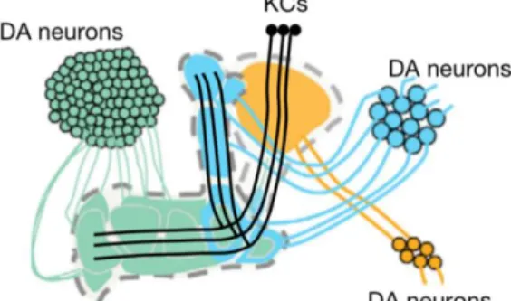
Dopamine signaling in the central nervous system is crucial for memory and behavioral adaptation. In addition to well-described roles in motivation and convey reinforcement signals (Busto et al., 2010; Puig et al., 2014), dopamine signaling has been shown to stabilize nascent memory traces in Drosophila, by several clusters of the dopamine neurons (DANs) that innervate the KCs (Fig 3., modified from Aso et al., 2012). How is it accomplished?
2.1 Dopamine neurons in memory stabilization
By blocking the neurotransmission from broad DANs, it was demonstrated that dopamine modulation is required for the aversive short-term memory (Schwaerzel et al., 2003). Likewise, photo-activation of the same population of DANs together with odor exposure creates
Figure 3. Schematic showing several
clusters of dopamine neurons that innervate the axons of the kenyon cells. (modified from Aso et al., 2012)
aversive memory (Claridge-Chang et al., 2009). Further study identified a subset of DANs in the protocerebral anterior medial (PAM) cluster can signal to substitute the appetitive reinforcement, and thus artificially creates appetitive memory (Liu et al., 2012).
The role of memory stabilization is shown to be conserved across animal phyla ranging from arthropoda to chordata (Plaçais et al., 2012; Musso et al., 2015; Rossato et al., 2009; O’Carroll et al., 2006; Takeuchi et al., 2016). In Drosophila, paired presentations of odor and electric shocks increase the activity of specific dopamine neurons (DANs) named MP1 (Fig. 2B, adopted from Aso et al., 2014) with oscillating intracellular calcium concentrations during consolidation (Plaçais et al., 2012). Consistently, the blockade of these MP1 DANs in the early retention period impedes long term memory (LTM) stabilization, while activation of them facilitates it (Plaçais et al., 2012; Plaçais et al., 2017). Intriguingly, blocking of the MP1 DANs during the time window of the first 45min after training impedes the sugar-rewarded LTM, indicating that they are also engaged in the stabilization of appetitive LTM with activity oscillation (Musso et al., 2015; Pavlowsky et al., 2018). Similarly, blocking the DANs named PAM-1 (Fig. 2C, adopted from Aso et al., 2014) during the first 30min after learning impedes the appetitive LTM, while the STM is not affected when they are blocked during the training, implying their roles in stabilizing memory (Ichinose et al., 2015).
2.2 Dopamine receptors in memory stabilization
Preceding introduction of dopamine signaling raises a question: which dopamine receptor in the KCs is responsible for learning and memory. There are four types of dopamine receptors in fly, all expressed in KCs (Croset et al., 2018): Dop1R1 and Dop2R, the functional counterparts of mammalian dopamine D1 and D2 receptors, respectively (Sugamori et al., 1995; Hearn et al., 2002); and two other invertebrate specific receptors, Dop1R2 and DopEcR (Mustard 2005; Srivastava et al., 2005). Dop1R1 is known to mediate reinforcement signaling (Kim et al., 2007), while receptors that have been reported to function for consolidated memories include Dop1R1 (Qin et al., 2012; Ichinose et al., 2015; Krüttner et al., 2015), Dop1R2 (Plaçais et al., 2017; Musso et al., 2015) and Dop2R (Scholz-Kornehl and Schwärzel, 2016). DopEcR is found to react to the hormone during courtship learning (Ishimoto et al., 2013), but never being reported for a role in olfactory memory.
3. Recapitulation of the work
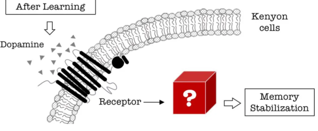
As stated above (2.1), dopamine signaling to KCs is indispensable in memory stabilization. Nevertheless, how dopamine regulates intracellular signaling in KCs to stabilize memory remains sparsely understood (Fig. 4). I here examined the role of different dopamine
receptors in KCs for appetitive memory. Furthermore, with combined biochemical, genetic and behavioral characterization, I found Dop1R2 stabilizes appetitive memory through the Raf and mitogen-activated protein kinase (MAPK) pathway in KCs. As MAPK is a well-characterized hub molecule to mediate synaptic plasticity, cytoskeleton reorganization or transcriptional change, this work provides a critical link between the extracellular dopaminergic modulation and the intracellular signaling in the context of LTM processing.
II. Materials and Methods
Key Resources TableREAGENT or RESOURCE SOURCE IDENTIFIER
Antibodies
Rabbit anti-Phospho-p44/42 MAPK Cell Signaling Technology Cat# 4370 Rabbit anti-p44/42 MAPK Cell Signaling Technology Cat# 4695 Goat anti-rabbit Alexa Fluor 680 antibody Thermo Fisher Scientific Cat# A20984
Rabbit anti-GFP Thermo Fisher Scientific Cat# A11122
Alexa Fluor 488 goat anti-rabbit Thermo Fisher Scientific Cat# A11034 Alexa Fluor 568 goat anti-rabbit Thermo Fisher Scientific Cat# A11036
Chemicals
4-methylcyclohexanol (MCH) Sigma-Aldrich 153095
octan-3-ol (OCT) Merck KGaA 222-226-0
PhosSTOP Sigma-Aldrich PHOSS-RO
Proteinase inhibitor cocktail Sigma-Aldrich S8830
RU486 Sigma-Aldrich M8046
Blocking one-P Nacalai Tesque 05999-84
Drosophila Strains
MB010B-GAL4 Janelia Farm Research Campus 2135061
P{MB-switch}12-1 Gift from Minoru Saitoe N/A
UAS-IVS-mCD8::GFP Bloomington Drosophila Stock Center BDSC_32189
GMR57C10-GAL4 Bloomington Drosophila Stock Center BDSC_39171
GMR13F02-GAL4 Bloomington Drosophila Stock Center BDSC_48571
UAS-Dop1R1.RNAi (P{TRiP.HMC02344}) Bloomington Drosophila Stock Center BDSC_55239
UAS-Dop1R2.RNAi (P{TRiP.HMC02893}) Bloomington Drosophila Stock Center BDSC_51423
UAS-Dop2R.RNAi (P{TRiP.HMC02988}) Bloomington Drosophila Stock Center BDSC_50621
UAS-DopEcR.RNAi (P{TRiP.JF03415}) Bloomington Drosophila Stock Center BDSC_31981
UAS-Raf.RNAi [1] (P{TRiP.HMC04133}) Bloomington Drosophila Stock Center BDSC_55863
UAS-Raf.RNAi [2] (P{TRiP.HMC03854}) Bloomington Drosophila Stock Center BDSC_55679
UAS-MAPK.RNAi (P{TRiP.HMS00173}) Bloomington Drosophila Stock Center BDSC_34855
UAS-Raf.GOF Kyoto Stock Center DGRC_106635
Raf-Venus Gift from Shu Kondo N/A
Dop1R1-Venus Gift from Shu Kondo N/A
Dop2R-Venus Gift from Shu Kondo N/A
DopEcR-Venus Gift from Shu Kondo N/A
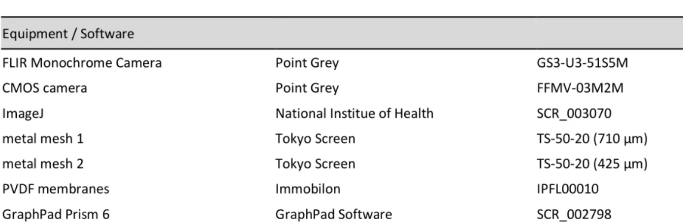
Equipment / Software
FLIR Monochrome Camera Point Grey GS3-U3-51S5M
CMOS camera Point Grey FFMV-03M2M
ImageJ National Institue of Health SCR_003070
metal mesh 1 Tokyo Screen TS-50-20 (710 µm)
metal mesh 2 Tokyo Screen TS-50-20 (425 µm)
PVDF membranes Immobilon IPFL00010
GraphPad Prism 6 GraphPad Software SCR_002798
1. GAL4/UAS binary system
In this work, I extensively used the transgenic GAL4/UAS system, for expressing the desired genes in certain cell populations. This system consists of two components: GAL4 gene, which codes yeast transcription activator proteins, and UAS (upstream activation sequence). The GAL4 gene is inserted randomly into the genome, therefore expresses the transcription factor GAL4 protein in certain cell populations. An optimal GAL4 binding site (upstream activation sequence) acting as enhancer, behind which a desired flanking gene is usually subcloned (Brand and Perrimon, 1993) (Fig. 5).
Figure 5. Schematic of how GAL4/UAS binary system works. After the copulation of transgenic flies
who carry GAL4 random insertion and who carry UAS, desired flanking gene (Gene-X) is expressed in the progeny.
In order to temporally restrict the function of this binary system, we also utilized a GAL4-progesterone receptor chimera, known as gene switch (Roman et al., 2001). This chimeric
protein contains a ligand-binding domain of the progesterone receptor, a p65 activation domain and a GAL4 DNA binding domain. Only when the ligand mifepristone (RU486) is present, this gene switch system is activated to execute its function to bind to UAS.
2. RNA interference
RNA interference (RNAi) was first identified in C. elegans to repress the gene expression. Once the RNAi reagents go into the cells, they would be recognized by dicer that processes them into small interfering RNA (siRNA). Subsequently siRNA will be incorporated into the RNA-induced silencing complex, to mediate gene silencing by degrade the target mRNA. In this work, short/small hairpin RNA based transgenic flies are used, which exhibit relatively better RNAi performance (Ni et al., 2011).
3. Animal culture and strains
Flies were cultured on the standard cornmeal medium at 24°C in a 12hr-12hr light-dark cycle. Canton-S was used as wild-type fly strain. MB010B-GAL4 (Aso et al., 2014) was obtained from Janelia Farm Research Campus. MBSW-GAL4 is the P{MB-switch}12-1 line (Mao et al., 2004). Fly strains obtained from the Bloomington Drosophila Stock Center (BDSC) include:
UAS-IVS-mCD8::GFP (#32189) (Pfeiffer et al., 2010), GM117C10-GAL4 (#39171), GMR13F02-GAL4 (#48571) (Jenett et al., 2012), UAS-Dop1R1.RNAi (P{TRiP.HMC02344},
#55239), UAS-Dop1R2.RNAi (P{TRiP.HMC02893}, #51423), UAS-Dop2R.RNAi (P{TRiP.HMC02988}, #50621), UAS-DopEcR.RNAi (P{TRiP.JF03415}, #31981), UAS-Raf.RNAi [1] (P{TRiP.HMC04133}, #55863), UAS-Raf.RNAi [2] (P{TRiP.HMC03854}, #55679) and UAS-MAPK.RNAi
(P{TRiP.HMS00173}, #34855). UAS-Raf.GOF (#106635, Brand and Perrimon, 1994) was acquired from the Kyoto Stock Center. Venus-tagged endogenous dopamine receptor strains were generated as described (Kondo et al., 2020).
4. Behavioral assay
Female and male flies aged from 5 to 9 days after eclosion were subjected to behavioral assay. Flies were wet-starved before and after conditioning so that the mortality reaches around 10% at the test.
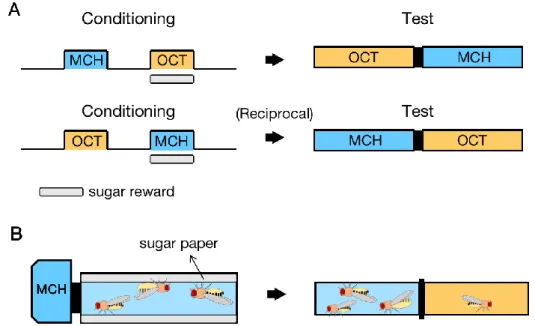
Appetitive differential conditioning: Unless specially stated, appetitive differential
conditioning was performed, during which, one odor (conditioned stimulus, CS+) was reinforced by dried sucrose reward, while the other odor (CS-) was not. Exposure of CS+ or CS- lasted for 1 min, with a 1 min interval in between. 4-methylcyclohexanol (MCH; Sigma-Aldrich) and octan-3-ol (OCT; Merck KGaA, Darmstadt, Germany), diluted in paraffin wax oil to 2% and
1.2%, respectively, were used as odors (Fig. 6A). Apparatus for conditioning was simplified as the schematics (Fig. 6B). The flies were enclosed into a cylindrical tube that contained a dried sucrose paper or dried clean paper, where they were exposed to certain odors driven from a perforated odor cup.
Test: During the test, flies were transferred into a T-maze, where two odors were sucked
from the two ends. Flies were allowed to freely explore between two arms and were recorded by cameras (FFMV-03M2M and GS3-U3-51S5M, Point Grey, Richmond, Canada) for 2 mins. Fly number in each arm was automatically counted using an Image J (National Institute of Health) custom-made macro. Based on these numbers, learning index (Tempel et al., 1983) was calculated for every second, based on the ensuing formula. An average of learning indices in the last 60 secs, when the performance usually reaches the plateau (Ichinose and Tanimoto, 2016), was used as a single data point.
Learning index = [(fly number in CS+) - (fly number in CS-)] / (total fly number)
Other types of conditioning: Besides differential conditioning stated above, we also
introduced (1) single-odor learning, where the protocol followed exactly that of differential learning, except that 1.2% OCT was replaced with the pure paraffin wax oil during both the conditioning and test phase; (2) unpaired stimuli presentation, where sucrose reward was presented first for 1 min, followed by a rest interval of 2 mins. Then 2% MCH and 1.2% OCT were presented each for 1 min, with another 1 min interval in between.
Figure 6. Appetitive differential conditioning. A, protocol of differential conditioning and testing. B,
5. Western blot
Starved flies were subjected to appetitive differential conditioning described above and were deep-frozen by liquid nitrogen at the indicated time points afterwards. The “naive” groups were similarly starved and mock-trained without odors and sugar reward, and subsequently were frozen. Around 200 frozen flies were put into a tube and were vortexed to separate heads and bodies. The mixture was poured onto a prechilled metal mesh (TS-50-20, 710 µm aperture, Tokyo Screen, Tokyo, Japan) to sieve the bodies, and subsequently onto another mesh (TS-50-20, 425 µm aperture, Tokyo Screen, Tokyo, Japan) to sieve the heads. The frozen heads were ground to powder by using Multibeads shocker (Yasui Kikai, Osaka, Japan), and were resuspended in Lysis buffer (20mM Tris/HCl, pH 7.4, 1mM EDTA, 1mM dithiothreitol, 150mM NaCl, 1% igepal CA-630, PhosSTOP (Sigma-Aldrich, Missouri, USA), Proteinase inhibitor cocktail (Sigma-Aldrich, Missouri, USA)). The samples were sonicated and were centrifuged at 20,000 xg for 20 min. Supernatants were collected and used as the protein extracts. Concentrations of the protein extracts were measured by BCA assay (Wako, Miyazaki, Japan).
For western blot analysis, 15 μg of each sample was loaded to 10% acrylamide gel and subjected to SDS-PAGE. Separated proteins were transferred to PVDF membranes (Immobilon-FL, Millipore, Bedford, MA, USA). The membranes were blocked with Blocking one-P (Nacalai Tesque, Kyoto, Japan) for 1h and probed with primary antibodies (Rabbit anti-Phospho-p44/42 MAPK (CST #4370), or rabbit anti-p44/42 MAPK (CST #4695)). Goat anti-rabbit Alexa Fluor 680 antibody (Thermo Fisher Scientific Cat# A20984) was used as a secondary antibody. Bands of antibody binding were detected and quantified using Odyssey CLx imaging system (LI-COR Biosciences, Nebraska, USA).
6. Immunohistochemistry
MAPK, Venus-tagged dopamine receptor and Venus-tagged Raf immunohistochemistry: For
Venus-tagged dopamine receptors, Venus-tagged Raf and MAPK immunohistochemistry, brains were dissected in PBS, followed by the fixation in 4% paraformaldehyde in PBS for 2 hrs at room temperature. Brains were subsequently washed three times and incubated in blocking solution (PBS with 3% goat serum and 0.1% Triton X-100). Then brains were incubated with antibodies in PBS containing 1% goat serum and 0.1% Triton X-100. Following primary antibodies were used at the indicated dilution: rabbit anti-GFP (1:1000; Thermo Fisher Scientific Cat# A11122), rabbit anti-p44/42 MAPK (1:500; CST #4695). Alexa Fluor 488 goat anti-rabbit (1:1000; Thermo Fisher Scientific Cat# A11034) was used as the secondary antibody.
pMAPK immunohistochemistry: For immunohistochemistry of pMAPK (Fig. 15), flies were
paraformaldehyde and 15% saturated picric acid solution). Then brains were fixed in the preceding fixative for 2hrs on ice. Washing and blocking were carried out similarly as stated above, but on ice instead. Rabbit anti-Phospho-p44/42 MAPK (1:500, CST #4370) and Alexa Fluor 568 goat anti-rabbit (1:1000, Thermo Fisher Scientific Cat# A11036) were used as primary and secondary antibodies respectively at the indicated dilution. Images were obtained using Olympus FV1200 confocal microscope, acquired at the same time periods under the identical microscope settings. pMAPK positive cell counting was performed in a blinding strategy: samples were randomized by a third experimenter so that they could not be identified by the one who counted the cells. The average number from the two brain hemispheres was used as a single data point.
7. Drug administration
RU486 (mifepristone, Sigma-Aldrich) was administrated with food for two days, then with water during food-deprivation. RU486 was removed for the last 2 hrs before conditioning and after conditioning to avoid any nonspecific effects (Mao et al., 2004). RU486 was dissolved in ethanol (10mg/mL) and mixed with melted food or water in a final concentration of 200 µM (Mao et al., 2004). The same amount of ethanol was added to the food or water for the control groups.
8. Data analysis and statistics
Statistical analyses were performed on GraphPad Prism 6 (GraphPad Software). Data were analyzed with parametric statistics: one-sample t-test or one-way analysis of variance (ANOVA) followed by Sidak’s multiple comparisons, when the assumption of normal distribution (Shapiro-Wilk normality test) and homogeneity of variance (Bartlett’s test) were not violated. Otherwise nonparametric statistics: Kruskal-Wallis test followed by Dunn’s multiple comparisons were performed. The significance level of statistical tests was set to 0.05.
III. Results
1. Dopamine receptor Dop1R2 in KCs stabilizes appetitive memory
To examine the role of the dopamine receptors for appetitive memory retention, I
systematically characterized the requirement of them from short term memory (STM) to LTM. To this end, I knocked down each of them in KCs by transgenic RNAi, using a KC-specific split-GAL4 strain MB010B-GAL4 that labels /, ’/’ and KCs (Vogt et al., 2014).
To verify the effectiveness of the RNAi strains I used here, I introduced the transgenic flies with endogenous dopamine receptors tagged with the Venus yellow fluorescent proteins (Kondo et al., 2020), abbreviated here as Dop1R1-Venus, Dop1R2-Venus, Dop2R-Venus and
DopEcR-Venus flies. In conjunction with R13F02-GAL4 (KC-labeling) and corresponding RNAi
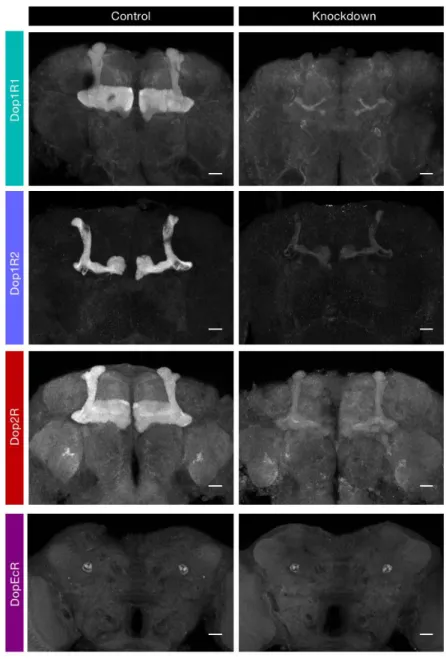
strains, my collaborator Shun Hiramatsu managed to visualize the knockdown effect (Fig. 7). It manifests that the transgenic RNAi effectively reduced the level of Dop1R1, Dop1R2 and Dop2R, but in all likelihood not DopEcR, in the MB.
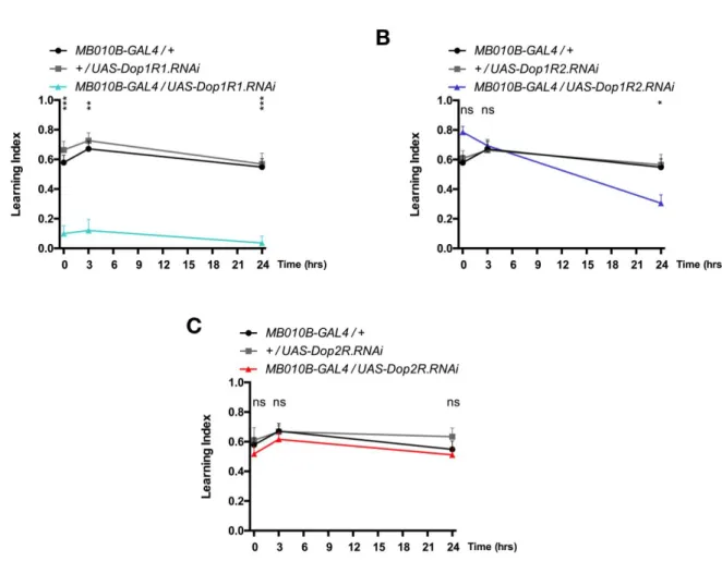
Subsequently, I tested the sugar-rewarded memory at 5 mins (STM), 3 hrs (often referred to as middle term memory, MTM) and 24 hrs (LTM, Krashes and Waddell, 2008; Colomb et al., 2009) after conditioning (Fig. 8). As a result, attenuation of Dop1R1 expression in KCs severely impaired appetitive memory irrespective of the retention time tested here (Fig. 8A). In contrast, attenuation of Dop1R2 expression in KCs left STM and MTM intact but impaired LTM (Fig. 8B), consistent with previous observation (Musso et al., 2015). I did not observe significant
differences when Dop2R expression was attenuated (Fig. 8C), and DopEcR cannot be verified as the UAS-DopEcR.RNAi does not seem to be effective.
These results suggest distinct roles of dopamine receptors: Dop1R1 and Dop1R2 for
mediating dopaminergic appetitive reinforcement and stabilizing memory, respectively; Dop2R may not be required for appetitive memory, at least by using the current RNAi strain.
Figure 7. Verification of the transgenic RNAi. Each dopamine receptor is knocked down in Kenyon cells
and the endogenous protein is visualized by tagging the Venus yellow fluorescent protein. The Venus protein is stained using an antibody described in Materials and Methods. Genotypes in the control and the knockdown groups are: (1st) Dop1R1-Venus,R13F02-GAL4/+ and
Dop1R1-Venus,R13F02-GAL4/UAS-Dop1R1.RNAi; (2nd) Dop1R2-Venus, R13F02-GAL4/+ and Dop1R2-Venus, R13F02-GAL4/UAS-Dop1R2.RNAi; (3rd) Dop2R-Venus/+;;R13F02-GAL4/+ vs Dop2R-Venus/+;; R13F02-GAL4/UAS-Dop2R.RNAi; (4th) DopEcR-Venus, R13F02-GAL4/+ and
DopEcR-Venus, R13F02-GAL4/UAS-DopEcR.RNAi. R13F02-GAL4 labels /, ’/’ and KCs. Z-projection images containing the horizontal and vertical lobes are shown. Scale bars, 20 m.
Figure 8. Differential engagement of dopamine receptors in short- and long- term olfactory appetitive
memory. Each of the four dopamine receptors is knocked down in KCs and appetitive memory is measured at different retention times. A, Attenuation of Dop1R1 expression severely impairs 5-min, 3-hr or 24-hr appetitive memory (MB010B-GAL4/UAS-Dop1R1.RNAi vs GAL4 and UAS controls: 5 min: p < 0.0001 to both; 3 hr: p = 0.0022 and p = 0.0002, respectively; 24 hr: p < 0.0001 to both). B,
Attenuation of Dop1R2 expression impairs 24-hr memory but leaves 5-min and 3-hr memory intact (MB010B-GAL4/UAS-Dop1R2.RNAi vs controls: 5 min: p = 0.1887 and p = 0.3635, respectively; 3 hr: p > 0.9 to both; 24 hr: p = 0.0216 and p = 0.0087, respectively). C, Attenuation of Dop2R expression does not significantly alter the memory. (MB010B-GAL4/UAS-Dop2R.RNAi vs controls: 5 min: p > 0.9 to both; 3 hr: p > 0.9 to both; 24 hr: p > 0.9 and p = 0.6906, respectively). n = 8-12. Bars and error bars, mean ± SEM, respectively. *p < 0.05, **p < 0.01, ***p < 0.001. ns, not significant (p > 0.05).
2. The Raf/MAPK pathway in KCs stabilizes appetitive memory 2.1 Raf is required for LTM processing
To probe into intracellular signaling under these dopamine receptors, I referred to the results of a phosphoproteomic analysis, where they investigated the dopamine receptor downstream effectors in the mouse striatum. This analysis identified many up- or down- regulated phosphorylation sites in response to the application of dopamine receptor agonists (Nagai, Nakamuta et al., 2016; Nagai, Yoshimoto et al., 2016). Emergence of numerous putative MAPK phosphorylation target proteins, along with Raf (MAPKKK) inspired me to hypothesize that the Raf/MAPK pathway as an effector of dopamine signaling in Drosophila as well.
To visualize the expression pattern of Raf, similar to above, I introduced the transgenic flies with endogenous Raf tagged with the Venus yellow fluorescent proteins, abbreviated here as
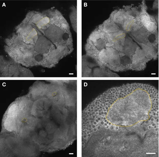
Raf-Venus, a gift from Shu Kondo. I confirmed that the ubiquitously expressed Raf does exist in
the KCs (Fig. 9).
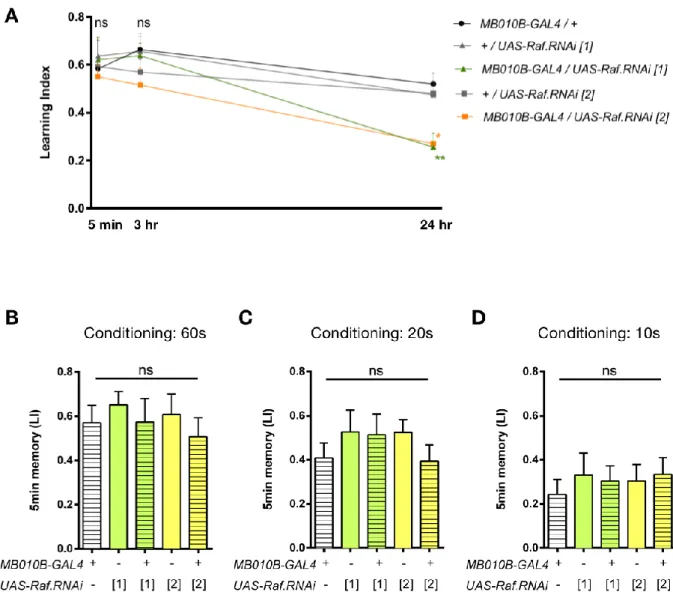
To investigate the role of Raf in appetitive memory maintenance, I downregulated Raf expression in KCs and tested memory retention. Attenuation of Raf expression using two independent RNAi insertions caused a selective deficit in 24-hr memory, while leaving 5-min and 3-hr memories intact (Fig. 10A), suggesting the selective role of Raf in LTM. As one may concern that the behavioral deficit resulted from lower basal performances, but not memory retention, I shortened the training duration from 60 to 20 and 10 seconds and the STM was still intact (Fig. 10B-D).
The evolutionary conserved oncogene Raf acts as a core component in multiple cellular processes during development (Leicht et al., 2007). To address the role of Raf in adult but not in developing KCs, I utilized the RU486-inducible transgenic expression in the MB (MBSW-GAL4) to restrict the Raf knockdown spatially and temporally (Mao et al., 2004; Roman et al., 2001). Before that, I first verified the effectiveness of MBSW-GAL4, using UAS-mCD8::GFP as the reporter (Fig. 11). Then I found the adult-specific Raf attenuation impaired 24-hr memory (Fig. 12B), in contrast to the intact STM (Fig. 12A) or LTM in the control group without RU486 application (Fig. 12C).
To confirm the effectiveness of the knockdown using MBSW-GAL4/UAS-Raf.RNAi, I utilized the Raf-Venus flies, and managed to verify the knockdown effect by RU486 administration (Fig. 12D).
Figure 9. Expression of endogenous Raf covers the KCs. Expression of Raf in all the MB lobes is confirmed, visualized by Raf.Venus staining. Outlined regions are lobes (A), lobes (B), peduncle (C) and calyx (D). Scale bars, 20 m.
Figure 10. Raf in the KCs is specifically required for appetitive long term memory. A, Attenuation of Raf
expression in KCs impairs 24-hr, but not 5-min or 3-hr memory (24 hr: F(4, 71) = 6.581, p = 0.0001;
MB010B-GAL4/UAS-Raf.RNAi[1] vs GAL4 and UAS controls: p = 0.0013 and p = 0.0062, respectively; MB010B-GAL4/UAS-Raf.RNAi[2] vs controls: p = 0.0031 and p = 0.0239, respectively, n = 11-17; 5 min: F(4, 44) = 0.3068, p = 0.8719; 3 hr: F(4, 45) = 1.091, p = 0.3723, n = 9-12). B-D, 5-min memory is intact when
conditioning duration is shortened to restrain the basal learning performance (B: F(4, 31) = 0.3555, p =
0.8382; C: F(4, 30) = 0.6884, p = 0.6057; D: F(4, 35) = 0.2163, p = 0.9276; n = 7-8). Bars and error bars, mean
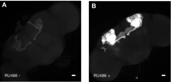
Figure 11. Validation of the treatment of drug administration in MBSW-GAL4 system. The MB GFP signal
is boosted in MBSW-GAL4/UAS-mCD8::GFP flies when they are fed with RU486 (B) comparing with non-fed group (A). Scale bars, 20 m.
Figure 12. Adult-specific Raf attenuation impairs long-term memory. A and B, Knockdown of Raf in adult
KCs using RU486-induced MBSW-GAL4 exhibits intact 5-min memory (A: F(4, 35) = 0.919, p = 0.4640, n =
8), while showing 24-hr memory defect (B: F(4, 53) = 6.173, p = 0.0004; MBSW-GAL4/UAS-Raf.RNAi[1] vs
controls: p = 0.0191 and p = 0.0150, respectively; MBSW-GAL4/UAS-Raf.RNAi[2] vs controls: p = 0.0113 and p = 0.0033, respectively, n = 9-14). C, Flies in control group without RU486 feeding attain intact 24-hr memory (F(4, 47) = 0.01589, p > 0.9, n = 9-11). D, Raf.Venus expression is suppressed in the MB
when the fly is treated with drug (right panel, RU486+), compared to the control (left panel, RU486-). lobes are outlined. Scale bars, 20 m. Bars and error bars, mean ± SEM, respectively. *p < 0.05. ns, not significant (p > 0.05).
2.2 MAPK is required for LTM processing
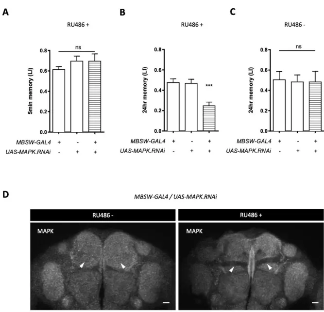
Subsequently, I questioned if the downstream effector MAPK is also required for LTM. Consistent with the selective LTM impairment of the Raf knockdown, attenuating the expression of the rolled (rl) gene encoding MAPK in adult KCs significantly impaired 24-hr memory, without perturbing 5-min memory (Fig. 13A-C).
To exclude the concern arousing by RNAi effectiveness, Shun Hiramatsu helped me perform immunohistochemistry of MAPK and verified the reduction of the protein in the MB (Fig. 13D). Collectively, these results reveal that the Raf/MAPK pathway is required in KCs for appetitive memory stabilization.
Figure 13. MAPK is specifically required for appetitive long term memory. A and B, Knockdown of MAPK
in adult KCs leaves 5-min memory intact (A: F(2, 21) = 0.8162, p = 0.4556, n = 8), while impairs 24-hr
memory (B: F(2, 42) = 11.39, p = 0.0001, n = 15). C, Flies without RU486 feeding show intact 24-hr
memory (F(2, 21) = 0.019, p > 0.9, n = 8). D, MAPK expression is suppressed in the MB when the RNAi is
induced in adult KCs (right panel, RU486+), compared to the control (left panel, RU486-). Arrowheads point to the lobes. Scale bars, 20 m. Bars and error bars, mean ± SEM, respectively. *p < 0.05, **p < 0.01, ***p < 0.001. ns, not significant (p > 0.05).
3. Appetitive conditioning activates MAPK in a Dop1R2-dependent manner
The consistent behavioral phenotype given by the downregulation of Dop1R2, Raf or MAPK (Fig. 8, 12, 13) inspired me to investigate on if there is any interaction between them. As no convenient and available tools to evaluate the activity of Raf in vivo, I chose to measure the activity of MAPK.
3.1 Conditioning event activates MAPK transiently in fly heads
Electric shock conditioning was reported to induce MAPK diphosphorylation, thereby activating the kinase (Pagani et al., 2009; Moressis et al., 2009; Miyashita et al., 2018; Zhang et al., 2018). To examine if appetitive training induces MAPK activation, my collaborator Tomoki Nishioka monitored the time course of phosphorylated MAPK (pMAPK) by sampling wild-type fly heads in different time points after appetitive conditioning (Fig. 14A). Quantification of the pMAPK ratio to the total MAPK (tMAPK) revealed significant increases immediately after learning (i.e. 1, 3 and 10 min), which returned to the basal level after 30 min. Interestingly, this experience-dependent increase of MAPK phosphorylation disappeared by neuronal knockdown of Dop1R2, whereas the genotypic controls attained MAPK activation (Fig. 14B).
3.2 Associative learning activates MAPK in KCs in a Dop1R2-dependent manner
In order to monitor learning-dependent MAPK activation in KCs, I next performed immunohistochemistry of pMAPK. The presentation of unconditioned stimuli may also activate MAPK, such as in water snails it was shown that food can induce MAPK phosphorylation (Ribeiro et al., 2005), here I introduced another control group, unpaired presentation of odors and sucrose (often referred as unpaired conditioning, see Materials and Methods), in contrast with associative conditioning.
By counting the number of pMAPK positive KCs, I found that the paired presentation of sugar reward and an odor induces MAPK phosphorylation in KCs, comparing to the unpaired group (Fig. 15A). Strikingly, associative training failed to induce MAPK phosphorylation in KCs where
Dop1R2 expression is downregulated (Fig. 15B). These results revealed that the association of
Figure 14. Experience-induced MAPK phosphorylation requires Dop1R2. A, Phosphorylation of MAPK
before and after appetitive conditioning in wild-type flies. Representative western blot shows phosphorylated MAPK (pMAPK) and total MAPK (tMAPK) in naive (N) and conditioned flies at different time points (from 1 min to 3 hr) after training. Ratio of pMAPK to tMAPK is plotted in the bar graph. pMAPK ratio elevates within the first 10 mins, then returns to basal level after 30 mins (1 min: t(16) =
2.859, p = 0.0114; 3 min: t(12.97) = 2.638, p = 0.0205; 10 min: t(12.81) = 2.628, p = 0.0211; 30 min: t(14.71) =
0.3083, p = 0.7622; 1 hr: t(15.59) = 0.2725, p = 0.7888; 3 hr: t(15.98) = 0.4946, p = 0.6276, n = 8-9). B, The
experience-dependent MAPK activation disappears in Dop1R2-attenuated flies. Western blot shows pMAPK and tMAPK in naive (N) and conditioned (3 min and 10 min after conditioning) transgenetic flies. Ratio of pMAPK to tMAPK is shown. pMAPK ratio relatively increases after conditioning in genetic control flies, but not in the Dop1R2-attenuated flies (R57C10-GAL4/+: F(2, 21) = 6.438, p = 0.0066;
+/UAS-Dop1R2.RNAi: F(2, 21) = 9.013, p = 0.0015; R57C10-GAL4/UAS-Dop1R2.RNAi: F(2, 21) = 0.007, p > 0.9,
n = 8). Bars and error bars, mean ± SEM, respectively. Each bars representing conditioned flies are
compared with the corresponding naive controls. *p < 0.05, **p < 0.01, ***p < 0.001. ns, not significant (p > 0.05).
Figure 15. Paired presentation of sugar reward and an odor induces Dop1R2-dependent MAPK
phosphorylation in KCs. A, pMAPK immunohistochemistry at the KC soma region (outlined) is shown in naive (left), 3 min after the unpaired presentation of sugar reward and an odor (middle) or 3 min after the paired presentation (right). KCs are labeled by mCD8::GFP and Dop1R2 is knocked down in KCs (Control in upper panels, UAS-mCD8::GFP/+;MB010B-GAL4/+. Dop1R2 knockdown in lower panels,
UAS-mCD8::GFP/+;MB010B-GAL4/UAS-Dop1R2.RNAi). B, The number of pMAPK positive KCs (pMAPK
counts) increases in a coincidence and Dop1R2 dependent manner. Unpaired presentation of sugar reward and an odor increases pMAPK counts compared to the naive in both control (F(5, 42) = 47.2, p <
0.0001; Naive vs Unpaired: p < 0.0001) and the Dop1R2 knockdown flies (Naive vs Unpaired: p < 0.0001). Further increase of pMAPK in paired group is observed in control flies (Paired vs Unpaired: p = 0.0009), but not in Dop1R2 knockdown flies (Paired vs Unpaired: p = 0.3865). A significant difference of pMAPK counts between the control and the Dop1R2 knockdown flies is detected after paired presentation (Control vs Dop1R2 knockdown: p < 0.0001). n = 7-9. pMAPK counts per hemispheres is shown. The sample images with median pMAPK counts in each group are selected for panel A. Bars and error bars, mean ± SEM, respectively. *p < 0.05, **p < 0.01, ***p < 0.001. ns, not significant (p > 0.05).
4. Dop1R2 and Raf interact during LTM processing
If the Raf/MAPK pathway is the intracellular target of Dop1R2 signaling for appetitive LTM, activation of Raf could surrogate Dop1R2 signaling.
I introduced a truncated form of Raf lacking the regulatory domain, gain-of-function Raf (Raf.GOF; Brand and Perrimon, 1994), for Raf overactivation, and expressed Raf.GOF together with Dop1R2-knockdown inside the KCs. Surprisingly, the 24-hr memory deficit in Dop1R2 knockdown flies was rescued by overexpressing Raf.GOF (Fig. 16, MBSW-GAL4,
UAS-Raf.GOF/UAS-Dop1R2.RNA). In contrast, the impairment due to the Dop1R1 knockdown
was not rescued by Raf.GOF expression (Fig. 16, MBSW-GAL4, UAS-Raf.GOF/UAS-Dop1R1.RNA). Taken together, these observations reveal that Raf/MAPK is a pivotal downstream effector of Dop1R2, but not Dop1R1, during appetitive memory stabilization.
Figure 16. Expression of constitutively active Raf rescues the LTM deficit caused by the Dop1R2
knockdown. Attenuation of Dop1R2 or Dop1R1 expression, as well as overexpression of active Raf (Raf.GOF) in adult KCs impairs 24-hr memory (F(8, 125) = 14.68, p < 0.0001;
MBSW-GAL4/UAS-Dop1R2.RNAi vs GAL4 and UAS controls: p = 0.0011 and p = 0.0083, respectively; MBSW-GAL4/UAS-Dop1R1.RNAi vs controls, p < 0.0001 to both; MBSW-GAL4/UAS-Raf.GOF vs controls: p = 0.0004 and p = 0.0034). Deficit in 24-hr memory caused by Dop1R2-attenuation is rescued by the
overexpression of Raf.GOF, whereas that by Dop1R1-attenuation is not (MBSW-GAL4,
UAS-Raf.GOF/UAS-Dop1R2.RNAi vs MBSW-GAL4/UAS-Dop1R2.RNAi: p = 0.0275; MBSW-GAL4, Raf.GOF/Dop1R1.RNAi vs MBSW-GAL4/Dop1R1.RNAi: p > 0.9). n = 12-17. Bars and error bars, mean ±
5. Over-activation and down-regulation of Raf impair LTM in distinct mechanisms
In the course of the experiments, I noticed that overexpression of Raf.GOF selectively impairs LTM (Fig. 16), similar situation happens to the knockdown of Raf (Fig. 10). Here I hypothesize that over-activation and down-regulation of Raf affect LTM in distinct ways.
5.1 Over-activating Raf selectively impairs LTM
To further study the impact of over-activating Raf to memory, I also introduced an active form of Ras, RasV12S35, which preferentially activates the Raf/MAPK pathway (Jiang and Edgar, 2009). Over-expression of Raf.GOF and RasV12s35 in the MB was achieved two days right before the experiments, which led to a selective LTM defect (Fig. 17 A-C). Similarly,
over-expression of Raf.GOF triggered by heat-shock also impaired LTM (Fig. 17 D). Apparently over-activation of Raf does not help LTM processing, one possible explanation will be provided in the following section.
5.2 A simpler learning paradigm for ruling out the odor discrimination
Notice that in the preceding experiments, I used the standard differential conditioning protocol (Fig. 6 or Fig. 18A), in which one of the two odors presented during conditioning was not paired with the sugar reward, and therefore serves as the reference odor (Tempel et al., 1983). Presentation of an unpaired odor during training has been shown to be critical for odor
discrimination (Schleyer et al., 2018; Barth et al., 2014). Therefore, I firstly simplified the paradigm by presenting only one odor
in training and test, referred as single-odor learning here (Fig. 18B), to rule out the odor discrimination effect (schematics shown as Fig. 18C).
Interestingly, overexpression of Raf.GOF in KCs did not lead to 24-hr memory deficit in single-odor learning (Fig. 19), while knockdown of Raf or Dop1R2 did (Fig. 20BC, 21BC), as in differential learning. Still consistently, expression of Raf.GOF in KCs significantly rescued the memory impairment of transient Dop1R2 knockdown in single-odor conditioning as well (Fig. 20D, 21D).
These observations imply that activation of Dop1R2 and Raf is critical for associative reward memory, whereas the over-activation of Raf broadly inside KCs may likely disrupt the odor discrimination in previous differential conditioning.
5.3 Over-activation of Raf disrupts the odor discrimination during conditioning phase Preceding experiments imply that the over-activation of Raf broadly inside KCs may likely disrupt the odor discrimination in differential conditioning. The difference between single odor
conditioning and differential conditioning (Fig. 18) is that whether there exists a
second/reference odor during training and testing phase. To further specify how the reference odor affect the behavior, I introduced the reference odor only in testing or training phase.
When the second odor was presented only to testing phase, over-activation of Raf did not impair LTM (Fig. 22), similar to the case of single odor conditioning (Fig. 19). Nevertheless, presentation of the reference odor only to training phase impaired LTM (Fig. 23), similar to the case of differential conditioning (Fig. 17).
To recapitulate, the Dop1R2/Raf/MAPK signaling cascade is critical in LTM processing, though it seems that over-activation of Raf broadly in the MB does not facilitate LTM, but instead disturbs the odor discrimination in training phase (Fig. 24).
Figure 17. Over-activating Raf selectively impairs LTM. A and B, Knockdown of MAPK in adult KCs leaves
5-min memory intact (A: F(4, 46) = 0.443, p = 0.7767, n = 9-11), while impairs 24-hr memory (B: F(4, 65) =
5.336, p = 0.0009; MB010B-GAL4/UAS-Raf.GOF vs GAL4 and UAS controls: p = 0.0148 and p = 0.0148, respectively; MB010B-GAL4/UAS-Ras.V12S35 vs controls: p = 0.0101 and p = 0.0052, respectively n = 13-15). C, Flies without RU486 feeding show intact 24-hr memory (F(4, 43) = 0.6813, p = 0.6088, n = 9-10).
D, Heat shock induced Raf.GOF overexpression impairs 24hr memory (F(3, 32) = 12.6, p < 0.0001;
Hsp.Raf.GOF/+ vs control: heat shock presented, p = 0.0053; heat shock absent, p = 0.9452; n = 8-10).
Bars and error bars, mean ± SEM, respectively. *p < 0.05, **p < 0.01. ns, not significant (p > 0.05).
Figure 18. Experimental design to simplify the learning protocol by using only one odorant. Comparing
to the differential learning (A), in single-odor learning (B) only one certain odor
Figure 19. Over-activation of Raf does not impair LTM in single-odor learning paradigm. A, When MCH
was used, overexpression of Raf.GOF does not impair 24-hr memory (F(2, 32) = 0.8472, p = 0.4380, n =
13-14). B, When OCT was used, overexpression of Raf.GOF does not impair 24-hr memory (F(2, 22) =
Figure 20. Expression of active Raf rescues the LTM deficit caused by the Dop1R2 knockdown in
single-odor (MCH) conditioning. A, Schematics of single-odor learning by using only
4-methylcyclohexanol (MCH). (Note that all preceding figures show the results of differential learning using two odors, MCH and octan-3-ol). B, C, In single-odor learning, knockdown of Raf or Dop1R2 impairs 24hr memory (B: p = 0.0062, n = 8-12; C: F(4, 36) = 6.789, p = 0.0004;
MBSW-GAL4/UAS-Raf.RNAi[1] vs controls: p = 0.0008 and p = 0.008, respectively;
MBSW-GAL4/UAS-Raf.RNAi[2] vs controls: p =0.0140 and p = 0.0311, respectively. n = 8-12). D,
Overexpression of Raf.GOF rescues the impaired 24-hr memory caused by Dop1R2-attenuation in KCs.
(F(5, 8) = 7.077, p < 0.0001; MBSW-GAL4, UAS-Raf.GOF/UAS-Dop1R2.RNAi vs
MBSW-GAL4/UAS-Dop1R2.RNAi: p = 0.0222; MBSW-GAL4/UAS-Raf.GOF vs controls: p > 0.9 and p =
0.4995, respectively; MBSW-GAL4/UAS-Dop1R2.RNAi vs controls: p = 0.0004 and p = 0.0011, respectively, n = 8-11). Bars and error bars, mean ± SEM, respectively. *p < 0.05, **p < 0.01, ***p < 0.001. ns, not significant (p > 0.05).
Figure 21. Expression of active Raf rescues the LTM deficit caused by the Dop1R2 knockdown in
single-odor (OCT) conditioning. A, Schematics of single-odor learning by using only
4-methylcyclohexanol (OCT). (Note that all preceding figures show the results of differential learning using two odors, MCH and octan-3-ol). B, C, In single-odor learning, knockdown of Raf or Dop1R2 impairs 24hr memory (B: F(2, 22) = 0.2033, p = 0.007; C: F(4, 37) = 9.74, p < 0.0001;
MBSW-GAL4/UAS-Raf.RNAi[1] vs controls: p = 0.0111 and p = 0.068, respectively;
MBSW-GAL4/UAS-Raf.RNAi[2] vs controls: p =0.0001 and p = 0.0008, respectively. n = 8-10). D,
overexpression of Raf.GOF rescues the impaired 24-hr memory caused by Dop1R2-attenuation in KCs.
(F(5, 57) = 7.0, p < 0.0001; MBSW-GAL4, UAS-Raf.GOF/UAS-Dop1R2.RNAi vs
MBSW-GAL4/UAS-Dop1R2.RNAi: p = 0.0467; , n = 9-12). Bars and error bars, mean ± SEM, respectively.
Figure 22. Overexpression of Raf.GOF does not alter 24hr memory when the reference odor is
presented only during test phase. A, Presence of the reference odor (OCT) in test phase does not impair 24hr memory (F(2, 31) = 0.145, p = 0.8657, n = 11-12). B, Presence of the reference odor (MCH) in test
phase does not impair 24hr memory (F(2, 31) = 1.42, p = 0.2577, n = 11-12). Bars and error bars, mean ±
Figure 23. Overexpression of Raf.GOF impairs 24hr memory when the reference odor is presented only
during training phase. A, Presence of the reference odor (OCT) in conditioning phase impairs 24hr memory (F(2, 21) = 16.2, p < 0.0001; MBSW-GAL4/UAS-Raf.GOF vs GAL4 and UAS control: p = 0.0004 and
p < 0.0001, respectively. n = 8). B, Presence of the reference odor (MCH) in conditioning phase impairs
24hr memory (F(2, 21) = 5.87, p < 0.0095; MBSW-GAL4/UAS-Raf.GOF vs GAL4 and UAS control: p = 0.0159
and p = 0.0121, respectively. n = 8). Bars and error bars, mean ± SEM, respectively. *p < 0.05, ***p < 0.001.
Figure 24. The hypothesis that overactivation of Raf in KCs broadly disturbs the odor discrimination in
IV. Perspective
To reveal the dopamine signaling pathway in memory stabilization, I chose to study it in the context of the appetitive learning in Drosophila, as the memory could be rapidly stabilized after one single training. I firstly studied the roles of the dopamine receptors and found that Dop1R2 is engaged in memory stabilization (Fig. 8). Similarly, I demonstrated that kinase Raf and MAPK are also required for memory stabilization (Fig. 12, 13), and showed that MAPK is transiently activated after associative learning (Fig. 14, 15). The common function of Dop1R2 and Raf/MAPK to stabilize memory inspired me that they might on the same pathway and I indeed gave the evidence from the perspective of biochemistry (Fig. 15) and behavior (Fig. 16). Taken together, these findings support the idea that Dop1R2 signaling through the Raf/MAPK pathway in KCs is critical in stabilizing appetitive memory (Fig. 25). Meanwhile, I investigated on how the overactivation of Raf in KCs impairs LTM, and hypothesized that overactivation of Raf in KCs broadly disturbs the odor discrimination in training phase (Fig. 24). Nevertheless, there are still some puzzles remain to be tackled, here listed the potential perspectives that may shed light on the future work.
Figure 25. Model of Dop1R2/Raf/MAPK pathway in appetitive memory stabilization. 1. Dop1R2 signaling in learning and memory
How is post-training Dop1R2 signaling triggered in this context? Accumulating evidence implies that Dop1R2 detects the basal dopamine release after learning (Berry et al., 2012; Musso et al., 2015; Ichinose et al., 2017). In aversive olfactory learning, the post-training enhancement of the oscillatory activity of MB-projecting DANs (MB-MP1 and MB-MV1) underlies LTM consolidation (Plaçais et al., 2012), and Dop1R2 in KCs is responsible for detecting the
enhanced dopamine signals (Plaçais et al., 2017). This signaling is also reported to mediate forgetting early labile memory (Berry et al., 2012), suggesting distinct neural mechanisms to regulate memories with different temporal dynamics. In appetitive learning, Dop1R2 is suggested to be the mediator of the oscillating DANs, which represent the energy value of the reward and consolidate LTM (Musso et al., 2015; Pavlowsky et al., 2018). Collectively, post-conditioning Dop1R2 signaling upon specific reinforcement input is a conserved mechanism to stabilize LTM. As MB-projecting DANs are also engaged in conveying reward information during memory acquisition (Liu et al., 2012; Burke et al., 2012; Yamagata et al., 2015), the Dop1R2/Raf/MAPK pathway might additionally be involved during acquisition of LTM.
2. Intracellular molecular mechanism of Dop1R2/Raf/MAPK pathway
In contrast to well-characterized receptor tyrosine kinase signaling, it is rather unexpected to find the Raf/MAPK pathway as a downstream target of Dop1R2, a G-protein coupled receptor (Fig. 16, 20D, 21D). Dop1R2 was recently shown to have a preferential affinity to the Gq subunit to elicit a robust intracellular Ca2+ increase upon ligand stimulation in KCs (Himmelreich et al., 2017; Handler et al., 2019). There are multiple lines of biochemical evidence suggesting that Gq-dependent Ca2+ signals could trigger several pathways, such as small GTPase Rap1, protein kinase C or Ras, to activate Raf (Goldsmith and Dhanasekaran, 2007; Liebmann, 2001). Furthermore, some reports suggested that calcium influx through N-methyl-D-aspartate receptor induces transient MAPK phosphorylation (English and Sweatt, 1996; Atkin et al., 1998). Hence, intracellular Ca2+ might be the key second messenger system to link Dop1R2 and Raf/MAPK in appetitive LTM.
3. Raf/MAPK in stabilization of memory
I found that MAPK has a pivotal role to stabilize appetitive memory in KCs (Fig. 13). MAPK signaling is known to regulate different cellular processes ranging from cytoskeletal dynamics to transcriptional modulation (Pullikuth and Catling, 2007; Giese and Mizuno, 2013). In
Drosophila, a recent work unveiled that MAPK stabilizes presynaptic structural changes in KCs
upon associative training with electric shocks, reportedly by changing the activity of an actin cytoskeleton regulator (Zhang et al., 2018). Such MAPK-induced cytoskeletal change might also occur in appetitive learning. Alternatively, a recent study showed that LTM consolidation involves MAPK translocation to the nuclei in KCs (Li et al., 2016). Consistently, it is reported that MAPK activates transcription factors like c-Fos and cAMP-response element binding protein
(CREB) in KCs to form aversive LTM (Miyashita et al., 2018). Appetitive LTM is also dependent on CREB in KCs (Krashes and Waddell, 2008; Widmer et al., 2018). Collectively, I propose that MAPK stabilizes appetitive memory by regulating these transcription factors. Future investigation on the downstream of the MAPK pathway should reveal the newly transcribed genes for memory stabilization.
4. Dopamine receptors other than Dop1R2 in memory stabilization
Although Dop1R1 is required for appetitive LTM (Fig. 8), it is well-known for driving second-messenger cAMP cascade and mediating aversive/appetitive reinforcement signaling (Kim et al., 2007; Liu et al., 2012). Moreover, I did not observe any tendency of rescue by Raf over-activation (Fig. 16), which implies that probably Dop1R1 and Dop1R2 are driving distinct second-messenger pathways and thus act different roles. Nevertheless, I cannot deny the connection between Dop1R1 and Raf, as for example, Dop1R1 in gamma lobes is found to be responsible for STM, while in alpha/beta lobes is required for LTM (Ichinose et al., 2015; Qin et al., 2012), but my manipulations did not distinguish the lobes.
Despite the knockdown of Dop2R does not look as effective as that of Dop1R1 (Fig. 7), indeed I have used this strain for aversive learning and observed 3-hr memory impairment (data not shown), consistent with previous observation (Scholz-Kornehl and Schwärzel, 2016). Still, further work could leverage more potent tools to study its function in appetitive memory.
DopEcR, a receptor for ecdysteroid and dopamine, is known to mediate various fly behaviors including ethanol sedation, sleep/awake circadian, stress response and so on. Unfortunately, I failed to acquire effective RNAi impact, judging from the DopEcR-Venus expression level (Fig. 7). Although so far there is no report on its function towards olfactory memory, it is known to mediate cAMP signaling in the MB, modulated by a steriod hormone named 20-hydroxy-ecdysone, and thus indispensable for courtship memory (Ishimoto et al., 2013). Since cAMP signaling is greatly engaged in both olfactory and courtship memory, further work could leverage an effective DopEcR-RNAi strain to disclose its function in distinct types of memories.
5. Dop1R2 and MAPK phosphorylation
The western-blot results indicate a potential interaction with Dop1R2 and MAPK. Notwithstanding, the basal pMAPK in Dop1R2 downregulation flies seemed not to be at the similar level of controls (Fig. 14B), which even sounds contradictory to my hypothesis. This could be resulted from the low precision by sampling the whole heads, and/or low resolution of pan-neuronal manipulation of Dop1R2. Dop1R2 couples with Gq in the MB, facilitating
calcium response when being activated (Himmelreich et al., 2017). On the other hand, Dop1R2 in fan-shape body is indicated to couple with Gi/o and impose inhibitory effects (Pimentel et al., 2016). Hence, function of Dop1R2 may differ depends on its locations. Still, these western blot results offer me with suggestive signs and prompted me to study it with better resolution.
Bibliography
Aso Y, Hattori D, Yu Y, Johnston RM, Iyer NA, Ngo TT, Dionne H, Abbott LF, Axel R, Tanimoto H, Rubin GM (2014) The neuronal architecture of the mushroom body provides a logic for associative learning. Elife 3:e04577.
Atkins CM, Selcher JC, Petraitis JJ, Trzaskos JM, Sweatt JD (1998) The MAPK cascade is required for mammalian associative learning. Nat Neurosci 1:602-9.
Barth J, Dipt S, Pech U, Hermann M, Riemensperger T, Fiala A (2014) Differential associative training enhances olfactory acuity in Drosophila melanogaster. J Neurosci 34:1819-37. Beck CDO, Schroeder B, Davis RL (2000) Learning Performance of Normal and Mutant
Drosophilia after Repeated Conditioning Trials with Discrete Stimuli. J Neurosci 20:2944– 2953.
Berry JA, Cervantes-Sandoval I, Nicholas EP, Davis RL (2012) Dopamine is required for learning and forgetting in Drosophila. Neuron 74:530-42.
Brand AH, Perrimon N (1993) Targeted gene expression as a means of altering cell fates and generating dominant phenotypes. Development 118:401-15.
Brand AH, Perrimon N (1994) Raf acts downstream of the EGF receptor to determine dorsoventral polarity during Drosophila oogenesis. Genes Dev 8:629-39.
Burke CJ, Huetteroth W, Owald D, Perisse E, Krashes MJ, Das G, Gohl D, Silies M, Certel S, Waddell S (2012) Layered reward signalling through octopamine and dopamine in Drosophila. Nature 492:433-7.
Busto GU, Cervantes-Sandoval I, Davis RL (2010) Olfactory Learning in Drosophila. Physiology (Bethesda) 25: 338–46.
Claridge-Chang A, Roorda RD, Vrontou E, Sjulson L, Li H, Hirsh J, Miesenböck G (2009) Writing memories with light-addressable reinforcement circuitry. Cell 139:405-15.
Colomb J, Kaiser L, Chabaud MA, Preat T (2009) Parametric and genetic analysis of Drosophila appetitive long-term memory and sugar motivation. Genes Brain Behav 8:407-15.
Croset V, Treiber CD, Waddell S (2018) Cellular diversity in the Drosophila midbrain revealed by single-cell transcriptomics. Elife 7:e34550.
English JD, Sweatt JD (1996) Activation of p42 mitogen-activated protein kinase in hippocampal long term potentiation. J Biol Chem 271:24329-32.
Giese KP, Mizuno K (2013) The roles of protein kinases in learning and memory. Learn Mem 20:540-52.
Goldsmith ZG, Dhanasekaran DN (2007) G protein regulation of MAPK networks. Oncogene 26:3122-42.
Dopamine Receptor Pathways Underlie the Temporal Sensitivity of Associative Learning. Cell 178:60-75.e19.
Hearn MG, Ren Y, McBride EW, Reveillaud I, Beinborn M, Kopin AS (2002) A Drosophila dopamine 2-like receptor: Molecular characterization and identification of multiple alternatively spliced variants. Proc Natl Acad Sci U S A 99:14554-9.
Himmelreich S, Masuho I, Berry JA, MacMullen C, Skamangas NK, Martemyanov KA, Davis RL (2017) Dopamine Receptor DAMB Signals via Gq to Mediate Forgetting in Drosophila. Cell Rep 21:2074-2081.
Ichinose T, Aso Y, Yamagata N, Abe A, Rubin GM, Tanimoto H (2015) Reward signal in a recurrent circuit drives appetitive long-term memory formation. Elife 4:e10719.
Ichinose T, Tanimoto H (2016) Dynamics of memory-guided choice behavior in Drosophila. Proc Jpn Acad Ser B Phys Biol Sci 92:346-357.
Ichinose T, Tanimoto H, Yamagata N (2017) Behavioral Modulation by Spontaneous Activity of Dopamine Neurons. Front Syst Neurosci 11:88.
Ishimoto H, Wang Z, Rao Y, Wu CF, Kitamoto T (2013) A Novel Role for Ecdysone in Drosophila Conditioned Behavior: Linking GPCR-mediated Non-Canonical Steroid Action to cAMP Signaling in the Adult Brain. PLoS Genet 9:e1003843.
Jenett A et al. (2012) A GAL4-driver line resource for Drosophila neurobiology. Cell Rep 2:991-1001.
Kondo S, Takahashi T, Yamagata N, Imanishi Y, Katow H, Hiramatsu S, Lynn K, Abe A,
Kumaraswamy A, Tanimoto H (2020) Neurochemical organization of the Drosophila brain visualized by endogenously tagged neurotransmitter receptors. Cell Rep 30:284-29. Krashes MJ, Waddell S (2008) Rapid consolidation to a radish and protein synthesis-dependent
long-term memory after single-session appetitive olfactory conditioning in Drosophila. J Neurosci 28:3103-13.
Krüttner S, Traunmüller L, Dag U, Jandrasits K, Stepien B, Iyer N, Fradkin LG, Noordermeer JN, Mensh BD, Keleman K (2015) Synaptic Orb2A Bridges Memory Acquisition and Late Memory Consolidation in Drosophila. Cell Rep 11:1953-65.
Leicht DT, Balan V, Kaplun A, Singh-Gupta V, Kaplun L, Dobson M, Tzivion G (2007) Raf kinases: function, regulation and role in human cancer. Biochim Biophys Acta 1773:1196-212. Li Q, Zhang X, Hu W, Liang X, Zhang F, Wang L, Liu ZJ, Zhong Y (2016) Importin-7 mediates
memory consolidation through regulation of nuclear translocation of training-activated MAPK in Drosophila. Proc Natl Acad Sci U S A 113:3072-7.
Liebmann C (2001) Regulation of MAP kinase activity by peptide receptor signalling pathway: paradigms of multiplicity. Cell Signal 13:777-85.
Liu C, Plaçais PY, Yamagata N, Pfeiffer BD, Aso Y, Friedrich AB, Siwanowicz I, Rubin GM, Preat T, Tanimoto H (2012) A subset of dopamine neurons signals reward for odour memory in Drosophila. Nature 488:512-6.
Mao Z, Roman G, Zong L, Davis RL (2004) Pharmacogenetic rescue in time and space of the rutabaga memory impairment by using Gene-Switch. Proc Natl Acad Sci U S A
101:198-203.
Miyashita T, Kikuchi E, Horiuchi J, Saitoe M (2018) Long-Term Memory Engram Cells Are Established by c-Fos/CREB Transcriptional Cycling. Cell Rep 25:2716-2728.
Moressis A, Friedrich AR, Pavlopoulos E, Davis RL, Skoulakis EM (2009) A dual role for the adaptor protein DRK in Drosophila olfactory learning and memory. J Neurosci 29:2611-25. Musso PY, Tchenio P, Preat T (2015) Delayed dopamine signaling of energy level builds
appetitive long-term memory in Drosophila. Cell Rep 10:1023-31.
Mustard JA, Beggs KT, Mercer AR (2005) Molecular biology of the invertebrate dopamine receptors. Arch Insect Biochem Physiol 59(3):103-17.
Nagai T, Nakamuta S, et al. (2016) Phosphoproteomics of the Dopamine Pathway Enables Discovery of Rap1 Activation as a Reward Signal In Vivo. Neuron 89:550-65.
Nagai T, Yoshimoto J, Kannon T, Kuroda K, Kaibuchi K (2016) Phosphorylation Signals in Striatal Medium Spiny Neurons. Trends Pharmacol Sci 37:858-871.
Ni JQ, Zhou R, Czech B, Liu LP, Holderbaum L, Yang-Zhou D, Shim HS, Tao R, Handler D,
Karpowicz P, Binari R, Booker M, Brennecke J, Perkins LA, Hannon GJ, Perrimon N (2011) A genome-scale shRNA resource for transgenic RNAi in Drosophila. Nat Methods 8:405-7. O'Carroll CM, Martin SJ, Sandin J, Frenguelli B, Morris RG (2006) Dopaminergic modulation of
the persistence of one-trial hippocampus-dependent memory. Learn Mem 13:760-9. Pagani MR, Oishi K, Gelb BD, Zhong Y (2009) The phosphatase SHP2 regulates the spacing effect
for long-term memory induction. Cell 139:186-98.
Pavlowsky A, Schor J, Plaçais PY, Preat T (2018) A GABAergic Feedback Shapes Dopaminergic Input on the Drosophila Mushroom Body to Promote Appetitive Long-Term Memory. Curr Biol 28:1783-1793.e4.
Pfeiffer BD, Ngo TB, Hibbard KL, Murphy C, Jenett A, Truman JW, Rubin GM (2010) Refinement of tools for targeted gene expression in Drosophila. Genetics 186:735-55.
Pimentel D, Donlea JM, Talbot CB, Song SM, Thruston AJF, Miesenbock G (2016) Operation of a homeostatic sleep switch. Nature 536: 333-337.
Plaçais PY, de Tredern É, Scheunemann L, Trannoy S, Goguel V, Han KA, Isabel G, Preat T (2017) Upregulated energy metabolism in the Drosophila mushroom body is the trigger for long-term memory. Nat Commun 8:15510.