Abbreviations: BTB, bromothymol blue; CFU, colony forming unit; MIC, minimum inhibitory concentration; SMA, sodium mercaptoacetic acid
The Clinical Aspects of
β
-Lactam-Resistant
Stenotro-phomonas maltophilia
Daisuke Kataoka and Yoshinori Tanaka
Division of Microbiology, Department of Microbiology and Pathology, School of Medicine, Tottori University Faculty of Medicine, Yonago 683-8503 Japan
Recent papers argue that major increases in the isolation of multidrug-resistant Gram-negative bacteria have been of some concern in clinical practice. Among these bacteria, Stenotrophomonas maltophilia is an intrinsic producer of β-lactamases, and has been recognized as a nosocomial pathogen. But few reports are available on the impact of the potential risk of mixed infections. The goal of this review is to explore that impact. S. maltophilia is often isolated from the respiratory tract together with other Gram-negative species, and yields at least two β-lactamases. The enzymes show the capacity to hydrolyze a large amount of imipenem and ceftazidime, and exhibit a susceptibility to aztreonam in combination with cefozopran. The last section elaborates on the idea that S. maltophilia can assist in the survival of other imipenem-susceptible bacteria such as Serratia marcescens and Pseudomonas aeruginosa. This theory is also valid for ceftazidime-susceptible P. aeruginosa. The present review confirms the potential threat of S. maltophilia as an indirect pathogen, and brings an often ignored fact to light: even originally fragile bacteria can live through a strong antimicrobial attack in the presence of a helper bacteria such as S. maltophilia. Although it is sometimes difficult to attribute a causative role to this bacterium, in fact, the existence of S. maltophilia is worthy of attention.
Key words: indirect pathogenicity; β-lactamase; Stenotrophomonas maltophilia
Gram-negative bacilli as a cause of hospital-acquired infection has recently been receiving in-creasing attention, and its resistance to multidrug therapy baffles medical workers (Avison et al., 2001; Livermore, 2002). Hanberger et al. (2001) reported that immunodeficiency is the most im-portant factor in infections by multidrug-resistant strains, and in addition, improper usage of broad-spectrum antimicrobial agents accompanied with prolonged hospital stays may contribute to this phenomenon (Denton and Kerr, 1998; Geiger et al., 2001). Among Gram-negative bacilli, multidrug-resistant Pseudomonas aeruginosa has been solely
designated as a notifiable organism in Japan since 1999. Nonetheless, this step is apparently incom-plete because the resistant mechanism differs from species to species (Kataoka et al., 2001). Clinicians need to recognize Stenotrophomonas maltophilia as an important nosocomial pathogen because this bacterium retains a noticeable resistance to multi-drug treatment and disinfectants. This review focuses on studies about the clinical aspects of β -lactam-resistant S. maltophilia.
The plan of the review is as follows: Section I outlines the significance of S. maltophilia among Gram-negative bacilli; Section II considers clinical
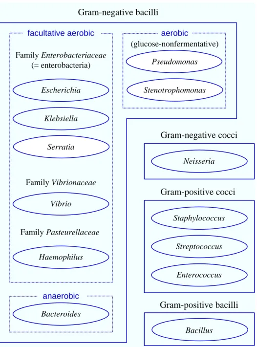
Streptococcus Enterococcus Staphylococcus Neisseria Bacteroides Vibrio Haemophilus Pseudomonas Stenotrophomonas Escherichia Klebsiella Serratia
Gram-positive cocci
Gram-negative cocci
anaerobic aerobic facultative aerobic Family Enterobacteriaceae (= enterobacteria) Family Vibrionaceae Family Pasteurellaceae BacillusGram-positive bacilli
(glucose-nonfermentative)Gram-negative bacilli
Fig. 1. Schema of bacteriological classification. Solid lines, grouping by Gram stain. Dotted lines, grouping by metabolism. Ellipse, typical genus.
properties including habitat and antimicrobial resistance. In Section III, we present differential activities of β-lactamases, such as hydrolytic activi-ty and susceptibiliactivi-ty to aztreonam as an inhibitor. Section IV describes indirect pathogenicity, that is, hidden risks in mixed infections.
I. Significance of S. maltophilia among Gram-Negative Bacilli
The emergence of β-lactam-resistant Gram-nega-tive bacilli has increasingly been recognized on the grounds that β-lactam agents came to be used
ex-Table 1. Number (%) of broad-spectrum β -lactam-resistant* strains for bacterial species
Number of strains 176 (100.0) Bacterial species Stenotrophomonas maltophilia† 96 (54.5) Pseudomonas aeruginosa† 34 (19.3) Chryseobacterium indologenes† 10 (5.7) Chryseobacterium meningosepticum† 7 (4.0) Serratia marcescens‡ 6 (3.4) Acinetobacter spp.† 4 (2.3) Other species 19 (10.8)
* Resistant to imipenem, ceftazidime and aztreonam. † Glucose-nonfermentative bacteria.
‡ Enterobacteria.
Table 2. Classification of β-lactamases
Molecular class Main bacterial species Serine-β-lactamase A (penicillinase) Haemophilus influenzae
Klebsiella oxytoca Klebsiella pneumoniae Proteus mirabilis
Stenotrophomonas maltophilia C (cephalosporinase) Citrobacter freundii
Enterobacter cloacae Morganella morganii Pseudomonas aeruginosa Serratia marcescens D (oxacillinase) Pseudomonas aeruginosa Metallo-β-lactamase B (carbapenemase) Aeromonas hydrophila
Bacillus cereus Bacteroides fragilis Burkholderia cepacia
Stenotrophomonas maltophilia From Kuga and Inoue, 2002 with some modifications.
tensively in wards (Livermore, 1995). β-Lactam agents are classified into several groups according to chemical structure. In general, penicillins are still effective against Gram-positive cocci, while they are weak in fighting Gram-negative bacilli. The newer the cephalosporins and cephamycins, the wider the antimicrobial spectrum. On one hand, the antimicrobial spectrum of monobactams is lim-ited to Gram-negative bacilli in contrast to peni-cillins. On the other hand, the spectrum of carba-penems covers almost all Gram-negative bacteria (Kataoka et al., 2002a).
The antimicrobial susceptibilities of Gram-negative bacilli were surveyed at Tottori University Hospital in 2001. As a matter of course, Gram-negative cocci and Gram-positive bacteria includ-ing Staphylococcus spp., Streptococcus spp., Entero-coccus spp. and Bacillus spp. are not intended infec-tions (Fig. 1). The survey showed that 176 strains (6.3%) out of 2785 were resistant to all 3 broad-spectrum β-lactam agents, that is, imipenem, ceftazidime and aztreonam. Most of the 176 strains are not enterobacteria but glucose-nonfermentative and aerobic bacteria such as P. aeruginosa, Chryseo-bacterium spp. and S. maltophilia in particular
(Table 1). These bacteria occupy ecological niches both inside and outside hospitals, and cause oppor-tunistic infection or iatrogenic transmission. The growth of these bacteria is slower than that of enterobacteria; however, glucose-nonfermentative bacteria are often of resistant to multidrug treatment and disinfectants (Mori, 2000). Classical resistance to β-lactam agents in these bacterial species is usu-ally due to an alternation of penicillin-binding proteins, changes in outer membrane permeability to β-lactams and most fre-quently, intrinsic β-lactamase production (Gál et al., 2000; Fang et al., 2002). There is a likelihood of invalidity with standard chemotherapy be-cause of hydrolysis by the β -lactamases.
The exact opposite of glucose-nonfermentative bac-teria, most enterobacteria are naturally susceptible to β -lac-tams (Kataoka et al., 2002a). However, an overdose of β -lactams during hospitalization causes an emergence of AmpC β-lactamase, whose gene is commonly found on the chro-mosomes of several entero-bacteria species and is typically induced by ceftazidime or
imi-Table 3. Acid production from sugars Bacterial species
Sugar S. maltophilia S. marcescens P. aeruginosa
Glucose – F O
Lactose – – –
Maltose O F –
F, fermentation; O, oxidation; –, negative of acid pro-duction. From Kataoka et al., 2003b with some modifi-cations.
Fig. 2. Acidity of bacterial colony. Both media contain glucose and bromothymol blue (BTB), which is a pH-indicator. On the left plate, colorless colonies (S. maltophilia) are observed because it does not pro-duce any acid from glucose. S. maltophilia needed to metabolize pep-tone for growth, and some alkalis made from peppep-tone have turned the appearance of colonies bluish. On the right plate, yellow colonies (S. marcescens) are grown with a fair amount of acidity by fermentation of glucose.
penem (Coudron et al., 2000; Kataoka et al., 2002b). Therefore, the treatment against enterobacteria should be con-sidered carefully. In passing, AmpC is grouped into class C (cephalospori-nase) of Ambler’s prestigious classi-fication (Table 2) (Ambler, 1980). AmpC β-lactamase of Serratia mar-cescens is presented in Section IV once again.
II. Clinical Properties Although S. maltophilia is a little-known microbe, it was the 5th Gram-negative species according to the number of isolates found at Tottori University Hospital in 2001, after P. aeruginosa, Escherichia coli, Klebsi-ella pneumoniae and S. marcescens (Kataoka et al., 2002a). S. maltophilia is a Gram-negative aerobic bacillus
(Fig. 1), having a strictly respiratory type of metabolism with oxygen as the terminal electron acceptor (Park, 1967; Snell and Lapage, 1971). As Hugh and Ryschenkow (1961) noted, a key distinguishing feature of S. maltophilia is acid production not from glucose but from maltose (Fig. 2 and Table 3). The term “maltophilia” seems to come from the bacterium’s preference for maltose. It goes without saying that all enterobacteria including S. marcescens ferment glucose, and produce some acid and/or gas (Sakazaki, 1992).
S. maltophilia is found in a wide variety of en-vironments, and has been recovered from nosoco-mial water sources including ice-making machines, sink traps and nebulizers (Sattler, 2000; Rogues et al., 2001). Despite the fact that almost all strains of S. maltophilia are isolated from inpatients, Denton et al. (1998) maintain that person-to-person trans-mission is an infrequent occurrence in the nosoco-mial setting. Epidemiological research of S. malto-philia also highlights that babies and elderly people are more likely to get this bacterium than those in the generations in between. As to the sources, they are also usually isolated from the respiratory tract such as sputum and the broncho-alveolar lavage.
The respiratory tract is the most common site of S. maltophilia’s isolation in hospitalized pa-tients; however, most patients are colonized rather than infected at this site (Geiger et al., 2001). As Rogues et al. (2001) have stated: A ventilator can be colonized with the patient’s endogenous flora and becomes a hotbed of secondary infections. In our investigation, more than 10 strains of S. maltophilia were actually separated from superinfections of
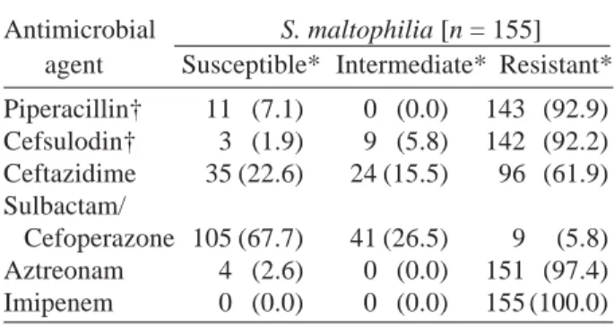
Table 4. Antimicrobial susceptibility (%) of S. maltophilia
Antimicrobial S. maltophilia [n = 155] agent Susceptible* Intermediate* Resistant* Piperacillin† 11 (7.1) 0 (0.0) 143 (92.9) Cefsulodin† 3 (1.9) 9 (5.8) 142 (92.2) Ceftazidime 35 (22.6) 24 (15.5) 96 (61.9) Sulbactam/ Cefoperazone 105 (67.7) 41 (26.5) 9 (5.8) Aztreonam 4 (2.6) 0 (0.0) 151 (97.4) Imipenem 0 (0.0) 0 (0.0) 155 (100.0) * Categories are recommended by the National
Commit-tee for Clinical Laboratory Standards (2000). † No data with 1 isolate.
pneumonia, bacteremia or cholangitis (Kataoka et al., 2003b). S. maltophilia has been associated with an expansive spectrum of clinical syndromes: endocarditis, conjunctivitis, keratitis, urethritis, ulcerative colitis and Crohn’s disease (Denton and Kerr, 1998).
S. maltophilia has been reported to be resistant to extended-spectrum cephalosporins, carbapenems and aminoglycosides (Denton et al., 1999; Barbier-Frebourg et al., 2000). The medical community has increasingly become dependent on carbapenems, because of their high affinity for penicillin-binding proteins, excellent penetration across bacterial out-er membranes, and resistance to hydrolysis by the majority of serine-based β-lactamases (Payne et al., 1997; Rasmussen and Bush, 1997; Yin et al., 2003). Therefore, carbapenems are so-called the final trumps. But all strains really revealed resistance to imipenem grouped into carbapenems (Table 4), and this peculiarity coincides with former documents (Hanberger et al., 2001; Simm et al., 2002). Cefta-zidime appeared to possess reasonable activity against some strains (Table 4), but as resistance is highly variable, ceftazidime cannot be used in empiric chemotherapy (Geiger et al., 2001). β -Lactamase inhibitor such as sulbactam was excep-tionally effective, so inhibition of β-lactamases could be a key to the problem. This hypothesis leads to the Discussion in the next Section.
This Section argues that S. maltophilia can live through the selective pressure of standard chemotherapy using β-lactams, and get its chance in the limelight. For this reason, clinicians should attach more importance on information from the clinical laboratory in order not to overlook this so-called “reserved” organism.
III. Differential Activities of β-Lactamases The most common theory is that S. maltophilia chromosomally yields at least 2 inducible β -lac-tamases, a class B metallo-β-lactamase (carbapene-mase) L1 and a class A active-site serine-β -lac-tamase (penicillinase) L2 (Table 2) (Ambler, 1980). Both enzymes mediate an essential resistance to β -lactam agents as a result of cutting the carbon-nitrogen bond of β-lactam rings off (Ullah et al., 1998; Avison et al., 2001; Spencer et al., 2001). There are still no clinically useful inhibitors of metallo-β -lactamases in contrast with serine-β-lactamases (Simm et al., 2002). The expression of L1 and L2 has been shown by coinducibility under the exis-tence of a single β-lactam agent as an inducer, and also by some single site mutations resulting in over-expression of both genes (Avison et al., 2002).
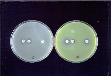
In recent years, the sodium mercaptoacetic acid (SMA) disk was developed for the screening of metallo-β-lactamase-producing Gram-negative bacilli (Arakawa et al., 2000). Though the main target of this thiol compound is originally IMP-1-producing P. aeruginosa or S. marcescens, this con-venient test can be applied to L1-producing S. malt-ophilia. Even the imipenem-resistant P. aerugino-sa strain indicated no reaction, whereas S. malto-philia showed good susceptibility to SMA (Fig. 3). The resistance to imipenem of that P. aeruginosa strain is not due to production of carbapenemase, but to mutational impermeability of up-regulated MexEF-OprN and reduced OprD (Livermore, 2002).
The hydrolytic activities of L1 and L2 β -lac-tamases have already been measured (Kataoka et al., 2003a). Decrease in UV absorption at an
appro-70 80 90 100 Inactivation time 1 Absorbance 2 3 4 0 (%) Imipenem Ceftazidime (h)
Fig. 4. Hydrolysis of β-lactam agents by S. maltophilia. The absorbances of the β-lactam rings were measured by UV spectrophotometry. Closed circle, imipenem; closed square, ceftazidime.
Fig. 3. Inhibition by sodium mercaptoacetic acid (SMA). On the left plate (S. maltophilia), a considerable expansion in zone size around an imipenem (IPM) disk close to SMA is revealed, in contrast to IPM without SMA. That means positive of carbapenemase. On the right plate (P. aeruginosa, which is yellow-green due to the fluorescent pig-ment), no expansions between the two IPM disks are shown. That naturally means negative of carbapenemase despite resistance to IPM. From Kataoka et al., 2003b.
priate wavelength means hydrolysis of the β-lactam ring (Samuni, 1975). By inactivation for 4 h, an av-erage reduction of approximately 20% was shown in both absorbances of imipenem and ceftazidime (Fig. 4). Taking into consideration the affinity, the hydrolysis of imipenem is performed by L1, and that of ceftazidime is largely related to L2. It ap-pears that the hydrolytic function of L2 is equiva-lent to that of an extended-spectrum β-lactamase.
The notion of coordinated expression allowed Krueger et al. (2001) to point out that the bacterial primary resistance to β-lactams is not overcome until both β-lactamases are inhibited. Although β -lactam agents are thought to be unrealistic tools against infections with S. maltophilia, this bacte-rium is useful as an extreme model of a β-lactamase producer. Our inhibition test notes that imipenem and cefpirome are predominantly hydrolyzed by L1, whereas ceftazidime is predominantly hydro-lyzed by L2 (Kataoka et al., 2003a). Incidentally, aztreonam grouped into monobactams has been reported as an inhibitor of extracellular β-lactamase
(Lister et al., 1998; Song et al., 2003). These reports are in harmony with the proposal of Nishida et al. (1999) that a monobactam derivative can indicate considerable synergy with cefpirome. Upon further examination, no synergy of aztreonam with imipenem is exhibited. These results also explain that aztreonam tends not to inhibit carbapenemases, because imipenem should be broken down only by carbapenemase L1. Therefore, the synergy of aztreonam with cefpirome was probably due to the aztreonam’s inhibition of penicillinase L2.
The inhibitory activity of aztre-onam against the penicillinases of S. maltophilia has been explored (Kataoka and Tanaka, in press). All 5 strains of S. maltophilia had already been con-firmed susceptible to clavulanic acid so they were identified possessors of L2 β-lactamase (Fang et al., 2002; Coudron et al., 2003). In spite of little synergy of imipenem to all 5 strains, a considerable synergy of aztreonam with cefozopran was found in 4 strains out of 5 (Table 5). Sulbactam is definitely known as a potent inhibitor of penicillinases (Mahgoub and Aly, 1998), and actually showed an excellent
Table 5. MICs against S. maltophilia
MIC (µg/mL)
Strain Aztreonam Imipenem Aztreonam/ Cefozopran Aztreonam/
Imipenem Cefozopran S. maltophilia TOT8 256 512 512/512 256 256/256 S. maltophilia TOT16 1024 512 512/512 256 128/128 S. maltophilia TOT19 1024 256 256/256 256 128/128 S. maltophilia TOT43 256 256 256/256 128 64/64 S. maltophilia TOT57 512 256 256/256 128 64/64
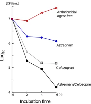
MIC, minimum inhibitory concentration. From Kataoka and Tanaka, in press with some modifications. 4 5 6 7 0 2 4 6 (h) Aztreonam Aztreonam/Cefozopran Cefozopran Log 10 Incubation time Antimicrobial agent-free (CFU/mL)
Fig. 5. Colony forming unit (CFU) count of S.
malto-philia in culture treated with antimicrobial agents as fol-lows: Cross, antimicrobial-agent free; closed circle, aztreonam; open square, cefozopran; closed square, aztreonam and cefozopran. From Kataoka and Tanaka, in press with some modifications.
synergy with cefoperazone (not cefozopran) against S. maltophilia (Table 4). These findings support the idea that aztreonam as well as sulbactam inhibits penicillinases instead of performing aztreonam’s own antimicrobial activity. This account is defended by both the high resistant frequency of aztreonam and the high MICs of aztreonam alone against S. maltophilia (Tables 4 and 5). Figure 5
presents the antimicrobial effect of the combination of aztreonam and cefozopran. The colony forming unit (CFU) counts at 6 h in cultures treated with aztreonam-cefozopran were approximately 1 log lower than those in cultures treated with cefozopran alone, and 2 logs lower than those treated with aztreonam alone.
Aztreonam is additionally suitable in combi-nation chemotherapy for Gram-negative infections. One reason is that monobactams have no antimicro-bial activity against Gram-positive bacteria: The risk of superinfection by Gram-positive bacteria, for instance enteritis caused by Staphylococcus aureus, is estimated to be low. Improper usage of broad-spectrum antimicrobial agents might prepare a hotbed of multidrug-resistant strains; therefore, the minimum usage of narrow-spectrum ones has recently been recommended for the control of hospital-acquired infections. We hope that a reduc-tion in the dosage of cefozopran by employing aztreonam comes to not only obtain antimicrobial synergy and minimize toxicity but also to prevent the emergence of penicillinase producers.
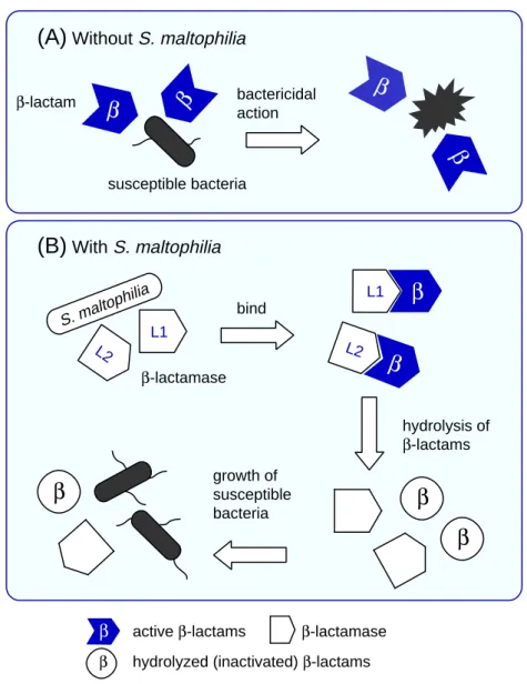
IV. Indirect Pathogenicity
A popular idea exists nowadays that S. maltophilia is of strictly limited pathogenicity (Denton and Kerr, 1998), while this organism has the capacity to hy-drolyze a variety of β-lactam antimicrobial agents and the possibility of assisting the survival of other
β
β
β
(A)
Without S. maltophiliaβ
(B)
With S. maltophilia S. maltophilia bind bactericidal action hydrolysis of β-lactams growth of susceptible bacteria β-lactam susceptible bacteria β-lactamase L1 L2 L1 L2β
β
β
β active β-lactams β-lactamase β hydrolyzed (inactivated) β-lactams
β
β
Fig. 6. Schema of indirect pathogenicity.
A: Without S. maltophilia, susceptible bacteria are killed by β-lactam agents.
B: With S. maltophilia, β-lactamases leaking from S. maltophilia hydrolyze β-lactam agents, so even susceptible bacteria can survive.
susceptible pathogens. S. maltophilia is apparently a β-lactamase supplier rather than a direct cause of disease where it is confronted with β-lactam agents. It is generally isolated from severely immuno-suppressed hosts because of its weak virulence, and has frequently been isolated together with other Gram-negative species in our observations (Kataoka et al., 2003b). It looks like broad-spectrum anti-microbial therapy takes aim at the major opportu-nistic pathogens such as P. aeruginosa and S.
mar-cescens in most cases of Gram-negative infection. As it turns out, the existence of S. maltophilia is apt to be less reflected in the choice of antimicrobial agents. However, broad-spectrum β-lactam agents could act as a sieve of multidrug-resistant strains such as S. maltophilia and serve to induce their own β-lactamases (Kataoka et al., 2001, 2002b). That is why we propose the concept of indirect pathogenicity (Fig. 6): A chain of the preceding processes can result in the encouragement of mixed
Fig. 7. Mixed culture of S. marcescens and S. maltophilia. On the top left plate, an extremely large number of yellow colonies (S. marcescens) are seen (none for imipenem was supplemented). On the top right plate, the number of yellow colonies is obviously decreased because of 16 µg/mL of imipenem in the medium. On the bottom plate, however, a fairly large number of yellow colonies and colorless colo-nies (S. maltophilia) are indicated even under exposure to 16 µg/mL imipenem (from Kataoka et al., 2003b).
3 4 5 6
Concentration of imipenem added in culture
4 16
**
Log 10 (CFU/mL) (µg/mL)**
Fig. 8. Colony forming unit (CFU) count of S.
marces-cens in culture which contains imipenem. Pale bar, pure culture of S. marcescens; dark bar, mixed culture of S. marcescens with S. maltophilia. **P < 0.01 with t-test. The bars represent means plus SD. From Kataoka et al. (2003b) with some modifications.
infections caused by another pathogen, even if S. maltophilia originally colonizes in the patient’s res-piratory tract.
The indirect pathogenicity of S. maltophilia has already been confirmed (Kataoka et al., 2003b). The CFU of the yellow colonies (S. marcescens and P. aeruginosa) was counted for each plate, because both S. marcescens and P. aeruginosa form yellow colonies by acid production from glucose in contrast with colorless colonies of S. maltophilia (Figs. 2 and 7).
Figure 8 demonstrates that S. marcescens ex-posed to imipenem increased in the existence of S. maltophilia. This concept is also applicable to a mixed culture of P. aeruginosa with S. maltophilia (Kataoka et al., 2003b). On the other hand, the CFU count of S. marcescens seemed not to be influenced in cases when it was faced to ceftazidime instead of imipenem (Kataoka et al., 2003b), because S. marcescens is a potential owner of AmpC β -lactamase (Coudron et al., 2000). An induction of AmpC makes the strain highly resistant to
ceftazidime (Raimondi et al., 2001). We consider that both β-lactamases of L2 and AmpC were yielded during exposure to ceftazidime in mixed culture, and have assumed that L2 leaking from S. maltophilia hid itself behind S. marcescens’ own AmpC. As proof of the output of L2, P. aerugi-nosa confronted with a large quantity of ceftazidime could grow under the support of S. maltophilia (Kataoka et al., 2003b). As Livermore (2002) has demonstrated, P. aeruginosa also har-bors AmpC β-lactamase, but unlike S. marcescens the level of resistance de-pends on the degree of depression. Following this thesis, the P. aerugino-sa strain does not seem to have pro-duced enough β-lactamases to protect itself from ceftazidime.
Though a multidrug-resistant strain is not always relevant to indirect pathogenicity, at least our positive findings can be thought of as a formal explanation for some potential threat: Even an ordinary bacterium can endure antimicrobial attack
by the assistance of β-lactamases from an indirect pathogen, namely S. maltophilia.
Conclusion
Carbapenems and cephalosporins are among the most frequently administered antimicrobial agents due to their excellent antimicrobial activity and low toxicity; therefore, these compounds are often prescribed for severe Gram-negative infections (Coudron et al., 2003; Yin et al., 2003). The present study upholds the theory that overuse of these anti-microbial trumps has a profound effect on a selec-tion pressure on account of the inducselec-tion of β -lactamases, and results in the appearance of an antibiotic-free area around S. maltophilia. In other words, sulbactam, minocycline and trimethoprim-sulfamethoxazole, relatively unpopular but effec-tive drugs, should be selected more in a case where mixed infection occurs (Kataoka et al., 2002a). Although it is sometimes difficult to attribute a causative role to S. maltophilia when the organism is isolated from respiratory secretions alone (Sattler, 2000), throughout the course of this essay we establish that S. maltophilia is worthy of atten-tion. Thus, a full understanding of the indirect patho-genicity in vivo awaits future study.
Acknowledgments: We are grateful to Mr. Hiromitsu Fujiwara and Ms. Ayako Tanimoto, Division of Clinical Laboratory, Tottori University Hospital, for their techni-cal assistance. We also appreciate the advice and exper-tise of Professor Shiro Ikawa, Division of Clinical Labo-ratory Medicine, Department of Pathophysiological and Therapeutic Science, School of Medicine, Tottori Uni-versity Faculty of Medicine.
References
1 Ambler RP. The structure of β-lactamases. Phil Trans R Soc Lond B 1980;289:321–331.
2 Arakawa Y, Shibata N, Shibayama K, Kurokawa H, Yagi T, Fujiwara H, et al. Convenient test for screening metallo-β-lactamase-producing
gram-negative bacteria by using thiol compounds. J Clin Microbiol 2000;38:40–43.
3 Avison MB, Higgins CS, von Heldreich CJ, Bennett PM, Walsh TR. Plasmid location and molecular heterogeneity of the L1 and L2 β-lactamase genes of Stenotrophomonas maltophilia. Antimicrob Agents Chemother 2001;45:413–419.
4 Avison MB, Higgins CS, Ford PJ, von Heldreich CJ, Walsh TR, Bennett PM. Differential regulation of L1 and L2 β-lactamase expression in Stenotropho-monas maltophilia. J Antimicrob Chemother 2002; 49:387–389.
5 Barbier-Frebourg N, Boutiba-Boubaker I, Nouvellon M, Lemeland JF. Molecular investigation of Steno-trophomonas maltophilia isolates exhibiting rapid emergence of ticarcillin-clavulanate resistance. J Hosp Infect 2000;45:35–41.
6 Coudron PE, Moland ES, Thomson KS. Occurrence and detection of AmpC beta-lactamases among Escherichia coli, Klebsiella pneumoniae, and Pro-teus mirabilis isolates at a Veterans Medical Center. J Clin Microbiol 2000;38:1791–1796.
7 Coudron PE, Hanson ND, Climo MW. Occurrence of extended-spectrum and AmpC beta-lactamases in bloodstream isolates of Klebsiella pneumoniae: isolates harbor plasmid-mediated FOX-5 and ACT-1 AmpC beta-lactamases. J Clin Microbiol 2003;41: 772–777.
8 Denton M, Kerr KG. Microbiological and clinical aspects of infection associated with Stenotrophomo-nas maltophilia. Clin Microbiol Rev 1998;11:57– 80.
9 Denton M, Todd NJ, Kerr KG, Hawkey PM, Littlewood JM. Molecular epidemiology of Stenotro-phomonas maltophilia isolated from clinical specimens from patients with cystic fibrosis and associated en-vironmental samples. J Clin Microbiol 1998;36:1953– 1958.
10 Denton M, Keer V, Hawkey PM. Correlation between genotype and β-lactamases of clinical and environmental strains of Stenotrophomonas maltophilia. J Antimicrob Chemother 1999;43:555– 558.
11 Fang H, Edlund C, Hedberg M, Nord CE. New findings in beta-lactam and metronidazole resistant Bacteroides fragilis group. Int J Antimicrob Agents 2002;19:361–370.
12 Gál Z, Szabó D, Kovács P, Hernádi F, Tóth-Martinez B, Rozgonyi F. β-Lactam susceptibility patterns and investigation of cephalosporin hydro-lysing β-lactamases of Gram-negative extraintesti-nal clinical isolates. Int J Antimicrob Agents 2000; 16:395-400.
13 Geiger AM, Hogardt M, Heesemann J. Burkho-lderia/Stenotrophomonas. Contrib Microbiol 2001; 8:20–34.
14 Hanberger H, Diekema D, Fluit A, Jones R, Struelens M, Spencer R, et al. Surveillance of anti-biotic resistance in European ICUs. J Hosp Infect 2001;48:161–176.
15 Hugh R, Ryschenkow E. Pseudomonas maltophilia, an Alcaligenes-like species. J Gen Microbiol 1961; 26:123-132.
16 Kataoka D, Kawakami T, Fujiwara H, Tanaka Y, Tanimoto A, Ikawa S, et al. The antibiotic resistance of gram-negative bacilli isolated in Tottori Univer-sity Hospital. Tottori Igaku Zasshi 2001;29:161– 167 (in Japanese).
17 Kataoka D, Fujiwara H, Tanimoto A, Tanaka Y. The results of antibiotic susceptibility of gram-negative rods at Tottori University Hospital: the possibility of the sensible use of narrow-spectrum antimicrobial agents. Kansenshogaku Zasshi 2002a;76:542–549 (in Japanese with English abstract).
18 Kataoka D, Fujiwara H, Tanimoto A, Tanaka Y. Relation between hospitalization and the resistance of enterobacteria to β-lactam antimicrobial agents. Kansenshogaku Zasshi 2002b;76:1045–1047. 19 Kataoka D, Fujiwara H, Tanimoto A, Ikawa S,
Tanaka Y. The differential β-lactamase activity of Stenotrophomonas maltophilia. J Hosp Infect 2003a;54:246–248.
20 Kataoka D, Fujiwara H, Kawakami T, Tanaka Y, Tanimoto A, Ikawa S, et al. The indirect pathogenicity of Stenotrophomonas maltophilia. Int J Antimicrob Agents 2003b;22:601–606.
21 Kataoka D, Tanaka Y. The combination of aztreonam and cefozopran against Stenotrophomonas maltophi-lia. J Infect Chemother (in press).
22 Krueger TS, Clark EA, Nix DE. In vitro suscepti-bility of Stenotrophomonas maltophilia to various antimicrobial combinations. Diagn Microbiol Infect Dis 2001;41:71–78.
23 Kuga A, Inoue M. Antimicrobial capacity. Rinsho To Biseibutsu 2002;29:3–11 (in Japanese).
24 Lister PD, Sanders WE, Sanders CC. Cefepime-aztreonam: a unique double β-lactam combination for Pseudomonas aeruginosa. Antimicrob Agents Chemother 1998;42:1610–1619.
25 Livermore DM. β-Lactamases in laboratory and clinical resistance. Clin Microbiol Rev 1995;8:557– 584.
26 Livermore DM. Multiple mechanisms of antimicro-bial resistance in Pseudomonas aeruginosa: our worst nightmare? Clin Infect Dis 2002;34:634–640. 27 Mahgoub H, Aly FA. UV-spectrophotometric
deter-mination of ampicillin sodium and sulbactam sodi-um in two-component mixtures. J Pharm Biomed Anal 1998;17:1273–1278.
28 Mori T. Identification of Gram-negative bacilli. In: Oguri T, ed. Clinical microbiology handbook. Tokyo: Miwa Syoten; 2000. p. 82–96 (in Japanese). 29 National Committee for Clinical Laboratory Stan-dards. Methods for dilution antimicrobial suscepti-bility tests for bacteria that grow aerobically. In: Approved standard, 5th ed. NCCLS document M7-A5. Wayne (PA): National Committee for Clinical Laboratory Standards; 2000.
30 Nishida K, Kunugita C, Uji T, Higashitani F, Hyodo A, Unemi N, et al. In vitro and in vivo activities of Syn2190, a novel β-lactamase inhibitor. Antimicrob Agents Chemother 1999;43:1895–1900.
31 Park RWA. A comparison of two methods for de-tecting attack on glucose by Pseudomonads and Acromobacters. J Gen Microbiol 1967;46:355–360. 32 Payne DJ, Bateson JH, Gasson BC, Proctor D, Khushi T, Farmer TH, et al. Inhibition of metallo-β -lactamases by a series of mercaptoacetic acid thiol ester derivatives. Antimicrob Agents Chemother 1997;41:135–140.
33 Raimondi A, Sisto F, Nikaido H. Mutation in Serratia marcescens AmpC β-lactamase producing high-level resistance to ceftazidime and cefpirome. Antimicrob Agents Chemother 2001;45:2331–2339. 34 Rasmussen BA, Bush K. Carbapenem-hydrolyzing
β-lactamases. Antimicrob Agents Chemother 1997;41:223–232.
35 Rogues AM, Maugein J, Allery A, Fleureau C, Boulestreau H, Surcin S, et al. Electronic ventilator temperature sensors as a potential source of respira-tory tract colonization with Stenotrophomonas mal-tophilia. J Hosp Infect 2001;49:289–292.
36 Sakazaki R. Definition of the family Enterobacteri-aceae. In: Sakazaki R, Tamura T, eds. Enterobacteria. Tokyo: Kindai Syuppan; 1992. p. 6–17 (in Japanese). 37 Samuni A. A direct spectrophotometric assay and
determination of Michaelis constants for the β -lactamase reaction. Anal Biochem 1975;63:17–26. 38 Sattler CA. Stenotrophomonas maltophilia infection
in children. Pediatr Infect Dis J 2000;19:877–878. 39 Simm AM, Higgins CS, Carenbauer AL, Crowder
MW, Bateson JH, Bennett PM, et al. Characteriza-tion of monomeric L1 metallo-β-lactamase and the role of the N-terminal extension in negative coopera-tivity and antibiotic hydrolysis. J Biol Chem 2002; 277:24744–24752.
40 Snell JJS, Lapage SP. Comparison of four methods for demonstrating glucose breakdown by bacteria. J Gen Microbiol 1971;68:221–225.
41 Song W, Woo HJ, Kim JS, Lee KM. In vitro activity of β-lactams in combination with other antimicrobial agents against resistant strains of Pseudomonas aeruginosa. Int J Antimicrob Agents 2003;21:8–12. 42 Spencer J, Clarke AR, Walsh TR. Novel mechanism of hydrolysis of therapeutic β-lactams by Stenotro-phomonas maltophilia L1 metallo-β-lactamase. J Biol Chem 2001;276:33638–33644.
43 Ullah JH, Walsh TR, Taylor IA, Emery DC, Verma CS, Gamblin SJ et al. The crystal structure of the L1 metallo-β-lactamase from Stenotrophomonas
malto-philia at 1.7 Å resolution. J Mol Biol 1998;284:125– 136.
44 Yin XH, Mikamo H, Tamaya T. Nosocomial infec-tious potency of imipenem-resistant Pseudomonas aeruginosa isolated from obstetric and gynecologic infections. J Infect Chemother 2003;9:97–100.
Received September 9, 2003;accepted October 23, 2003 Corresponding author: Yoshinori Tanaka, MD, PhD