Prognostic Impact of Specific Molecular Profiles in Pediatric
1
Acute Megakaryoblastic Leukemia in Non-Down Syndrome
2
3
Yusuke Hara1,2,3, Norio Shiba1,2,3, Kentaro Ohki1,4, Ken Tabuchi5, Genki Yamato1,2,3,
4
Myoung-ja Park1, Daisuke Tomizawa6, Akitoshi Kinoshita7, Akira Shimada8,
5
Hirokazu Arakawa2, Akiko M. Saito3, Nobutaka Kiyokawa4, Akio Tawa9, Keizo
6
Horibe3, Takashi Taga10, Souichi Adachi11, Tomohiko Taki12*, and Yasuhide
7
Hayashi1,3,13*
8 9
1Department of Hematology and Oncology, Gunma Children's Medical Center, Shibukawa, Japan 10
2Department of Pediatrics, Gunma University Graduate School of Medicine, Maebashi, Japan 11
3Clinical Research Center, National Hospital Organization Nagoya Medical Center, Nagoya, 12
Japan 13
4Department of Pediatric Hematology and Oncology Research, National Research Institute for 14
Child Health and Development, Tokyo, Japan 15
5Department of Pediatrics, Tokyo Metropolitan Komagome Hospital, Tokyo, Japan 16
6Division of Leukemia and Lymphoma, Children’s Cancer Center, National Center for Child 17
Health and Development, Tokyo, Japan 18
7Department of Pediatrics, St Marianna University School of Medicine, Kawasaki, Japan 19
8Department of Pediatrics, Okayama University Hospital, Okayama, Japan 20
9Department of Pediatrics, National Hospital Organization Osaka Medical Center, Osaka, Japan 21
10Department of Pediatrics, Shiga University of Medical Science, Otsu, Japan 22
11Department of Human Health Sciences, Kyoto University Graduate School of Medicine, Kyoto, 23
Japan 24
12Department of Molecular Diagnostics and Therapeutics, Kyoto Prefectural University of 25
Medicine, Graduate School of Medical Science, Kyoto, Japan 26
13Gunma Red Cross Blood Center, Maebashi, Japan 27
28
*Correspondence to:
29
Yasuhide Hayashi, MD, Department of Hematology/Oncology, Gunma Children's
1
Medical Center, 779, Shimohakoda, Hokkitsu, Shibukawa, 377-8577 Gunma, Japan.
2
Tel.: +81-279-52-3551, Fax: +81-279-52-2045, E-mail: [email protected]
3 4
Tomohiko Taki, MD, Department of Molecular Diagnostics and Therapeutics, Kyoto
5
Prefectural University of Medicine, Graduate School of Medical Science, 465,
6
Kajii-cho, Kawaramachi-Hirokoji, Kamigyo-ku, Kyoto, 602-8566, Kyoto, Japan. Tel.:
7
+81-75-251-5659, Fax: +81-75-251-5659, E-mail: [email protected]
8 9
Supported by: Cancer Research grant, a grant for Research on Children and Families,
10
and Research on Intractable Diseases, Health and Labor Sciences Research Grants
11
from the Ministry of Health, Labor, and Welfare of Japan; a Grants-in-Aid for
12
Scientific Research B (24390268), C (25461611, 26461598, and 26461599), and
13
Exploratory Research (25670482) from the Ministry of Education, Culture, Sports,
14
Science and Technology of Japan; the Grant of the National Center for Child Health
15
and Development (26-20).
16 17
Running title: Molecular Features of Pediatric AMKL
18
Tables/Line figures/Color figures: 5/1/1
1
Abstract Word Count: 245
2
Word count: 3,528
3
Reference: 33
4
Key words: pediatric leukemia, acute myeloid leukemia, gene rearrangements,
5
prognostic factors
6 7 8
Abstract
1
Pediatric acute megakaryoblastic leukemia with non-Down syndrome (AMKL) is a
2
unique subtype of acute myeloid leukemia (AML). Novel CBFA2T3-GLIS2 and
3
NUP98-KDM5A fusions recurrently found in AMKL were recently reported as poor
4
prognostic factors. However, their detailed clinical and molecular characteristics in
5
patients treated with recent improved therapies remain uncertain. We analyzed
6
molecular features of 44 AMKL patients treated on two recent Japanese AML
7
protocols, the AML99 and AML-05 trials. We identified CBFA2T3-GLIS2,
8
NUP98-KDM5A, RBM15-MKL1, and KMT2A rearrangements in 12 (27%), 4 (9%), 2
9
(5%), and 3 (7%) patients, respectively. Among 459 other AML patients,
10
NUP98-KDM5A was identified in 3 patients, whereas CBFA2T3-GLIS2 and
11
RBM15-MKL1 were only present in AMKL. GATA1 mutations were found in 5
12
patients (11%). Four-year overall survival (OS) and event-free survival (EFS) rates of
13
CBFA2T3-GLIS2-positive patients in AMKL were 41.7% and 16.7%, respectively.
14
Three-year cumulative incidence of relapse in CBFA2T3-GLIS2-positive patients was
15
significantly higher than that of CBFA2T3-GLIS2-negative patients (75.0% vs 35.7%,
16
P = 0.024). In multivariate analyses, CBFA2T3-GLIS2 was an independent poor
17
prognostic factor for OS (HR, 4.34; 95% CI, 1.31–14.38) and EFS (HR, 2.95; 95% CI,
18
1.20–7.23). Furthermore, seven (54%) of 13 infant AMKL patients were
19
CBFA2T3-GLIS2-positive. Notably, out of 7 CBFA2T3-GLIS2-positive infants, six
20
(86%) relapsed and five (71%) died. Moreover, all of CBFA2T3-GLIS2-positive
21
patients who experienced induction failure (n = 3) were infants, indicating worse
22
prognosis of CBFA2T3-GLIS2-positive infants. These findings indicated the
23
significance of CBFA2T3-GLIS2 as a poor prognostic factor in AMKL patients,
24
particularly in infants.
1 2
Introduction
1
Pediatric acute megakaryoblastic leukemia in non-Down syndrome (AMKL) is a
2
clinically and biologically distinct, FAB M7 subtype of acute myeloid leukemia
3
(AML), accounting for approximately 5–15% of all pediatric AML patients (Athale et
4
al., 2001; Dastugue et al., 2002; Reinhardt et al., 2005). AMKL was shown to have a
5
poor prognosis with a survival rate of less than 40% (Athale et al., 2001; Barnard et al.,
6
2007), however, recent advances in diagnostic techniques and intensive chemotherapy
7
have led to improved long-term survival rates of over 60% (Hama et al., 2008;
8
Schweitzer et al., 2015). Although the t(1;22)(p13;q13)/RBM15-MKL1 molecular
9
marker was repeatedly detected in 10%–25% of AMKL (Ma et al., 2001; Mercher et
10
al., 2001; Inaba et al., 2015), information on cytogenetic and molecular pathogenesis in
11
most AMKL patients was limited until the recent identification of novel cryptic
12
translocations, inv(16)(p13.3q24.3) and t(11;12)(p15;p13) that encode
13
CBFA2T3-GLIS2 and NUP98-KDM5A fusion genes, respectively (Gruber et al., 2012;
14
Thiollier et al., 2012; de Rooij et al., 2013). The frequencies of these fusion genes in
15
AMKL patients were reported to be 13%–27% and 8%–10%, respectively (Gruber et
16
al., 2012; Thiollier et al., 2012; de Rooij et al., 2013). Whereas CBFA2T3-GLIS2 was
17
demonstrated to be a poor prognostic factor in AMKL patients (Gruber et al., 2012),
18
the correlation between NUP98-KDM5A and AMKL prognosis was unclear. However,
19
a recent intergroup study has reported a poor prognosis with these fusion genes (de
20
Rooij et al., 2016).
21
New biological insights into AMKL have been gradually accumulating; however,
22
the prognostic significance and detailed characteristics of such novel fusion genes in
23
patients treated with improved therapies in recent clinical trials have not been reported,
24
partially because of the small numbers of patients. Thus, in this study, we investigated
1
the molecular and clinical features of 44 AMKL patients treated on two recent
2
Japanese clinical trials, AML99 and AML-05.
3 4
Material and Methods
5
Patients and Samples
6
This present retrospective cohort study enrolled patients younger than 18 years who
7
were diagnosed with de novo AML and participated in one of the two recent clinical
8
trials in Japan, the AML99 trial by the Japanese Childhood AML Cooperative Study
9
between January 2000 and December 2002 and the AML-05 trial by the Japanese
10
Pediatric Leukemia/Lymphoma Study Group (JPLSG) between November 2006 and
11
December 2010 (Tsukimoto et al., 2009; Tomizawa et al., 2013). The AML-05 trial is
12
registered with UMIN Clinical Trials Registry (UMIN–CTR, URL:
13
http://www.umin.ac.jp/ctr/index.htm), number UMIN000000511. A total of 503
14
patients whose leukemic samples were available were included in the present study;
15
134 from a total of 280 patients in the AML99 trial and 369 from a total of 443 patients
16
in the AML-05 trial were eligible for this study, and patients with Down syndrome and
17
acute promyelocytic leukemia were excluded. Among the eligible patients, 44 patients
18
(9%) (10 from AML99 and 34 from AML-05) were diagnosed with AMKL; the
19
remaining 459 patients were diagnosed with other FAB subtypes of AML (referred to
20
as other AML). Extensive details on the diagnosis, risk-stratification, and treatment in
21
these protocols were previously reported (Tsukimoto et al., 2009; Tomizawa et al.,
22
2013; Kinoshita et al., 2014). Morphological, immunological, cytogenetic, and
23
molecular characteristics of patients in the AML-05 trial were centrally reviewed,
24
whereas the diagnosis of patients in the AML99 trial was made by each hospital.
1
Treatment protocols and data and sample collections in both clinical trials were
2
approved by the institutional review boards of each participating institution after
3
written informed consent was obtained from patients or their parents/guardians. The
4
present study was conducted in accordance with the Declaration of Helsinki and
5
approved by the institutional review board of Gunma Children’s Medical Center.
6 7
Cytogenetic and Molecular Characterization
8
Genomic DNA and total RNA were extracted from leukemic samples using the
9
ALLPrep DNA/RNA Mini Kit (Qiagen, Hilden, Germany) and were
10
reverse-transcribed to cDNA using the cDNA Synthesis Kit (GE Healthcare, Tokyo,
11
Japan). Mutation analyses of FLT3-ITD, NRAS, KRAS, KIT, WT1, NPM1, and GATA1
12
(Xu et al., 2003; Shimada et al., 2006; Sano et al., 2012; Shiba et al., 2013) and fusion
13
gene analyses including CBFA2T3-GLIS2, NUP98-KDM5A, KMT2A-MLLT3,
14
KMT2A-MLLT10, RBM15-MKL1, RUNX1-RUNX1T1, CBFβ-MYH11, NUP98-NSD1,
15
and FUS-ERG were performed using polymerase chain reaction (PCR)/reverse
16
transcription-PCR followed by Sanger sequencing, as previously reported (Gruber et
17
al., 2012; Thiollier et al., 2012; de Rooij et al., 2013; Shiba et al., 2013). In this study,
18
a complex karyotype was defined by three or more chromosome abnormalities (Slovak
19
et al., 2000; Byrd et al., 2002; Schoch et al., 2005).
20 21
Statistics
22
Survival rates were estimated using the Kaplan–Meier method and compared using the
23
log-rank test. Overall survival probability (OS) was defined as the time from diagnosis
24
to death by any cause, and event-free survival probability (EFS) was defined as the
1
time from diagnosis to relapse, death by any cause, or induction failure (Cheson et al.,
2
2003). Cumulative incidence of relapse (CIR) was defined as the time between
3
diagnosis and relapse (induction failure was attributed to an event on day 0) and was
4
analyzed by the Kalbfleisch and Prentice method that considered death and second
5
malignancy as competing events. Groups were compared using the Gray’s test. Data
6
related to hematopoietic stem cell transplantation (HSCT) were restricted to all 34
7
AMKL patients in the AML-05 trial and seven of 10 AMKL patients in the AML99
8
trial. Statistical analyses were performed using the Fisher’s exact test for categorical
9
variables and Mann–Whitney U test for continuous variables (i.e., age and white blood
10
cell [WBC] count). Independence of prognostic factors was examined using
11
multivariate Cox regression analysis using age, WBC count at diagnosis, fusion genes,
12
and gene mutations assessed in this study. For all analyses, P values of <0.05 were
13
considered statistically significant with two-tailed testing. All analyses were performed
14
using the SPSS® statistical package program version 22 (SPSS, Tokyo, Japan),
15
GraphPad Prism® Version 6 (GraphPad Software, Tokyo, Japan), and EZR® version
16
1.20 (Saitama Medical Center, Jichi Medical University, Saitama, Japan).
17
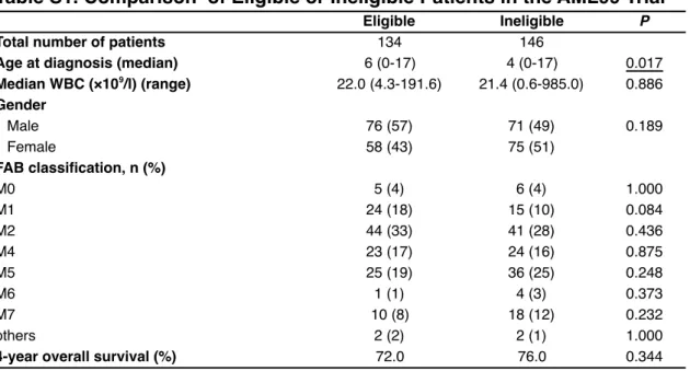
Comparison of clinical features between eligible and ineligible patients in the
18
AML99 trial is shown in Table S1, whereas that in the AML-05 trial was previously
19
reported (Shiba et al, 2016). No significant differences in any features other than age at
20
diagnosis (the AML 99 trial) and the frequency of RAEB-T (the AML-05 trial) were
21
observed between the groups.
22 23
Results
24
Identification of Cytogenetic and Molecular Features of AMKL Patients
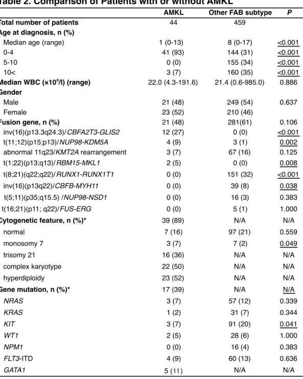
1
Among a total of 44 AMKL patients, five fusion gene patterns were identified in 21
2
patients (47.7%): CBFA2T3-GLIS2, NUP98-KDM5A, RBM15-MKL1, KMT2A-MLLT3,
3
and KMT2A-MLLT10 in 12 (27%), 4 (9%), 2 (5%), 2 (5%), and 1 (2%) patients,
4
respectively (Fig. 1). Although t(1;22)(p13;q13)/RBM15/MKL1 and
5
t(9;11)(p22;q23)/KMT2A-MLLT3 were found by conventional G-banding,
6
inv(16)(p13.3q24.3)/CBFA2T3-GLIS2, t(11;12)(p15;p13)/NUP98-KDM5A, and
7
t(10;11)(p12;q23)/KMT2A-MLLT10 were not identified. One
8
CBFA2T3-GLIS2-positive patient had a single-cell abnormality of t(15;16)(q24;q24),
9
whereas none of the NUP98-KDM5A-positive patients had cytogenetic abnormalities
10
involving 11p15 or 12p13 (Table 1). Detailed information on cytogenetic, molecular,
11
and clinical features of all AMKL patients are shown in Tables 1 and S2.
12
Gene mutations, including FLT3-ITD, NRAS, KRAS, KIT, WT1, and GATA1,
13
were detected in 17 patients (39%) (Fig. 1). GATA1 mutation was the most frequent
14
gene mutation (11.3%), whereas NPM1 mutation was not found in any of the patients.
15
Complex karyotype, acquired trisomy 21, and hyperdiploidy were found in 22
16
(50%), 16 (36%), and 23 (52%) of 44 patients, respectively (Fig. 1). Only two patients
17
did not have any cytogenetic features analyzed in the present study.
18
The differences in cytogenetic and molecular aberration frequencies between
19
AMKL and other AML patients are shown in Table 2. In fusion gene analyses,
20
CBFA2T3-GLIS2 and RBM15-MKL1 were only found in AMKL patients, whereas
21
NUP98-KDM5A was detected in both groups. The remaining three AML patients with
22
NUP98-KDM5A fusion gene were diagnosed with FAB M5, M6, and RAEB-T
23
subtypes. Core binding factor-AML, NUP98-NSD1, and FUS-ERG were found in only
24
other AML patients.
1 2
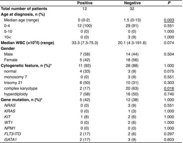
Correlation of Fusion Genes with Gene Mutations and Cytogenetic Abnormalities
3
Assessment of cytogenetic features of 12 CBFA2T3-GLIS2-positive patients revealed
4
that the complex karyotype was found in only two patients, which was significantly
5
lower than in CBFA2T3-GLIS2-negative patients (P = 0.016) (Table 3). The normal
6
karyotype, trisomy 21, and hyperdiploidy frequently coexisted with this fusion (33%,
7
50%, and 58%, respectively). Analysis of gene mutations in 12
8
CBFA2T3-GLIS2-positive patients identified FLT3-ITD, KIT, and GATA1 in 2, 1, and
9
2 patients, respectively (Fig. 1).
10
No gene mutations were observed in other fusion-positive patients. Although
11
NUP98-KDM5A was known as a cryptic fusion gene, three (75%) of the four patients
12
with this fusion had a complex karyotype (Table 1).
13 14
Prognostic Relevance of Cytogenetic and Molecular Markers
15
No significant difference was observed in the 4-year OS between the AML99 (n = 134)
16
and AML-05 (n = 369) trials (76.0% vs 66.9%, P = 0.202), whereas the 4-year EFS of
17
the AML99 trial was significantly higher than that of the AML-05 trial (64.2% vs
18
52.4%, P = 0.016). Among AMKL patients, the 4-year OS and EFS of the AML99 trial
19
were not significantly different than those of the AML-05 trial (60.0% vs 57.6%, P =
20
0.964, and 50.0% vs 30.9%, P = 0.305, respectively). Furthermore, 44 AMKL patients
21
had significantly lower 4-year OS and EFS rates than those of 459 other AML patients
22
(58.6% vs 71.8%, P = 0.019, and 36.6% vs 57.7%, P < 0.001, respectively). Analysis
23
of survival rates in AMKL patients who received HSCT (n = 28) determined that six
24
(67%) of the nine patients with first complete remission (CR), three (30%) of the 10
1
relapsed patients, and four (44%) of the nine patients with induction failure finally
2
survived.
3
CBFA2T3-GLIS2-positive patients (n = 12) tended to have lower 4-year OS and
4
EFS rates than CBFA2T3-GLIS2-negative patients (n = 32) (41.7% vs 66.4%, P =
5
0.193, and 16.7% vs 44.1%, P = 0.068, respectively) (Fig. 2A and 2B). When the
6
analysis was restricted to patients in the AML-05 trial (n = 34), the 4-year EFS of
7
CBFA2T3-GLIS2-positive patients (n = 11) was significantly lower than that of
8
CBFA2T3-GLIS2-negative patients (n = 23) (9.1% vs 41.9%, P = 0.030) (Fig. 2D).
9
Furthermore, the 3-year CIR of CBFA2T3-GLIS2-positive patients was significantly
10
higher than that of CBFA2T3-GLIS2-negative patients (75.0% vs 35.7%, P = 0.024)
11
(Fig. 2E). Only two (17%) CBFA2T3-GLIS2-positive patients survived without relapse,
12
and all of five CBFA2T3-GLIS2-positive patients who received chemotherapy alone in
13
intensification therapy relapsed (Table 1). Eventually, all CBFA2T3-GLIS2-positive
14
patients received HSCT, which was significantly more frequent than in
15
CBFA2T3-GLIS2-negative patients (100% vs 57%, P = 0.014). Specifically, three of
16
five CBFA2T3-GLIS2-positive patients who survived received HSCT at first CR.
17
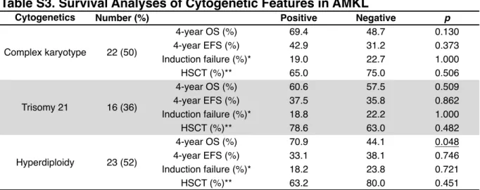
Analysis of other cytogenetic and molecular features for prognosis demonstrated
18
that patients with hyperdiploidy had a significantly better 4-year OS than those lacking
19
hyperdiploidy (P = 0.048) (Table. S3). Notably, three of the six patients with
20
hyperdiploidy who died were CBFA2T3-GLIS2-positive (Table 1). Patients with a
21
complex karyotype also tended to have favorable 4-year OS, although the difference
22
was not significant (P = 0.130) (Table S3). Among the 22 patients with a complex
23
karyotype, four had induction failure and received HSCT, and three (75%) of those
24
survived without relapse. The prognosis of patients with trisomy 21 was not
1
significantly different than that of those without trisomy 21 (Table S3).
2
3
Age Dependency of Molecular and Clinical Features in AMKL
4
The ages of AMKL patients were characterized by a bimodal distribution. When the
5
patients were divided into early-onset (n = 41, 0–4 years) and late-onset (n = 3, 12–13
6
years) groups, fusion genes were observed in only early-onset patients, whereas all
7
late-onset patients harbored gene mutations: two, one, and one patient with FLT3-ITD,
8
WT1, and KIT, respectively (Tables 1 and S2).
9
Out of 41 early-onset patients, 13 (32%) were less than 1 year old (i.e., infants)
10
and seven (54%) infants had CBFA2T3-GLIS2. Notably, among
11
CBFA2T3-GLIS2-positive infants, six (86%) relapsed and five (71%) died (Table 1).
12
Furthermore, all of CBFA2T3-GLIS2-positive patients who experienced induction
13
failure (n = 3) were infants, indicating a worse prognosis of CBFA2T3-GLIS2-positive
14
infants than CBFA2T3-GLIS2-positive older patients.
15
Finally, 3 late-onset patients tended to have a poor prognosis, although the
16
number of patients was small; among these, two patients had induction failure, two
17
patients relapsed, and all three patients died.
18
19
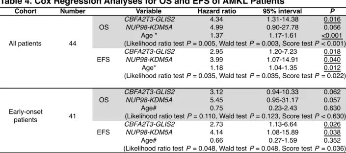
Cox Regression Analysis
20
Multivariate Cox regression analyses of OS and EFS (see Methods) in all AMKL
21
patients (n = 44) using WBC count and age as continuous variables identified that
22
CBFA2T3-GLIS2 was an independent prognostic factor for poor OS and EFS and that
23
NUP98-KDM5A was an independent prognostic factor for poor EFS (Table 4).
24
Whereas, multivariate Cox regression analyses of early-onset patients (n = 41),
1
using age as a categorical variable (infants vs older patients), revealed that
2
CBFA2T3-GLIS2 and NUP98-KDM5A were independent prognostic factors for poor
3
EFS (Table 4).
4 5
Discussion
6
Among 44 AMKL patients treated with two recent AML protocols in Japan,
7
CBFA2T3-GLIS2 fusion gene was the most frequently identified fusion gene (27%). In
8
addition, NUP98-KDM5A (9%), RBM15-MKL1 (5%), and KMT2A rearrangements
9
(7%) were recurrently found. Gene mutations in AMKL tended to be less frequent in
10
fusion-positive patients than in fusion-negative patients (14% vs 35%). Survival
11
analyses indicated that CBFA2T3-GLIS2 was a strong candidate for poor prognostic
12
factor in AMKL patients, even in those treated with recent improved chemotherapies.
13
Our study included patients consecutively treated with either of the two recent
14
clinical trials in Japan and revealed that AMKL had an improved OS at approximately
15
60%, which was consistent with a recent report by the Berlin-Frankfurt-Münster
16
(BFM) study group that reported the OS as 70% (Schweitzer et al., 2015). Thus, our
17
study might be able to identify relatively accurate frequencies as well as the clinical
18
impact of genetic features in AMKL patients who were treated in recent clinical trials.
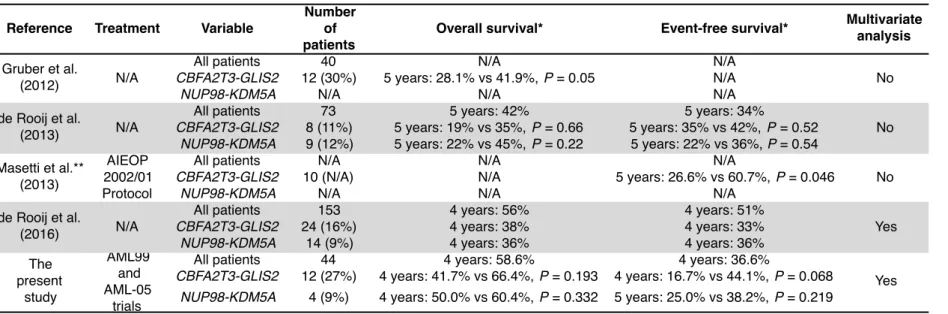
19
The frequency of CBFA2T3-GLIS2 in this study was either similar to or two-fold
20
higher than those reported in three previous studies (27%, 13%, and 16%); additionally,
21
the present study demonstrated that CBFA2T3-GLIS2-positive patients had a poor
22
prognosis, in agreement with previous studies (Table 5).
23
In prognostic analyses of our cohort, the 4-year EFS was lower and the 3-year
24
CIR was higher in CBFA2T3-GLIS2-positive patients than in
1
CBFA2T3-GLIS2-negative patients. One potential reason for this outcome is the high
2
relapse rate of CBFA2T3-GLIS2-positive patients treated with chemotherapy alone as
3
initial treatment. Among nine CBFA2T3-GLIS2-positive patients who achieved CR
4
after induction therapy, four received HSCT at first CR, three of whom survived. In
5
contrast, four of the remaining five patients who received chemotherapy alone as initial
6
treatment died after relapse. Although the patient number was limited in the present
7
study, and while a previous intergroup study reported that a benefit of HSCT for
8
AMKL patients could not be demonstrated (de Rooij et al., 2016), these results
9
indicated that HSCT at first CR should be considered in CBFA2T3-GLIS2-positive
10
patients to avoid relapse.
11
The tendency of very poor prognosis observed in CBFA2T3-GLIS2-positive
12
infants in this study raised the possibility that CBFA2T3-GLIS2-positive patients
13
should be stratified into risk groups by age. The prognosis of infant AML patients was
14
previously reported not to be poor, with a 5-year OS of 61%–75% and 5-year EFS of
15
44%–51% (Creutzig et al., 2012). Furthermore, in contrast to infant ALL (Pui et al.,
16
2002), KMT2A rearrangements in infant AML were not associated with a poor
17
prognosis (KMT2A-positive vs negative; 5-year OS, 71% vs 66%; 5-year EFS, 43% vs
18
52%) (Creutzig et al., 2012). High WBC counts (43.2 × 10⁹/l) and high induction
19
failure rate (43%) of CBFA2T3-GLIS2-positive infants in the present study might be
20
indicators of hyper-proliferation of leukemic cells. Thus, future studies with a larger
21
number of patients will be needed for further characterization of infant AMKL.
22
Although a previous intergroup study reported that NUP98-KDM5A was
23
associated with poor prognosis in AMKL (Table 5), the present study could not
24
determine the prognostic power of NUP98-KDM5A due to the small number of
1
NUP98-KDM5A-posiitve patients (n = 4). However, when prognostic analyses were
2
performed in all 503 AML patients across both trials, including four AMKL and three
3
other AML patients with NUP98-KDM5A (one of each with M5, M6, and RAEB-T
4
subtypes), the 4-year OS and EFS of a total of seven NUP98-KDM5A-positive patients
5
were significantly lower than those of NUP98-KDM5A-negative patients (28.6% vs
6
71.2%, P = 0.003; and 14.3% vs 56.9%, P < 0.001, respectively. data not shown).
7
Furthermore, several studies reported that AML patients with other
8
NUP98-rearrangements had a poor prognosis (Taketani et al., 2010; Hollink et al.,
9
2011; Shiba et al., 2013). Thus, all together, these findings suggested that
10
NUP98-KDM5A was potentially a poor prognostic factor in pediatric AML patients.
11
Hyperdiploidy was frequently observed in AMKL patients, consistent with a
12
previous report (Sandahl et al., 2014), and was a significantly good prognostic factor in
13
the present study. One reason for this finding was the high survival rate of patients
14
with hyperdiploidy after induction failure and/or relapse. Their 5-year EFS was not
15
significantly different than that of patients without hyperdiploidy (38.1% vs 33.1%, P
16
= 0.746) (Table S3), suggesting that patients with hyperdiploidy could be salvaged by
17
intensified chemotherapy instead of therapies used in intermediate-risk patients.
18
Although the first option for patients who experienced induction failure or relapse is
19
still HSCT, more intensified chemotherapy without HSCT might be a potential option
20
for patients with hyperdiploidy.
21
The biology of leukemogenesis in fusion-negative patients is predicted to be
22
heterogeneous. Interestingly, all late-onset patients were in the fusion-negative group
23
(Table S2). Gene mutations such as FLT3-ITD, WT1, and KIT were found in all
24
late-onset patients, whereas a complex karyotype, found in 60% of fusion-negative
1
patients, was not observed. Furthermore, cytogenetic aberrations that are frequently
2
found in adult AMKL patients (Dastugue et al., 2002), such as t(9;22)(q34;q11),
3
3q21q26 changes, and -5/del(5q), were not observed in late-onset patients. A recent
4
intergroup study reported the data from 82 pediatric AMKL patients lacking
5
CBFA2T3-GLIS2, NUP98-KDM5A, KMT2A rearrangements or monosomy 7 (de Rooij
6
et al., 2016). The age distribution of these patients was as follows: 0–4 years, n = 72
7
(88%); 5–7 years, n = 4 (5%); 8–10 years, n = 0; and 11–17, n = 6 (7%). Although
8
molecular details of these patients were not investigated, this age distribution was
9
similar to that observed in the present study and supported our findings. Thus, further
10
analysis in a larger cohort is necessary to understand the heterogeneity of
11
fusion-negative patients.
12
Molecular differences between AMKL and other AML were identified in the
13
present study. CBFA2T3-GLIS2 was not found in any of the 459 other AML patients,
14
including 97 patients with a normal karyotype, although this fusion gene was reported
15
in 4% (10/237) of other AML patients with a normal karyotype (Masetti et al., 2013).
16
This discrepancy might be partially explained by the racial difference between the
17
Japanese and American/European populations, which might be related to the relatively
18
higher frequency of this fusion gene in AMKL patients in the present study. Several
19
studies reported the possible differences of the relationship of FAB subtypes with
20
certain fusion genes, such as RUNX1-RUNX1T1 and CBFβ-MYH11, in the Japanese
21
population compared with the American/European populations. A previous study from
22
the BFM study group reported that all 57 RUNX1-RUNX1T1-positive patients and 41
23
of 42 CBFβ-MYH11-positive patients harbored FAB-M1/M2 and M4/M4Eo subtypes,
24
respectively (von Neuhoff et al., 2010). However, in the AML-05 trial, 3 of 86
1
RUNX1-RUNX1T1-positive patients and 5 of 30 CBFβ-MYH11-positive patients
2
harbored non-M1/M2 and non-M4/M4Eo subtypes, respectively (data not shown).
3
Otherwise, relatively small number of patients in the present study was associated with
4
this discrepancy. Additionally, only 24% of fusion-positive AMKL patients had gene
5
mutations, and all fusion-positive patients were early-onset. A recent study reported a
6
very low frequency of gene mutations in infant ALL patients with KMT2A
7
rearrangements (1.3 mutations/patient) (Andersson et al., 2015). Thus, the present
8
study suggested a similarity of leukemogenesis between fusion-positive AMKL and
9
infant ALL with KMT2A rearrangements.
10
In conclusion, the present study clarified the cytogenetic and molecular features
11
and their clinical impact in pediatric AMKL patients treated in recent clinical trials.
12
CBFA2T3-GLIS2 was the most frequently identified fusion gene and might be a strong
13
candidate for a poor prognostic factor in this disease, especially in infants. We propose
14
that these findings will enable clinicians to design and administer appropriate
15
risk-stratified therapies and develop new molecular-targeted therapies for this unique
16
pediatric AML subtype.
17 18
Acknowledgements
19
The authors would like to thank Yuki Hoshino for the experiments and Enago
20
(www.enago.jp) for the English language review.
21 22
Authorship Contributions
23
Y.Hara and Y.Hayashi designed the study. Y.Hara, N.S., G.Y. and K.O. performed the
1
experiments. H.A., T.Taki and Y.Hayashi supervised the work. Y.Hara and K.T.
2
analyzed the results. Y.Hara, K.T. and T.Taki constructed the figures. M.P., D.T, A.K,
3
A.M.S, N.K, A.T, K.H, T.Taga and S.A provided patient samples and data. Y.Hara,
4
N.S., T.Taki and Y.Hayashi wrote the paper and all the authors critically reviewed and
5
revised the manuscript.
6 7
Conflict of Interest
8
The authors declare that they have no conflict of interest.
9 10
References
1
Andersson AK, Ma J, Wang J, Chen X, Gedman AL, Dang J, Nakitandwe J, Holmfeldt
2
L, Parker M, Easton J, Huether R, Kriwacki R, Rusch M, Wu G, Li Y, Mulder H,
3
Raimondi S, Pounds S, Kang G, Shi L, Becksfort J, Gupta P, Payne-Turner D,
4
Vadodaria B, Boggs K, Yergeau D, Manne J, Song G, Edmonson M, Nagahawatte P,
5
Wei L, Cheng C, Pei D, Sutton R, Venn NC, Chetcuti A, Rush A, Catchpoole D,
6
Heldrup J, Fioretos T, Lu C, Ding L, Pui CH, Shurtleff S, Mullighan CG, Mardis
7
ER, Wilson RK, Gruber TA, Zhang J, Downing JR. 2015. The landscape of somatic
8
mutations in infant MLL-rearranged acute lymphoblastic leukemias. Nat Genet
9
47:330-337.
10
Athale UH, Razzouk BI, Raimondi SC, Tong X, Behm FG, Head DR, Srivastava DK,
11
Rubnitz JE, Bowman L, Pui CH, Ribeiro RC. 2001. Biology and outcome of
12
childhood acute megakaryoblastic leukemia: a single institution's experience. Blood
13
97:3727-3732.
14
Barnard DR, Alonzo TA, Gerbing RB, Lange B, Woods WG. 2007. Comparison of
15
childhood myelodysplastic syndrome, AML FAB M6 or M7, CCG 2891: report
16
from the Children's Oncology Group. Pediatr Blood Cancer 49:17-22.
17
Byrd JC, Mrozek K, Dodge RK, Carroll AJ, Edwards CG, Arthur DC, Pettenati MJ,
18
Patil SR, Rao KW, Watson MS, Koduru PR, Moore JO, Stone RM, Mayer RJ,
19
Feldman EJ, Davey FR, Schiffer CA, Larson RA, Bloomfield CD. 2002.
20
Pretreatment cytogenetic abnormalities are predictive of induction success,
21
cumulative incidence of relapse, and overall survival in adult patients with de novo
22
acute myeloid leukemia: results from Cancer and Leukemia Group B (CALGB
23
8461). Blood 100:4325-4336.
24
Cheson BD, Bennett JM, Kopecky KJ, Buchner T, Willman CL, Estey EH, Schiffer
1
CA, Doehner H, Tallman MS, Lister TA, Lo-Coco F, Willemze R, Biondi A,
2
Hiddemann W, Larson RA, Lowenberg B, Sanz MA, Head DR, Ohno R,
3
Bloomfield CD. 2003. Revised recommendations of the International Working
4
Group for Diagnosis, Standardization of Response Criteria, Treatment Outcomes,
5
and Reporting Standards for Therapeutic Trials in Acute Myeloid Leukemia. J Clin
6
Oncol 21:4642-4649.
7
Creutzig U, Zimmermann M, Bourquin JP, Dworzak MN, Kremens B, Lehrnbecher T,
8
von Neuhoff C, Sander A, von Stackelberg A, Schmid I, Stary J, Steinbach D,
9
Vormoor J, Reinhardt D. 2012b. Favorable outcome in infants with AML after
10
intensive first- and second-line treatment: an AML-BFM study group report.
11
Leukemia 26:654-661.
12
Dastugue N, Lafage-Pochitaloff M, Pages MP, Radford I, Bastard C, Talmant P,
13
Mozziconacci MJ, Leonard C, Bilhou-Nabera C, Cabrol C, Capodano AM,
14
Cornillet-Lefebvre P, Lessard M, Mugneret F, Perot C, Taviaux S, Fenneteaux O,
15
Duchayne E, Berger R. 2002. Cytogenetic profile of childhood and adult
16
megakaryoblastic leukemia (M7): a study of the Groupe Francais de Cytogenetique
17
Hematologique (GFCH). Blood 100:618-626.
18
de Rooij JD, Hollink IH, Arentsen-Peters ST, van Galen JF, Berna Beverloo H,
19
Baruchel A, Trka J, Reinhardt D, Sonneveld E, Zimmermann M, Alonzo TA,
20
Pieters R, Meshinchi S, van den Heuvel-Eibrink MM, Zwaan CM. 2013.
21
NUP98/JARID1A is a novel recurrent abnormality in pediatric acute
22
megakaryoblastic leukemia with a distinct HOX gene expression pattern. Leukemia
23
27:2280-2288.
24
de Rooij JD, Masetti R, van den Heuvel-Eibrink MM, Cayuela JM, Trka J, Reinhardt
1
D, Rasche M, Sonneveld E, Alonzo TA, Fornerod M, Zimmermann M, Pigazzi M,
2
Pieters R, Meshinchi S, Zwaan CM, Locatelli F. 2016. Recurrent genetic
3
abnormalities can be used for risk-group stratification in pediatric AMKL: results of
4
a retrospective intergroup study. Blood 127:3424-30
5
Gruber TA, Larson Gedman A, Zhang J, Koss CS, Marada S, Ta HQ, Chen SC, Su X,
6
Ogden SK, Dang J, Wu G, Gupta V, Andersson AK, Pounds S, Shi L, Easton J,
7
Barbato MI, Mulder HL, Manne J, Wang J, Rusch M, Ranade S, Ganti R, Parker M,
8
Ma J, Radtke I, Ding L, Cazzaniga G, Biondi A, Kornblau SM, Ravandi F,
9
Kantarjian H, Nimer SD, Dohner K, Dohner H, Ley TJ, Ballerini P, Shurtleff S,
10
Tomizawa D, Adachi S, Hayashi Y, Tawa A, Shih LY, Liang DC, Rubnitz JE, Pui
11
CH, Mardis ER, Wilson RK, Downing JR. 2012. An Inv(16)(p13.3q24.3)-encoded
12
CBFA2T3-GLIS2 fusion protein defines an aggressive subtype of pediatric acute
13
megakaryoblastic leukemia. Cancer Cell 22:683-697.
14
Hama A, Yagasaki H, Takahashi Y, Nishio N, Muramatsu H, Yoshida N, Tanaka M,
15
Hidaka H, Watanabe N, Yoshimi A, Matsumoto K, Kudo K, Kato K, Horibe K,
16
Kojima S. 2008. Acute megakaryoblastic leukaemia (AMKL) in children: a
17
comparison of AMKL with and without Down syndrome. Br J Haematol
18
140:552-561.
19
Hollink IH, van den Heuvel-Eibrink MM, Arentsen-Peters ST, Pratcorona M, Abbas S,
20
Kuipers JE, van Galen JF, Beverloo HB, Sonneveld E, Kaspers GJ, Trka J, Baruchel
21
A, Zimmermann M, Creutzig U, Reinhardt D, Pieters R, Valk PJ, Zwaan CM. 2011.
22
NUP98/NSD1 characterizes a novel poor prognostic group in acute myeloid
23
leukemia with a distinct HOX gene expression pattern. Blood 118:3645-3656.
24
Inaba H, Zhou Y, Abla O, Adachi S, Auvrignon A, Beverloo HB, de Bont E, Chang
1
TT, Creutzig U, Dworzak M, Elitzur S, Fynn A, Forestier E, Hasle H, Liang DC,
2
Lee V, Locatelli F, Masetti R, De Moerloose B, Reinhardt D, Rodriguez L, Van
3
Roy N, Shen S, Taga T, Tomizawa D, Yeoh AE, Zimmermann M, Raimondi SC.
4
2015a. Heterogeneous cytogenetic subgroups and outcomes in childhood acute
5
megakaryoblastic leukemia: a retrospective international study. Blood 126:1575-84.
6
Kinoshita A, Miyachi H, Matsushita H, Yabe M, Taki T, Watanabe T, Saito AM,
7
Tomizawa D, Taga T, Takahashi H, Matsuo H, Kodama K, Ohki K, Hayashi Y,
8
Tawa A, Horibe K, Adachi S. 2014. Acute myeloid leukaemia with myelodysplastic
9
features in children: a report of Japanese Paediatric Leukaemia/Lymphoma Study
10
Group. Br J Haematol 167:80-86.
11
Ma Z, Morris SW, Valentine V, Li M, Herbrick JA, Cui X, Bouman D, Li Y, Mehta
12
PK, Nizetic D, Kaneko Y, Chan GC, Chan LC, Squire J, Scherer SW, Hitzler JK.
13
2001. Fusion of two novel genes, RBM15 and MKL1, in the t(1;22)(p13;q13) of
14
acute megakaryoblastic leukemia. Nat Genet 28:220-221.
15
Masetti R, Pigazzi M, Togni M, Astolfi A, Indio V, Manara E, Casadio R, Pession A,
16
Basso G, Locatelli F. 2013. CBFA2T3-GLIS2 fusion transcript is a novel common
17
feature in pediatric, cytogenetically normal AML, not restricted to FAB M7 subtype.
18
Blood 121:3469-3472.
19
Mercher T, Coniat MB, Monni R, Mauchauffe M, Nguyen Khac F, Gressin L,
20
Mugneret F, Leblanc T, Dastugue N, Berger R, Bernard OA. 2001. Involvement of
21
a human gene related to the Drosophila spen gene in the recurrent t(1;22)
22
translocation of acute megakaryocytic leukemia. Proc Natl Acad Sci U S A
23
98:5776-5779.
24
Pui CH, Gaynon PS, Boyett JM, Chessells JM, Baruchel A, Kamps W, Silverman LB,
1
Biondi A, Harms DO, Vilmer E, Schrappe M, Camitta B. 2002. Outcome of
2
treatment in childhood acute lymphoblastic leukaemia with rearrangements of the
3
11q23 chromosomal region. Lancet 359:1909-1915.
4
Reinhardt D, Diekamp S, Langebrake C, Ritter J, Stary J, Dworzak M, Schrauder A,
5
Zimmermann M, Fleischhack G, Ludwig WD, Harbott J, Creutzig U. 2005. Acute
6
megakaryoblastic leukemia in children and adolescents, excluding Down's
7
syndrome: improved outcome with intensified induction treatment. Leukemia
8
19:1495-1496.
9
Sandahl JD, Kjeldsen E, Abrahamsson J, Ha SY, Heldrup J, Jahnukainen K, Jonsson
10
OG, Lausen B, Palle J, Zeller B, Forestier E, Hasle H. 2014. Ploidy and clinical
11
characteristics of childhood acute myeloid leukemia: A NOPHO-AML study. Genes
12
Chromosomes Cancer 53:667-675.
13
Sano H, Shimada A, Taki T, Murata C, Park MJ, Sotomatsu M, Tabuchi K, Tawa A,
14
Kobayashi R, Horibe K, Tsuchida M, Hanada R, Tsukimoto I, Hayashi Y. 2012.
15
RAS mutations are frequent in FAB type M4 and M5 of acute myeloid leukemia,
16
and related to late relapse: a study of the Japanese Childhood AML Cooperative
17
Study Group. Int J Hematol 95:509-515.
18
Schoch C, Kern W, Kohlmann A, Hiddemann W, Schnittger S, Haferlach T. 2005.
19
Acute myeloid leukemia with a complex aberrant karyotype is a distinct biological
20
entity characterized by genomic imbalances and a specific gene expression profile.
21
Genes Chromosomes Cancer 43:227-238.
22
Schweitzer J, Zimmermann M, Rasche M, von Neuhoff C, Creutzig U, Dworzak M,
23
Reinhardt D, Klusmann JH. 2015. Improved outcome of pediatric patients with
24
acute megakaryoblastic leukemia in the AML-BFM 04 trial. Ann Hematol
1
94:1327-1336.
2
Shiba N, Ichikawa H, Taki T, Park MJ, Jo A, Mitani S, Kobayashi T, Shimada A,
3
Sotomatsu M, Arakawa H, Adachi S, Tawa A, Horibe K, Tsuchida M, Hanada R,
4
Tsukimoto I, Hayashi Y. 2013a. NUP98-NSD1 gene fusion and its related gene
5
expression signature are strongly associated with a poor prognosis in pediatric acute
6
myeloid leukemia. Genes Chromosomes Cancer 52:683-693.
7
Shiba N, Ohki K, Kobayashi T, Hara Y, Yamato G, Tanoshima R, Ichikawa H,
8
Tomizawa D, Park MJ, Shimada A, Sotomatsu M, Arakawa H, Horibe K, Adachi S,
9
Taga T, Tawa A, Hayashi Y. 2016. High PRDM16 expression identifies a
10
prognostic subgroup of pediatric acute myeloid leukaemia correlated to FLT3-ITD,
11
KMT2A-PTD, and NUP98-NSD1: the results of the Japanese Paediatric
12
Leukaemia/Lymphoma Study Group AML-05 trial. Br J Haematol 172:581-591.
13
Shimada A, Taki T, Tabuchi K, Tawa A, Horibe K, Tsuchida M, Hanada R, Tsukimoto
14
I, Hayashi Y. 2006. KIT mutations, and not FLT3 internal tandem duplication, are
15
strongly associated with a poor prognosis in pediatric acute myeloid leukemia with
16
t(8;21): a study of the Japanese Childhood AML Cooperative Study Group. Blood
17
107:1806-1809.
18
Slovak ML, Kopecky KJ, Cassileth PA, Harrington DH, Theil KS, Mohamed A,
19
Paietta E, Willman CL, Head DR, Rowe JM, Forman SJ, Appelbaum FR. 2000.
20
Karyotypic analysis predicts outcome of preremission and postremission therapy in
21
adult acute myeloid leukemia: a Southwest Oncology Group/Eastern Cooperative
22
Oncology Group Study. Blood 96:4075-4083.
23
Taketani T, Taki T, Nakamura T, Kobayashi Y, Ito E, Fukuda S, Yamaguchi S,
24
Hayashi Y. 2010. High frequencies of simultaneous FLT3-ITD, WT1 and KIT
1
mutations in hematological malignancies with NUP98-fusion genes. Leukemia
2
24:1975-1977.
3
Thiollier C, Lopez CK, Gerby B, Ignacimouttou C, Poglio S, Duffourd Y, Guegan J,
4
Rivera-Munoz P, Bluteau O, Mabialah V, Diop M, Wen Q, Petit A, Bauchet AL,
5
Reinhardt D, Bornhauser B, Gautheret D, Lecluse Y, Landman-Parker J, Radford I,
6
Vainchenker W, Dastugue N, de Botton S, Dessen P, Bourquin JP, Crispino JD,
7
Ballerini P, Bernard OA, Pflumio F, Mercher T. 2012. Characterization of novel
8
genomic alterations and therapeutic approaches using acute megakaryoblastic
9
leukemia xenograft models. J Exp Med 209:2017-2031.
10
Tomizawa D, Tawa A, Watanabe T, Saito AM, Kudo K, Taga T, Iwamoto S, Shimada
11
A, Terui K, Moritake H, Kinoshita A, Takahashi H, Nakayama H, Kiyokawa N,
12
Isoyama K, Mizutani S, Hara J, Horibe K, Nakahata T, Adachi S. 2013. Appropriate
13
dose reduction in induction therapy is essential for the treatment of infants with
14
acute myeloid leukemia: a report from the Japanese Pediatric Leukemia/Lymphoma
15
Study Group. Int J Hematol 98:578-588.
16
Tsukimoto I, Tawa A, Horibe K, Tabuchi K, Kigasawa H, Tsuchida M, Yabe H,
17
Nakayama H, Kudo K, Kobayashi R, Hamamoto K, Imaizumi M, Morimoto A,
18
Tsuchiya S, Hanada R. 2009. Risk-stratified therapy and the intensive use of
19
cytarabine improves the outcome in childhood acute myeloid leukemia: the AML99
20
trial from the Japanese Childhood AML Cooperative Study Group. J Clin Oncol
21
27:4007-4013.
22
von Neuhoff C, Reinhardt D, Sander A, Zimmermann M, Bradtke J, Betts DR,
23
Zemanova Z, Stary J, Bourquin JP, Haas OA, Dworzak MN, Creutzig U. 2010.
24
Prognostic impact of specific chromosomal aberrations in a large group of pediatric
1
patients with acute myeloid leukemia treated uniformly according to trial
2
AML-BFM 98. J Clin Oncol 28:2682-2689.
3
Xu G, Nagano M, Kanezaki R, Toki T, Hayashi Y, Taketani T, Taki T, Mitui T, Koike
4
K, Kato K, Imaizumi M, Sekine I, Ikeda Y, Hanada R, Sako M, Kudo K, Kojima S,
5
Ohneda O, Yamamoto M, Ito E. 2003. Frequent mutations in the GATA-1 gene in
6
the transient myeloproliferative disorder of Down syndrome. Blood 102:2960-2968.
7 8
ID Protocol WBC (×109/l)
Age
(y) Mutation CK Hyper -diploidy
CR after
induction Risk group Relapse HSCT Outcome Cytogenetics
R-081 AML-05 48.2 0 - No Yes No N/A Yes Yes dead 47,XY,+21[9]/46,XY[11]
R-116 AML-05 7.3 1 FLT3-ITD No No Yes High Yes Yes dead 46,XY[20]
R-119 AML-05 11.2 1 FLT3-ITD No Yes Yes High Yes Yes alive 47,XX,+3[11]/46,XX[9]
R-144 AML-05 20.0 1 GATA1 No No Yes N/A Yes Yes dead 46,XY,t(15;16)(q24;q24)[1]/47,XY,+Y[1]/46,XY
R-159 AML-05 30.5 0 - No No Yes Intermediate Yes Yes dead 46,XY,[20]
R-192 AML-05 35.2 0 GATA1 No Yes Yes Intermediate Yes Yes alive 48,XX,+3,+21[9]/46,XX[11]
282-R AML-05 62.8 0 KIT Yes Yes Yes Intermediate Yes Yes dead 49,XY,+Y,+12,+21[2]/50,sl,+Y,+8,-12[18]
315-R AML-05 52.8 1 - No Yes Yes Intermediate No Yes alive 48,XY,+14,+21[20]
326-R AML-05 30.7 0 - No No No N/A No Yes dead 46,XX[20]
352-R AML-05 75.3 0 - No No No N/A Yes Yes alive 46,XY[20]
429-R AML-05 73.6 0 - Yes Yes Yes Intermediate Yes Yes dead #1
A159 AML99 10.5 2 - No Yes Yes Intermediate No Yes alive 46,XX[16]47,XX,+21[3]/48,ider,+4[1]
336-R AML-05 7.0 1 - Yes No Yes Intermediate Yes Yes dead #2
368-R AML-05 23.4 1 - Yes No Yes Intermediate No No alive #3
405-R AML-05 11.0 2 - Yes No No N/A No Yes alive #4
A262 AML99 12.5 1 - No No Yes Low Yes N/A dead 46,XY[20]
R-005 AML-05 12.0 0 - Yes Yes Yes Intermediate Yes Yes dead #5
R-162 AML-05 42.2 0 - No No Yes Intermediate No No alive #6
A093 AML99 24.1 2 - Yes Yes Yes Intermediate No Yes alive #7
A136 AML99 4.3 3 - No No Yes Intermediate No No alive 46XX,t(9;11)(q22;q23)[20]
A075 AML99 6.0 2 - Yes No Yes Intermediate No No alive #8
CK, complex karyotype; CR, complete remmision; HSCT, hematopoietic stem cell transplantation; N/A, not applicable
Outline of treatment was as follows: chemotherapy alone for low-risk in AML 99/AML-05 and intermediate-risk in AML-05; chemotherapy alone or HLA-matched- related HSCT for intermediate-risk in AML 99; allo-HSCT for high-risk in AML99/AML-05. R-144 withdrew AML-05 due to false positive FLT3-ITD. 315-R with intermediate-risk (AML-05) received HSCT at 1st CR due to doctor's decision.
#1: 48,XX,t(3;21)(q27;q22),+21,+21[11]/48,idem,der(19)(t(1;19)(q21;p13)[3]/90,idem×2,-4,-7,-9,-15,-18,-21[3]/46,XX[3]
#2: 45,XX,-15,add(18)(q21),add(19)(p13)[16]/46,sl,del(13)(q?)[2]/46,XX[2]
#3: 46,XX,add(6)(q23),der(8;15)(q10;q10),+mar[7]/46,XX,add(6)(q23),der(8;15)(q10;q10),del(13)(q12q14),+mar[4]/46,XX,add(11)(q13)[3]/46,XX[5]
#4: 46,XY,del(3)(q13.2),add(6)(p25),ins(11;?)(q13;?),ins(12;?)(q13;?),del(13)(q12q14)[10]/49,idem,+2,+9,+del(13)(q12q14),-17,+21[1]/46,XY[9]
#5: 61,XXX,der(1)t(1;22)(p13;q13),t(1;22)(p13;q13),-3,-4,-5,+7,-9,-11,-12,-13,-15,-18,+19,-22[10]/46,XX[10]
#6: 46,XY,der(1)t(1;22)(p13;q13)add(1)(q32),der(22)t(1;22)(add(1)(p22)[15]/46,XY[5]
#7: 61<2n>,XX,+2,+4,+6,+7,+8,+(9;11)(p22;q23),+10,+12,+14,+15,+17,+19,+20,+21,+22,+22[15/20]64,idem,+13,+15,+15,[1/20]46,XX [4/20]
Table 1. Clinical and Cytogenetic/Molecular Profiles of Fusion-Positive Patients
CBFA2T3-GLIS2
NUP98-KDM5A
RBM15-MKL1
KMT2A-MLLT3
KMT2A-MLLT10
AMKL Other FAB subtype P
Total number of patients 44 459
Age at diagnosis, n (%)
Median age (range) 1 (0-13) 8 (0-17) <0.001
0-4 41 (93) 144 (31) <0.001
5-10 0 (0) 155 (34) <0.001
10< 3 (7) 160 (35) <0.001
Median WBC (×109/l) (range) 22.0 (4.3-191.6) 21.4 (0.6-985.0) 0.886 Gender
Male 21 (48) 249 (54) 0.637
Female 23 (52) 210 (46)
Fusion gene, n (%) 21 (48) 281(61) 0.106
inv(16)(p13.3q24.3)/CBFA2T3-GLIS2 12 (27) 0 (0) <0.001
t(11;12)(p15;p13)/NUP98-KDM5A 4 (9) 3 (1) 0.002
abnormal 11q23/KMT2A rearrangement 3 (7) 67 (16) 0.125
t(1;22)(p13;q13)/RBM15-MKL1 2 (5) 0 (0) 0.008
t(8;21)(q22;q22)/RUNX1-RUNX1T1 0 (0) 151 (32) <0.001
inv(16)(p13q22)/CBFB-MYH11 0 (0) 39 (8) 0.038
t(5;11)(p35;q15.5) /NUP98-NSD1 0 (0) 16 (3) 0.383
t(16;21)(p11; q22)/FUS-ERG 0 (0) 5 (1) 1.000
Cytogenetic feature, n (%)* 39 (89) N/A N/A
normal 7 (16) 97 (21) 0.559
monosomy 7 3 (7) 7 (2) 0.049
trisomy 21 16 (36) N/A N/A
complex karyotype 22 (50) N/A N/A
hyperdiploidy 23 (52) N/A N/A
Gene mutation, n (%)* 17 (39) N/A N/A
NRAS 3 (7) 57 (12) 0.339
KRAS 1 (2) 31 (7) 0.344
KIT 3 (7) 91 (20) 0.041
WT1 2 (5) 28 (6) 1.000
NPM1 0 (0) 16 (4) 0.383
FLT3-ITD 4 (9) 60 (13) 0.636
GATA1 5 ( ) N/A N/A
Table 2. Comparison of Patients with or without AMKL
non-DS-AMKL, non-Down syndrome acute megakaryoblastic leukemia; other FAB subtype, M0-M6, excluding M3 and Down syndrome; WBC, white blood cell; FLT3-ITD, FLT3 internal tandem duplication; N/A, not applicable
*Number of patients who have any of these mutations.