Japan Advanced Institute of Science and Technology
JAIST Repository
https://dspace.jaist.ac.jp/
Title 遺伝暗号の修復のための人工デアミネーゼシステム
Author(s) BHAKTA, SONALI Citation
Issue Date 2020‑09
Type Thesis or Dissertation Text version ETD
URL http://hdl.handle.net/10119/17012 Rights
Description Supervisor:塚原 俊文, 先端科学技術研究科, 博士
Artificial deaminase system for restoration of genetic code
Sonali Bhakta
Japan Advanced Institute of Science and Technology
Doctoral Dissertation
Artificial deaminase system for restoration of genetic code
Sonali Bhakta
Supervisor: Professor Dr. Toshifumi Tsukahara
Graduate School of Advanced Science and Technology Japan Advanced Institute of Science and Technology
Materials Science
September, 2020
Doctoral Dissertation
Artificial deaminase system for restoration of genetic code
Main theme Advisor Professor Toshifumi Tsukahara Sub-theme Advisor Professor Kenjo Fuzimoto
Head of Committee Professor Dr. Toshifumi Tsukahara
Internal Member Professor Dr. Kenzo Fujimoto Internal Member Professor Dr. Takahiro Hosaka
Internal Member Associate Professor Dr. Hidekazu Tsutsui External Member Professor Dr. Yoshitsugo Aoki
Graduate School of Advanced Science and Technology
Japan Advanced Institute of Science and Technology Materials Science
September, 2020 SONALI BHAKTA
s1720427
Judging Committee
Table of Contents
I
No. Title of the contents Page No.
Abstract VI-VII
List of Figures VIII-XIV
List if Tables XV
Chapter I: General Introduction
1.1 Genetic engineering 2
1.2 Genome editing 2-3
1.3 RNA editing 3
1.4 Types of RNA editing 3
1.5 Advantages and Disadvantages of the genome and RNA editing 3-5
1.6 ADAR (Adenosine deaminases acting on RNA) 5-8
1.7 Apolipoprotein B mRNA editing enzyme, catalytic polypeptide 8
1.8 Types of the APOBEC and its function 9-11
1.9 Site Directed RNA editing methods 11-16
1.10 Mechanism of A to I editing 16-17
1.11 Mechanism of C to U editing 17-18
1.12 MS2 system 18-19
1.13 Mechanism of ApoB for RNA editing 20-21
1.14 Aim of the study 22
1.15 Expected impact of the study 22
1.16 References 23-33
Chapter II: A to I RNA editing by using ADAR1 artificial deaminase system for restoration of genetic code in Ochre (UAA) stop codon
2.1 Introduction 34-37
2.2 Materials and Methods 38-44
2.2.1 Preparation of the plasmid constructs 38
2.2.2 Construction of ADAR1 39
2.2.3 Preparing guideRNA for directing ADAR1 to target 39-40 2.2.4 Preparation of 1X MS2 on either side of the guideRNA 41
2.2.5 Cell culture 42
2.2.6 Transfection with Lipofectamine 2000 42
2.2.7 Cell expression of GFP 42
2.2.8 RNA extraction and cDNA synthesis 42-43
2.2.9 PCR-RFLP 43
2.2.10 Direct sequencing by Sanger’s method 43-44
2.3 Results 45-53
Table of Contents
II
2.3.1 Microscopic observation 45-46
2.3.2 Confirmation of the restoration by PCR-RFLP 47-48 2.3.3 Direct sequencing of restored EGFP mRNA 48-50 2.4 Application of the double MS2 on the either side of the guide with
ADAR1 deaminase
51-53 2.4.1 Juli Microscopic observation for confirmation of the
restoration
51 2.4.2 PCR-RFLP for confirmation of the restoration 52 2.4.3 Direct sequencing of restored EGFP mRNA 53
2.5 Discussions 54-56
2.6 Conclusions 57
2.7 References 58-60
Chapter III: Genetic code restoration in BFP (derivative of GFP) by artificial RNA editing using APOBEC 1 cytidine deaminase
3.1 Introduction 61-65
3.2 Materials and Methods 66-73
3.2.1 Target plasmid (mutated EGFP or BFP) construct preparation
66 3.2.2 APOBEC1 deaminase plasmid construction 66 3.2.3 Preparation of the gRNA to direct the deaminase to target 67-68
3.2.4 Culturing cells and transfection 68
3.2.5 Observation of fluorescence by confocal microscopy 69 3.2.6 RNA extraction and cDNA synthesis from transfected
cells
69 3.2.7 Confirmation of sequence restoration by PCR-RFLP 69-70
3.2.8 Sanger’s sequencing 70-71
3.2.9 Total RNA-sequencing (RNA-seq) 71-73
3.3 Results 74-84
3.3.1 Restoration of RNA using an artificial enzyme system 74
3.3.2 LSM confocal microscopy 74-76
3.3.3 Confirmation of the restoration by PCR-RFLP 77-79
3.3.4 Confirmation by Sanger sequencing 80-81
3.3.5 Confirmation of sequence restoration and observation of off-target effects by total RNA-sequencing (RNA-seq)
81-84
3.4 Discussion 85-89
3.5 Conclusions 90
Table of Contents
III
3.6 References 91-96
Chapter IV: A study on pol II, pol III promoters and single construct (made of combination of pol II and III promoter) for restoration efficacy in case of C-to-U editing
4.1 Introduction 97-101
4.1.2 Single construct having CMV and U6 promoter in combination
99-101
4.2 Materials and Methods 102-110
4.2.1 Target plasmid construct preparation 102
4.2.2 APOBEC 1 deaminase plasmid construct preparation 102 4.2.3 Preparation of the guide for directing the APOBEC 1 to
targeted site
102-106 4.2.3.1 Preparation of the guide under the control of pol II CMV
promoter
102-103 4.2.3.2 Preparation of the guide under the control of pol III U6
promoter
103 4.2.4 Preparation of the Single construct having APOBEC 1
deaminase under the pol II CMV promoter’s control and guideRNA under pol III U6 promoter’s control
104-105
4.2.5 Western blot analysis 106-108
4.2.6 Transfection into BFP stable transformed HEK 293 cells 108-109 4.2.7 Observation of the cells for fluorescence by confocal
microscope
109 4.2.8 RNA extraction and cDNA synthesis from the transfected
cells
109 4.2.9 Confirmation of the restoration results by PCR-RFLP 109-110
4.2.10 Sanger’s sequencing 110
4.3 Results 111-121
4.3.1 Western Blot analysis 111-112
4.3.2 Laser Confocal Microscopy for the restored green fluorescence observation
112-115 4.3.3 PCR-RFLP confirmation for the restoration of the
genetic code
115-118 4.3.4 Sanger’s sequencing confirmation of the genetic code
restoration
118-121
4.4 Discussion 122-125
Table of Contents
IV
4.5 Conclusion 126
4.6 References 127-131
Chapter V: Restoration of the genetic code from C to U in real model mouse (Macular Mouse)
5.1 Introduction 132-135
5.2 Materials and Methods 136-147
5.2.1 Rearing and caring of Macular mouse in laboratory 136
5.2.2 Collection of samples 136
5.2.3 RNA extraction and followed by cDNA synthesis from the collected samples
136-137 5.2.4 Identification of the Target mutated C in the ATP7A gene 137 5.2.5 Sequence confirmation of the mutation (T-to-C) in
ATP7A gene
137-138 5.2.6 APOBEC 1 deaminase enzyme preparation 139
5.2.7 Preparation of the guideRNA 139-140
5.2.8 Preparation of the Single construct having APOBEC 1 deaminase under the pol II CMV promoter’s control and guideRNA under pol III U6 promoter’s control
140-141
5.2.9 Preparation of 1X MS2 on either side of the guideRNA 141-143 5.2.10 Collection and culturing the Tail fibroblast cells 143-144 5.2.11 Electroporation of the cells for the editing 144-146 5.2.12 RNA extraction and cDNA synthesis from the
electroporated cells
146 5.2.13 PCR amplification of the synthesized cDNA and
Sanger’s sequencing
147 5.2.14 Sequence confirmation of the editing 147
5.3 Results 148-155
5.3.1 Body weight difference between the hemizygous male, heterozygous female and normal male littermate at 7, 10 and 14 days
148-149
5.3.2 Sanger’s sequencing confirmation of the genetic code restoration
150-153 5.3.3 Application of guideRNA with 1X MS2 on either side of
the guideRNA
154-155
5.4 Discussion 156-158
5.4.1 Body weight reduction in case of the Macular Mouse 156
Table of Contents
V
5.4.2 Sanger’s sequence confirmation of the RNA editing 156-158
5.5 Conclusion 159
5.6 References 160-162
Chapter VI: Final Discussion
6.1 Final Discussion 163-170
6.2 References 172-173
List of publications
Journal papers 174
Conferences (Domestic and International) 174-175
Awards 175
Acknowledgements XVI-XVII
VI
ABSTRACT
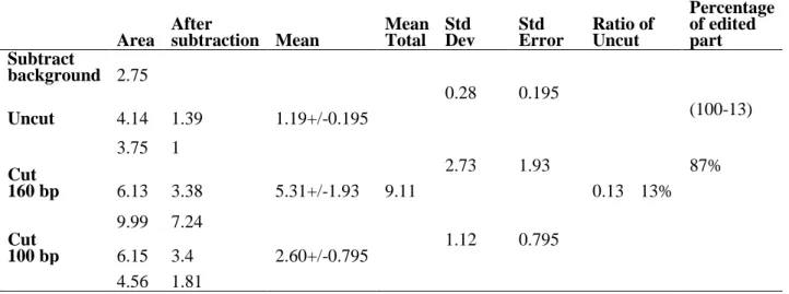
Site directed mutagenesis is an exceptionally viable way to deal with recode genetic information. Legitimate connecting of the synergist area of the RNA altering catalytic deaminase Adenosine Deaminase Acting on RNA (ADAR) or Cytidine Deaminase Acting on RNA (APOBEC) to an antisense direct RNA can change over explicit adenosines (As) to inosines (Is), with the last perceived as guanosines (Gs) during the translation procedure or Cytidines (Cs) to Uridines (Us). In this study, endeavors have been made to engineer the deaminase domain of ADAR1 and MS2 framework to target explicit A residues to reestablish G→A transformations. The target mRNA comprised of an ochre (TAA) stop codon, created from the TGG codon encoding amino acid 58 (Trp) of improved green fluorescent protein (EGFP). This framework had the capacity to change over the stop codon (TAA) to a decipherable codon (TGG), accordingly reestablishing fluorescence in a cell framework, as appeared by JuLi fluorescence and LSM confocal microscopy. The specificity of the editing was affirmed by Polymerase Chain Reaction-Restriction Fragment Length Polymorphism (PCR-RFLP), as the restored GFP mRNA could be cleaved into fragments of 160 and 100 base pairs, the absolute amplified length was 260 bp. Sanger’s sequencing illustration with both the sense and antisense primers indicated that the reclamation rate was higher for the 5'A than for the 3'A. This system might be very useful for treating genetic diseases that result from the G to A point mutations.
Further an artificial editase of RNA was engineered by combining the deaminase domain of APOBEC1 (apolipoprotein B mRNA editing catalytic polypeptide 1) with a guideRNA (gRNA) which is complementary to target mRNA. In this artificial enzyme system, gRNA is bound to MS2 stem-loop, and deaminase domain, which has the ability to convert mutated target nucleotide C-to-U, is fused to MS2 coat protein. As a target RNA, here RNA encoding Blue Fluorescent Protein (BFP) was used which is derivative of the gene encoding GFP by 199T>C mutation. Upon transient expression of both components (deaminase and gRNA), GFP fluorescence was observed by confocal microscopy, indicating that mutated 199th C in BFP had been converted to U, restoring original sequence of GFP. This result was confirmed by PCR-RFLP and Sanger’s sequencing using cDNA from transfected cells, revealing an editing efficiency of approximately 21%. Deep RNA sequencing result showed that off-target editing was sufficiently low in this system.
Later on, improving U6 promoter activity by CMV enhancer or promoter in target cells have been demonstrated to be a viable method to obtain satisfactory percentage of editing efficiency. The placement of a CMV enhancer nearby to U6 promoter or hybrid CMV-H1 promoter has been accounted for improving the efficiency of RNAi or shRNA delivery in vivo. From the experimental data it has been found that in case of the CMV promoter controlled process of RNA editing where both the deaminase and guideRNA constructs were prepared under the control of the pol II CMV promoter, the editing efficiency was lesser comparing to the U6 promoter containing guideRNA or in single construct having combined approach of CMV in deaminase domain and U6 promoter in guideRNA construct. From the PCR-RFLP (band intensity) data had also been observed that with the increase of the concentration of the deaminase or the guideRNA the restoration percentage had also increased. The editing efficiency has been calculated from the peak height of the Sanger’s sequencing data. After the calculation of the efficiency it was found that in case of the CMV controlled approach the rate was 21.02% whereas in case of the U6 controlled and in case of single construct the restoration rate was 39.37% and 41.65%, respectively.
VII
For performing the in vivo application of the developed artificial enzyme system the macular mouse model was chosen.
The mutation in the P type copper transporting ATPase (ATP7A) gene is responsible for the Menkes kinky hair disease, where T-to-C mutation happens. It was found from our data that all the heterozygous female (Ml/+), normal littermate male (+/y) and hemizygous male (Ml/y) had increased the body weight as usual up to 10 days of age. After that the body weight of heterozygous female (Ml/+) and normal littermate (+/y) increased significantly at 14 days as well but in case of the hemizygous male (Ml/y), its body weight significantly reduced at 14th day of age. The peak area and peak height from the Sanger’s sequencing analysis was measured by ImageJ (NIH) software. From the calculation it was found that by using the APOBEC1 deaminase and U6-21bp upstream-MS2-6X guideRNA 12.17% and 16.25% of the genetic code was restored in the macular mouse derived fibroblast cells by peak area and peak height, respectively.
Where the deaminase and guideRNA, were two different constructs. After that single construct was applied where the deaminase was controlled by pol II CMV vector and guideRNA was under the control of pol III U6 promoter, in the same plasmid vector. The peak area and peak height from the Sanger’s sequencing analysis were measured by using ImageJ (NIH) software. From the calculation we found that by using the APOBEC 1 deaminase and U6-MS2-6X-21bp upstream 27.20% and 26.09% of the genetic code was restored, respectively calculated from peak area and peak height.
Afterwards, the 1X MS2 on either side of guide sequence containing guideRNA construct was introduced along with the APOBEC 1 deaminase. Similarly the sample was sequenced for observing the editing rate. Editing rate was calculated both by peak area and peak height. I found that editing rate was 36.66% and 34%, respectively by peak area and peak height. For any developed system it is more important that the application could be achieved for the purpose of treatment. The developed artificial deaminase system for both the A-to-I and C-to-U editing could be applied to the through the viral vector (AAVs) easily into the host body for the therapeutic purpose. The proper application of the developed artificial deaminase system for the treatment of the patients who are suffering from such type of mutagenic diseases could open a new era in the field of genetic diseases.
Key words: Genetic code, RNA editing, Deaminase domain, Macular mouse, ATP7A gene
List of Figures
VIII Chapter
No.
Figure No. Name of the figure
Chapter I
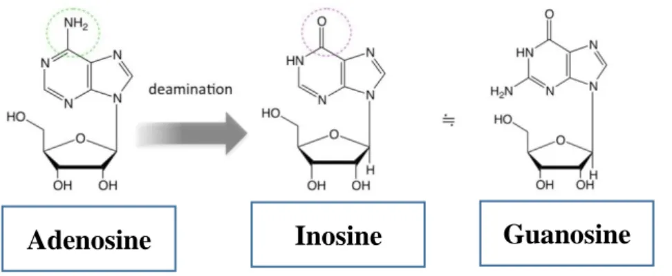
1 Chemical conversion of A to I and C to U
2 Chemical structural differences of Adenosine (A), Inosine (I) and Guanosine (G)
3 Different types of the ADAR deaminase
4 RNA editing by ADAR (Adenosine Deaminase Acting on RNA) 5 Mechanism of RNA editing by ADAR1 (A to I conversion) 6 Members of APOBEC family
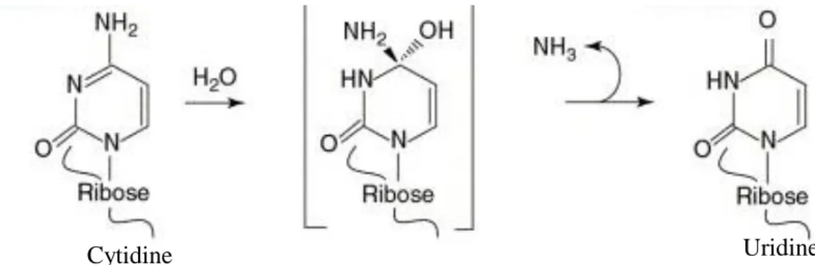
7 Chemical modification during the conversion of Cytosine to uracil, which happens through a deamination process
8 A. MS2-RNA stem-loop wild-type, B. change in few nucleotide result higher binding mutant, The numbering is from the first base of the AUG which is the initiation codon of the replicase mRNA C. Antiparallel oriented MS2 protein dimer with 10 β-sheets., The association between two MS2 coat protein monomers (red and grey). The β-sheets are named A to G, and the N- and C-termini of each monomer is indicated. C. Schematic representation of the MS2 system 9 C-to-U RNA editing of apolipoprotein B. The model for a4
35-nucleotide region of apoB RNA flanking the edited base (asterisk) is shown. A schematic representation illustrates apobec-1 (red) and ACF (blue) binding to RNA both 5 and 3 of the edited base and depicts the presence of additional proteins that may modulate assembly of the holoenzyme (green). Note that the stoichiometry of apobec-1 and ACF molecules with respect to the active enzyme is unknown. The model emphasizes the role of both cis-acting elements within the vicinity of the edited base (mooring sequence is bolded) and the requirement for an optimal structure, conferred by both 5 and 3 efficiency elements.
10 Mechanism for the RNA editing using APOBEC1 and guideRNA, having a mismatch at the target position in the guideRNA
Chapter II
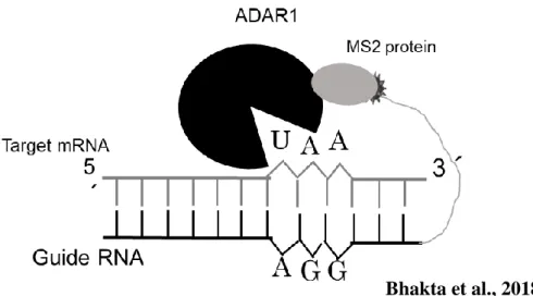
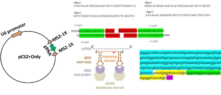
11 Schematic feature of the engineered ADAR1 (Black), MS2 protein (Grey), MS2 RNA (Dark ash) connected with Guide RNA. In the target RNA where the target Adenosine will be converted to Inosine (I) which is recognized as the Guanosine at the time of translation.
12 Sequence result of the target mRNA, EGFP containing Ochre stop codon
List of Figures
IX
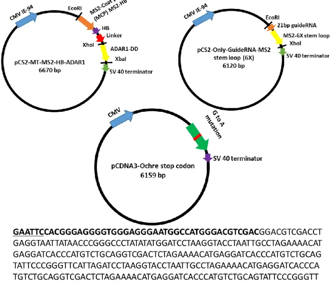
13 Schematic features of the construction of the three major editing factors.
Sequence result of the guideRNA-MS2-6X, Here Bold underline are the restriction sites. In the upstream EcoRI and downstream XhoI. The bold part is the 19bp guideRNA and rest are the MS2-6X part
14 Preparation of the MS2-1X stem loop (Double) on the either side of the 21 bp guideRNA in the pCS2+only plasmid vector under the control of U6 promoter
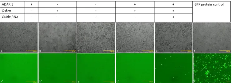
15 Transfection in HEK cell with wild type EGFP, 1 factor (Only EGFP containing ochre stop codon or Only ADAR1), 2 factors (EGFP containing Ochre stop codon + ADAR1 or EGFP containing Ochre stop codon + Guide RNA) and 3 factors (EGFP containing Ochre stop codon + ADAR1 + Guide RNA). Only the wild type EGFP and the 3 factors (EGFP containing Ochre stop codon +ADAR1+Guide RNA) express the fluorescence expression due to the presence of TGG in wild type and editing from TAA to TGG by the 3 factors (EGFP containing Ochre stop codon +ADAR1+Guide RNA) Imaging by JuLi smart fluorescence microscope.
16 Transfection in HEK cell with wild type EGFP, 2 factors (Ochre+ADAR1) or (Ochre+gRNA) and 3 factors (Ochre+ADAR1+Guide RNA). Only the wild type EGFP (a) and the 3 factors (Oche+ADAR1+Guide RNA) (d) express the fluorescence expression due to the presence of TGG in wild type and editing from TAA to TGG by the 3 factors (Oche+ADAR1+Guide RNA). However, the image of two factors (b & c) does not show any fluorescence expression as the TAA has not been restored by the two factors. *Ochre:
EGFP containing ochre stop codon
17 Representation of both the PCR and RFLP results, cut (wild type and restored) and uncut (single factor), after PCR-RFLP using BmgT120I restriction enzyme. For cut the band was found at 160 base pair and 100 base pair, whereas the uncut remain at 260 base pair as the only PCR result
18a Restored EGFP from ochre stop codon (TAA) to Normal codon TGG (Sense primer).
18b Restored EGFP from ochre stop codon (TAA) to Normal codon TGG (Anti-sense primer), Showing the restoration of the TAA to TGG.
19 Transfection in HEK 293 cell with wild type EGFP, 1 factor (Only
List of Figures
X
EGFP containing ochre stop codon or Only ADAR1), 2 factors (EGFP containing Ochre stop codon + ADAR1 or EGFP containing Ochre stop codon + Guide RNA) and 3 factors (EGFP containing Ochre stop codon + ADAR1 + Guide RNA). Only the wild type EGFP and the 3 factors (EGFP containing Ochre stop codon +ADAR1+Guide RNA) express the fluorescence expression due to the presence of TGG in wild type and editing from TAA to TGG by the 3 factors (EGFP containing Ochre stop codon +ADAR1+Guide RNA) Imaging by JuLi smart fluorescence microscope.
20 Representation of both the PCR and RFLP results, cut (wild type and restored) and uncut (single factor), after PCR-RFLP using BmgT120I restriction enzyme. For cut the band was found at 160 base pair and 100 base pair, whereas the uncut remain at 260 base pair as the only PCR result also showed that the amplified DNA was at 260 bp length. (n=3) 21 Restored EGFP from ochre stop codon (TAA) to Normal codon TGG,
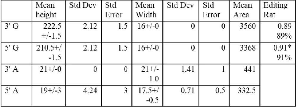
the double peak is there and the 5’A is more edited than the 3’.
According to the editing rate calculation, 26% of 5’A and 17% of 3’A was restored from TAA to TGG by peak area and peak height, respectively (n=3).
Chapter III
22 Schematic diagram of the MS2 system and the outcome of the experimental outline
23 Flowchart showing the workflow of the RNA-seq analysis. From the RNA samples to the final data, each step, including sample test, library preparation, and sequencing, influences the quality of the data, and data quality directly.
24 a. Schematic diagram of the MS2 system, The MS2 coat protein is attached to the APOBEC 1 catalytic domain and the MS2 stem loop is attached to the gRNA under the control of the CMV promoter. The MS2 coat protein and stem loop can bind to each other, enabling detection of a specific nucleotide sequence within the mRNA b. Stably BFP expressing HEK 293 cells were transfected with wild type of GFP, one factor (e.g., either APOBEC1or only guide RNA) or two factors (APOBEC1 + guide RNA). Green fluorescence expression was observed only when two factors were present, implying that both factors were necessary for C-to-U editing. Imaging was performed by LSM confocal microscopy. C. Calculation of the fluorescence intensity by
List of Figures
XI
using the Fluoviewer 10 software and for all data the statistical analysis (mean±SEM) was calculated, (n=5)
25 a. Schematic illustration of RFLP. The BtgI restriction enzyme can cut the BFP but not wild type of GFP or restored GFP (C to U converted) b.
PCR-RFLP of cDNA extracted from transfected cells (HEK 293 stably expressing BFP), restriction-digested with BtgI. BFP was cleaved into two fragments, 201 and 123 bp, whereas restored GFP was not cleaved and remained at 324 bp. c. For all data the statistical analysis (mean±SEM) was calculated, where n=5.
26 Confirmation of the restoration of BFP to GFP (C to U) by Sanger’s sequencing. a. Forward or Sense primer (CCA to CTA). In the sense primer the dual peaks were observed, which were due to the restoration of the genetic code from the C to U (BFP to GFP), after the application of the two editing factors (APOBEC1 deaminase and gRNA). C. Edit-R analysis of the Sense chromatogram height of the edited part of the peak and the statistical analysis (mean±SEM) has been done, where the n=5.
Edit R analysis has been done by using the Sanger’s sequencing Ab.1 file
27 APOBEC 1(DD)-MS2 system induces some off-target C-to-U RNA editing in HEK293 cells. a. Percentages of expressed genes with at least one edited cytosine (C-to-U or G-to-A) in total SNVs. b. Box plots showing rate of cytosines edited by APOBEC1(DD)-MS2 compared to control (mutated BFP target in HEK 293 cells), yellow line is median. c.
Jitter plots showing transcriptome-wide efficiencies of C-to-U or G-to-A edits (y-axis) identified from RNA-seq experiments in HEK293 cells modified by APOBEC1 (DD)-MS2 vs editing- negative control (BFP target, stably transformed in HEK 293 cells) . n: total number of modified cytosines identified
Whole BFP sequencing by placing different primers at different positions and also there were overlapped positions. In total 1109bp were amplified, among these at the position of 200-527 only at this position one off target event was found, which was located upstream of the targeted C which was to be edited
Chapter IV
28 Guide construct under control of the pol II CMV promoter 29 Guide construct under the control of the pol III U6
List of Figures
XII
30 APOBEC 1 deaminase construct under the control of pol II CMV promoter
31 Single construct, the guide portion under the control of the pol III U6 promoter and the APOBEC 1 deaminase portion under the control of pol II CMV promoter. Here CMV promoter is located upstream of the U6 promoter
32 Single construct, the guideRNA portion under the control of the pol III U6 promoter and the APOBEC 1 deaminase is under the control of the pol II CMV promoter. Here, CMV promoter is located at the downstream of the U6 promoter
33 Western blot analysis for checking the protein expression of APOBEC 1 in the HEK 293 cell lines. Beta actin was considered as the negative control without any enzyme which was observed at 42 kDa. The APOBEC1 expression was observed at 78 kDa, Lane 1,2:
MS2-HB-APOBEC1 in HEK 293 cells; Lane 3: Cell Lysate.
34 Confocal microscopic images for observation of the fluorescence after restoration. There are five panels where wild type EGFP is the positive control where there was green fluorescence and only BFP cells without enzyme or guideRNA is the negative control where there was only blue fluorescence but no green fluorescence. In case of the CMV containing guideRNA and APOBEC 1 there was appearance of the green fluorescence due to the restoration of the genetic code from C to U by editing and same happened in case of the U6 promoter and single construct. But the green fluorescence intensity differs from each other.
Although BFP was also observed in case of the restored samples as well because its not possible to restore 100% of target mRNA. But if from the negative control the BFP is compared with the BFP in restored samples the intensity was also reduced in case of the restored samples.
35 Restoration percentage according to Light intensity of BFP and GFP. In case of the CMV the restoration percentage is 30.65% whereas in case of U6 and Single construct the percentage is 45.31% and 51.53%, respectively. U6 gives better restoration percentage than CMV but Single construct prepared with the combination of the CMV promoter containing APOBEC1 and U6 promoter containing guideRNA was the best among the three.
36 Comparison between CMV and U6 promoter efficiency in respect to
List of Figures
XIII
restoration percentage at different concentrations during the time of transfection
37 Comparative sequence result of the different promoters (U6 and CMV) and Single construct (prepared from the U6 guide RNA and CMV promoter containing APOBEC 1)
38 Editing rate from the peak area of the different promoters (U6 and CMV) and Single construct (prepared from the U6 guide RNA and CMV promoter containing APOBEC 1) of the sequence result. Where, CMV promoter shows 21.02% and in case of U6 and single construct 39.37% & 41.65% respectively
Chapter V
39 PCR amplification of the ATP7A gene from the cDNA synthesized from the Macular mouse liver and spleen
40 Sequence confirmation of the T to C mutation in ATP7A gene, from the collected Liver and spleen samples.
41 Single construct, the guideportion under the control of the pol III u6 promoter and the APOBEC 1 deaminase portion under the control of the pol II CMV promoter. Here CMV promoter is located at the downstream of the U6 promoter
42 Schematic model of the 1X MS2 stem loop on either side of the guideRNA mediated editing by APOBEC 1 deaminase
43 Body weight change along with days according to the genotype. The graph shows that with the progress of the days the body weight of the heterozygous female (Ml/+) and normal littermate male (+/y) increased.
But in case of the hemizygous male (Ml/y), which is the macular mouse, the body weight increased upto 10 days but after that at 14th days it started to reduce which became even less than the normal bodyweight at 7th day.
44 Confirmation of the restoration of the genetic code where a. PCR amplification of the ATP7A gene from the synthesized cDNA of transfected cells b. Sanger’s sequencing showing the restoration of the genetic code. At the position of the mutated C there is a small peak of T (Arrow), which is due to the RNA editing, c. calculation of the editing efficiency from the peak area and peak height and d. graph showing that according to peak area and peak height 12.17 % and 16.25% has been restored, respectively
45 Confirmation of the restoration of the genetic code where by using
List of Figures
XIV
single construct a. Sanger’s sequencing showing the restoration of the genetic code. At the position of the mutated C there is a small peak of T (Arrow), which is due to the RNA editing, b. calculation of the editing efficiency from the peak area and peak height and c. graph showing that according to peak area and peak height 27.20 % and 26.09% has been restored, respectively.
46 a. Fluorescence image showing the transfection efficiency while the wild type GFP was transfected into macular mouse tail derived fibroblast cells having mutated ATP7A gene, a’. on the contrary when the APOBEC1 and guideRNA was transfected into the macular mouse tail derived fibroblast cells having mutated ATP7A gene there was no fluorescence expression as there was no fluorescence expressing gene, image was taken by Juli fluorescence microscope
b. PCR amplification of the targeted ATP7A gene from the predicted restored sample, transfected with APOBEC 1 deaminase and guideRNA c. Sanger’s sequencing confirmation of the C to U editing, by using the 1X MS2 guideRNA along with APOBEC 1 deaminase in macular mouse tail derived Fibroblast cells. a. Sequence data showing restored T peak at the position of mutated C peak.
d. calculation of the editing rate from the peak area and peak height, respectively.
List of Tables
XV
Chapter No. Table No. Title of table
Chapter I 1 Diseases caused by the T to C and G to A mutation
2 Human AID and APOBEC paralogs
Chapter II 3 Ratio of the edited and unedited from PCR-RFLP
4a Calculation of editing efficiency considering area by using sense primer
4b Calculation of editing efficiency considering area by using antisense primer
5a Calculation of the editing efficiency considering peak area by using antisense primer
5b Calculation of the editing efficiency considering peak area by using sense primer
Chapter IV 6 Different restoration percentage (average) in case of CMV and U6 promoter in relation to the increase of the concentration of the deaminase APOBEC1 or guideRNA:
Chapter V 7 Application of different pulse rate for electroporation 8 Weight measurement of the macular mouse at different ages
Chapter I Doctoral Thesis Sonali Bhakta
1
CHAPTER I
General Introduction
Chapter I Doctoral Thesis Sonali Bhakta
2
INTRODUCTION 1.1 Genetic engineering:
Genetic engineering is also termed as genetic modification or manipulation, is the direct manipulation of an organism's genes using biotechnology. It consists of a set of technologies which are used for regulating functions of intracellular target gene or expression has been widely used in basic research, medicinal and applications as therapy (1). It is also utilized for changing the genetic makeup of cells, including the transfer of genes within and across species boundaries to produce improved or novel organisms. New DNA is obtained by either isolating or replicating the hereditary material of interest utilizing recombinant DNA strategies or by artificially incorporating or synthesizing the DNA. Development of construct is generally made and used to embed this DNA into the host living being. The primary motivation behind the genetic engineering methodologies is to control the capacity of intracellular proteins associated with the organic procedures of intrigue.
1.2 Genome editing
Genome alteration or genome designing is a kind of genetic engineering where DNA is embedded, erased, adjusted or supplanted in the genome of a living organism. As of late progressions in genome altering innovations have generously improved the capacity to roll out exact improvements in the genomes of eukaryotic cells. In recent times several technologies of genome editing have made it possible to alter target genomic information (1-5). Notwithstanding to programmable regulation, one of the most significant highlights of the genome editing is that it can provide a permanent alteration to the targeted gene in the specific cell, while this perpetual modification can
Chapter I Doctoral Thesis Sonali Bhakta
3
successfully limit the target protein expressions. Such techniques may present wellbeing dangers if a blunder happens (6).
1.3 RNA editing
RNA editing is another significant mechanism for directing hereditary versatility through the age of elective protein items from a solitary basic quality. Substitutional RNA editing utilizes an assortment of genetic mechanisms, the biochemical premise of which has been explained following the advancement of in vitro measures that reiterate vital components of this procedure. For the most part two sorts of substitutional RNA exist in mammals, namely A-to-I and C-to-U RNA editing (7, 8), whereas another type U-to-C substitution is found mainly in lower plants. Important biochemical differentiations between these two procedures provide an informative basis for understanding the mechanisms of C-to-U RNA editing and the adjustments control the target particularity.
1.4 Types of RNA editing:
There are several types of RNA editing events in living organisms. Among these A-to-I, C-to-U and U-to-C are the most common types. A-to-I and C-to-U RNA editing are generally caused by ADARs and APOBEC-AID deaminase family respectively.
However, the enzymes responsible for U-to-C editing does not discover yet although it is the abundant phenomenon in lower plant species and rare in animals.
1.5 Advantages and disadvantages of the genome editing and RNA editing
The principle motto behind the genetic engineering techniques is to control the functional capacity of intracellular proteins engaged with the biological processes of intrigue. By recent times several genome editing technologies such as CRISPR-Cas system, TALEN, ZFN etc. have made it conceivable to control target genomic
Chapter I Doctoral Thesis Sonali Bhakta
4
information (10-14). In addition to programmable regulation, one of the most significant features of the genome editing is that it can provide a change to the targeted gene in cellular system, while this effect can control the expression or production of targeted protein efficaciously (15). However, genome editing has some drawbacks, as if any alteration at off target level occurs that will be permanent in case of the genome editing and will affect the genome sequence consequently, whereas mismatch will not be permanent in RNA editing; even not affect the genome sequence. RNA editing is much more for treatment purpose compared to genome editing. Particularly for very popular CRISPR-Cas genome editing system, homology-mediated repair system is needed to repair between cut and inserted DNA which is only present in dividing cells. In case of the non-dividing cells like nerve and muscle cells, this system facing challenges. In association with the technical challenges and bioethical issue, it’s still controversial regarding the clinical application of this technology .Therefore, I have chosen the RNA editing as my choice of approach to build up an artificial enzymatic system for restoration of genetic information.
RNA sequences can be edited with regarding their genome-encoded arrangement like how an editorial manager changes letters or words in an original copy. The principal recognized instance of such RNA editing was the post-transcriptional addition or erasure of non-encoded uridine (U) residues into the mitochondrial RNA of trypanosomes. This technique generates accurate, ‘readable’ open reading frames that cannot be reasoned from sequences of genomic arrangements. In higher eukaryotes, base alteration is the significant kind of RNA editing, with the best-portrayed model being deamination responses, in which cytidine (C) or adenosine (A) is converted to uracil (U) and inosine (I), respectively. Moreover, a couple of U-to-C and G-to-A
Chapter I Doctoral Thesis Sonali Bhakta
5
alterations have been accounted for, which may conceivably result from trans-amination reactions (Figure 2). If such base modifications happen in coding areas of mRNA, the amino-acid particularity of codons can be changed, bringing about the blend of protein isoforms not predictable from genome sequences. Be that as it may, in the event that it is happening in the anticodons of tRNAs, base deamination can increment or even change the codon-recognition capacity during translation.
1.6 ADAR (Adenosine deaminases acting on RNA) enzyme:
Adenosine deaminases acting on RNA (ADARs) are the enzymes for RNA editing which have the capacity to execute the hydrolytic deamination of the adenosine (A) in the context of RNA polymers, at the corresponding nucleotide positions converting to inosine (I) (22, 23, 24). This reaction has a an assortment of practical ramifications for the RNA substrates including modulating thermal stability of base-paired structures, modifying the significance of codons in mRNAs (recoding), changing the splicing patterns of pre-mRNAs, altering miRNA targeting within 3 untranslated regions (UTRs), etc. (25-31). With the recognition of the large numbers of inosine (I) locales in the human transcriptome this circumstance is widespread (32). Also, mis-regulation of A-to-I RNA editing is associated with human ailments (33–38, 39). To be precise, alteration of one of the human genes responsible for A-to-I RNA editing (ADAR1) is a reason for the acquired immune disease such as Aicardi–Goutieres syndrome (40).
Human cells express three distinct types of ADARs (adenosine deaminase acting on RNA), named ADAR1, 2, and 3, (Figure 3) which are possibly re-addressable for site-directed RNA editing (41, 42). ADAR1 and ADAR2 proteins have the well characterized activity of adenosine deamination (43). While ADAR3 is expressed in human brain however its capacity stays a mystery since no reactant action has been
Chapter I Doctoral Thesis Sonali Bhakta
6
accounted for the protein yet. The ADARs are particular in their structure with clearly identifiable RNA binding domains and also deaminase domains (Figure 1). In reality, RNA binding is largely dependent on double-stranded RNA binding domains (dsRBDs) present in each ADAR protein as multiple copies (44). dsRBDs tie up with any RNA duplex greater than ∼16 bp, in a sequence independent manner (45). The existance of dsRBDs in the ADAR structure is predictable with the prerequisite for duplex RNA in ADAR substrates for effective deamination to take place (Figure 4). However, ongoing studies intended to guide editing to new positions with fusion proteins containing ADAR deaminase domains propose these domains likewise require duplex RNA for effective responses, even in absence of dsRBDs (46-48). While the RNA-binding properties of dsRBDs of ADARs have been all around well archived (49, 50), scarcely any reports have concentrated on the RNA restricting necessities of the ADAR synergist areas.
Fig 1. Chemical conversion of A-to-I and C-to-U
Chapter I Doctoral Thesis Sonali Bhakta
7
Fig 4. RNA editing by ADAR (Adenosine Deaminase Acting on RNA) Fig 3. Different types of the ADAR deaminase
Slavov D et al., 2000
Fig 2. Chemical structural differences of Adenosine (A), Inosine (I) and Guanosine (G)
Slotkin et al., 2013
Adenosine Inosine Guanosine
https://image.slidesharecdn.com/rnaediting-171031063231/95/rna-editing -10-638.jpg?cb=1509431588
Chapter I Doctoral Thesis Sonali Bhakta
8
1.7 Apolipoprotein B mRNA editing enzyme, catalytic polypeptide (APOBEC):
The APOBEC group of cytidine deaminases has extended and veered although throughout vertebrate evolution and contemporarily consists of AID and five APOBECs, numbered from 1 to 5 (abbreviated A1 to A5). The family most likely developed from AID (or AICDA), which is thought to have been evolved from the Tad (tRNA adenosine deaminase)/ADAT2 (adenosine deaminase, tRNA Specific 2) tRNA-altering compounds before the vertebrate radiation.
Advancement has formed a quite certain capacity for APOBEC in adaptive immunity.
APOBEC is exceptionally communicated in enacted B cells and is fundamental for immunizer proclivity development and expansion (56). By DNA editing of the genomic immunoglobulin (Ig) loci APOBEC triggers particular downstream process:
class-switch recombination (CSR), somatic hypermutation (SHM), and, in certain vertebrates, gene alteration. Intriguingly, Ig hypermutation by APOBEC can likewise bring about an erasure of the entire substantial chain locus, causing B cell passing in a
RNA Editing (A to I) ATGCAGCAA ATGCI(G)GCAA
Hideyama et al., 2011
Fig 5. Mechanism of RNA editing by ADAR1 (A to I conversion)
Chapter I Doctoral Thesis Sonali Bhakta
9
procedure named locus suicidal recombination (57-59).
1.8 Types of the APOBEC and its function
APOBEC1, A1 is the first APOBEC to be found, works in humans as an RNA editor. A1 specifically C-to-U edits cytidine 6666 in ApoB mRNA, which encodes a key player in lipid transportation (60). This RNA editing, which happens explicitly in the small intestine in humans, makes an untimely stop codon that yields a short ApoB-48 peptide rather than the full-length 100 amino corrosive ApoB-100, the two of which are significant for lipid homeostasis (61). Thus, A1 operates similarly to ADAR enzymes by creating mRNAs with differential capacity from a single locus (62). Strikingly, A1 does not edit ApoB mRNA in amniotes, in spite of the fact that their A1 can alter genomic DNA when transfected to E. coli and confine retro-elements in ex vivo assays, proposing that these may reflect the genealogical function of A1. Like AID, this mutagenic capacity can be dangerous and overexpression of A1 can prompt to malignant growth.
APOBEC2, A2 is fundamentally expressed in heart and skeletal muscle in warm blooded animals (63, 64) and chickens (65). It is inessential for mouse development, survival, and fertility (66), but is needed for proper muscle growth (67, 68), for left–
right specification in lower vertebrate embryogenesis (69), and for retina and optic nerve regeneration (70). The molecular capacity of A2 has been tricky in light of the fact that it lacks any identifiable cytidine deaminase activity. Ongoing work on its role in retina regeneration (71) has demonstrated that the zinc-coordinating domain but not deaminase activity are required to stimulate the binding of the Pou6f2 (POU class 6 homeobox 2) transcription factor to DNA. Regardless of whether cytidine deamination by A2 takes place in explicit contexts in vivo, perhaps working in combination with vital cofactors, is yet to be resolved.
Chapter I Doctoral Thesis Sonali Bhakta
10
APOBEC3, A3s comprising of seven paralogs in the human genome (A3A–D, A3F–H), are DNA editors that are popular for their roles in innate immunity as potent inhibitors of viral infections and retro-elements. However, they are they are associated with invulnerability in different manners: A3G contributes to cellular immunity by enhancing natural killer (NK) cell recognition. A recent and novel finding is that, upon enlistment by inflammation-associated factors, A3A edits the mRNAs of several qualities of genes, some associated with viral pathogenesis, in monocytes and macrophages (72). A3s can also directly edit nuclear or mitochondrial DNA and transfected plasmids. APOBEC3 (regardless of its proposed function in restricting endogenous retrotransposition) are inessential for mouse development, survival, or fertility.
The physiological functions of A4 and A5 are yet to know. A4 is expressed in testicles (73) and does not deaminate DNA in E. coli and yeast assays (74). A5 is present in non-mammalian tetrapods and its similitude to AID and A5 suggests that it can edit DNA.
Fig 6. Members of APOBEC family
https://www.bing.com/images/search?vie w=detailV2
Chapter I Doctoral Thesis Sonali Bhakta
11 Name Genomic
location
Exons Deaminatio n domains
Expressio n
Cellular localization
Editing activity
Target
AID 12q13 5 1 Activated
B cells, testis
Mainly cytoplasmic, acts in the nucleus
DNA Immunoglob
ulin gene
APOBEC 1
12q13.1 5 1 Small
intestine
Cytoplasmic/
nuclear, acts
in the
nucleus
RNA, DNA
Apolipoprote in B m RNA
APOBEC 2
6p21 3 1 Skeletal
muscle, heart
Cytoplasmic/
nuclear
Unknow n
Unknown
APOBEC 3 A
22q13.1 5 1 Keratinoc
ytes, blood
Cytoplasmic/
nuclear
DNA Adeno-associ ated virus, retrotranspos ons
APOBEC 3B
22q13.1 8 2 Intestine,
Uterus, mammary gland, keratinoc ytes, other
Predominantl y nuclear
DNA Retroviruses, retrotranspos ons, HBV
APOBEC 3C
22q13.1 4 1 Many
tissues
Cytoplasmic/
nuclear
DNA Retroviruses, retrotranspos ons, HBV APOBEC
3DE
22q13.1 7 2 Thyroid,
spleen, blood
Unknown DNA Retroviruses
APOBEC3 F
22q13.1 8 2 Many
tissues
Cytoplasmic DNA Retroviruses, retrotranspos ons, HBV APOBEC
3 G
22q13.1 8 2 Many
tissues, T cells
Cytoplasmic DNA Retroviruses, retrotranspos ons, HBV APOBEC
3H
22q13.1 5 1 Blood,
thymus, thyroid, placenta
Unknown DNA Retroviruses
LOC19646 9*
12q23 1 2 Pseudoge
ne
- - -
APOBEC4 Iq25.3 2 1 Testis Unknown Unknow
n
Unknown
1.9 Site Directed RNA editing methods:
Site directed RNA editing is a method to recode genetic information at the RNA level Table 1. Human AID and APOBEC paralogs
Chapter I Doctoral Thesis Sonali Bhakta
12
(16). The approach is based on the enzymatic conversion of the adenosine (A) to inosine (I). In many biological or biochemical processes including translation, inosine are interpreted as guanosine (Figure 2), thus A-to-I RNA editing allows the recoding of amino acids, splice elements, miRNAs and miRNA binding sites among others (17).
Recently a new method has been developed named the site directed RNA editing that employs engineered deaminases in combination of the short guide RNAs to recode a single adenosine bases at specific sites in any user defined transcripts (18, 19). Due to the usage of the guide RNAs the target selection and specificity is easily and rationally programmed based on the simple Watson and Crick base pairing rules (21, 22). A single base change in the genome or mRNA can cause disease. Some genetic diseases caused by point mutations are shown in Table 1. If I can restore mutated RNAs, then these diseases can be treated.
Chapter I Doctoral Thesis Sonali Bhakta
13
No .
Disease state Gene Symbol
Base change
Amino acid
Cod on
Author Journal Vol Page Year
1. ADA deficiency ADA CTG-CCG Leu pro 107 Hirschhorn PNAS 87 6171 1990
2 ADA Deficiency ADA AAA-AGA Lys-Arg 80 Valeria EMBO J 5 113 1986
3 Adrenal Hyperplasia
CA21H B
AAC-AGC Asn-Ser 494 Rodrigues EMBO J 6 1653 1987
4 APRT Deficiency ART ATG-ACG Met-Thr 136 Hidaka JCI 81 945 1988
5 Albinism, Ocul (1) TYR GAC-GGC Asp-Gly 42 King MBM 8 19 1991
6 Albumin Komagome 2
ALB CAT-CGT His-Arg 128 Madison PNAS 88 9853 1991
7 Aldolase A defic. ALDA GAT-GGT Asp-Gly 128 Kishi PNAS 84 8623 1988
8 Amyloid Polyneur
PALB TAC-TGC Tyr-Cys 114 Ueno BBRC 169 143 1990
9 Amyloid Prealbumin
PALB GTG-GCG Val-Ala 30 Johns CLIN
GENET
41 70 82
10 Androgen insens.
syn.
AR TAC-TGC Tyr-Cys 761 McPhaul JCI 87 1413 1991
11 Antithrombin III def.
AT3 TTC-TCC Phe-Ser 402 Olds TH 65 670 1991
12 Antitrypsin ∝ 1 def.
PI CTC-CCG Leu-Pro 41 Takahashi JBC 263 1552
8
1988
13 Antitrypsin ∝1 def.
PI CTC-GCG Val-Ala 213 Nukiwa JBC 261 1598
9
1986
14 Chr. Granulomat.
dis.
CYBB9 1
CAT-CGT His-Arg 101 Bolscher BlOOD 77 2482 1991
15 Cystic Fibrosis CFR TAT-TGT Tyr-Cys 913 Vidaud HUM 85 446 1990
16 Elliptocytosis SPTA CTC-CCG Leu-Pro 207 Gallagher JCI 89 892 1992
17 Elliptocytosis SPTA AAG-AGG Lys-Arg 48 Floyd BLOOD 78 1364 1991
Table 2. T-to-C and G-to-A point mutated diseases
Chapter I Doctoral Thesis Sonali Bhakta
14 18 Epidermolysis
Bull
KRT14 CTG-CCG Leu-Pro 384 Bonfias Science 254 1202 1991
19 G6PD Deficiency G6PD CAC-CGC His-Arg 32 Chao NAR 19 6056 1991
20 G6PD Deficiency G6PD CTG-CCG Leu-pro 968 beutler BLOOD 74 2550 1989
21 Galactosaemia GALT CTG-CCG Leu-Pro 195 Reichardt GENOMI
CS
12 596 1992
22 Galactosaemia GALT CAG-CGG Gln-Arg 188 Reichardt AJHG 49 860 1991
23 Gangliosidosis GM1
GLB1 ATC-ACC Ile-Thr 51 Yoshida AJHG 49 435 1991
24 Gangliosidosis GM1
GLB1 TAT-TGT Tyr-Cys 316 Yoshida AJHG 49 435 1991
25 Gaucher’s disease (1)
GBA AAC-AGC Asn-Ser 370 Tsuji PNAS 85 2349 1988
26 Gaucher’s disease (2)
GBA CTG-CCG Leu-Pro 444 Tsuji PNAS 316 570 1987
27 Gyrate atrophy OAT CTT-CCT Leu-Pro 402 Mitchell PNAS 86 197 1989
28 HPRT deficiency HPRT CTA-CCA Leu-Pro 40 Davidson JCI 84 342 1989
29 HPRT deficiency HPRT ATT-ACT Ile-Thr 41 Davidson AJHG 48 951 1991
30 HPRT deficiency HPRT ATG-ACG Met-Thr 56 Skopeckn HUM
GENET
85 111 1990
31 HPRT deficiency HPRT TTG-TCG Leu-Ser 130 Gibbs PNAS 86 1919 1989
32 HPRT deficiency HPRT ATT-ACT Ile-Thr 131 Davidson AJHG 48 951 1991
33 HPRT deficiency HPRT GAT-GGT Asp-Gly 52 Lightfoot HUM
GENET
88 695 1992
34 HPRT deficiency HPRT ATT-ACT Ile-Thr 182 Tarle GENOMI
CS
10 499 1991
35 HPRT deficiency HPRT GAT-GGT Asp-Gly 200 Davidson JBC 264 20 1989
36 HPRT deficiency HPRT CAT-CGT His-Arg 203 Tarle GENOMI
CS
10 499 1991
37 Haemoglobin HBB CAT-CGT His-Arg 117 Kutlar HUM 86 591 1991
38 Haemolytic Anaemia
PGK CTG-CCG Leu-Pro 88 Maeda BLOOD 77 1871 1991
39 Haemophilia A F8 TTC-TCC Phe-Ser 293 Higuchi PNAS 88 7405 1991
40 Haemophilia A F8 TTG-TCG Leu-Ser 2166 Levinson AJHG 46 53 1990
41 Haemophilia A F8 GAA-CGA Glu-Gly 272 Youssoufia AJHG 42 867 1988
42 Haemophilia A F8 AAA-AGA Lys-Arg 425 Higuchi PNAS 88 7405 1991
Chapter I Doctoral Thesis Sonali Bhakta
15
43 Haemophilia A F8 TAT-TGT Tyr-Cys 473 Higuchi PNAS 88 7405 1991
44 Haemophilia A F8 GAT-GGT Asp-Gly 542 Higuchi PNAS 88 7405 1991
45 Haemophilia A F8 TAT-TGT Tyr-Cys 1680 Traystman GENOMI
CS
6 293 1990
46 Hepatic lipase def.
HL AAT-AGT Asn-Ser 193 Hegele BBRC 179 78 1991
47 Insulin Resistance INSR CTG-CCG Leu-Pro 233 Klinkham EMBO J 8 2503 1989
48 Isovaleric Acidaemia
IVD CTA-CCA Leu-Pro 13 VOckley AJHG 49 147 1991
49 LDLR deficiency LDLR TAT-TGT Tyr-Cys 807 Davis CELL 45 15 86
50 Laron dwarfism GHR TTT-TCT Phe-Ser 96 Amselem NEJM 321 989 1989
51 Leprechaunism INSR CAC-CGC His-Arg 209 Kadowaki JCI 86 254 1990
52 Leukocyte adhes.
Def.
LFA1 CTA-CCA Leu-Pro 149 Wardlaw JEM 172 335 1990
53 Lipoprt. lipase def.
LPL ATT-ACT Ile-Thr 194 Henderson JCI 87 2005 1991
54 Lipoprt. lipase def.
LPL GAT-GGT Asp-Gly 158 Ma JBC 267 1918 1992
55 LCAM deficiency LCAM AAT-AGT Asn-Ser 351 Nelson JBC 267 3351 1992
56 MCAD deficiency MCAD ATA-ACA Ile-thr 375 Yokota AJHG 49 1280 1991
57 Marfan syndrome COL1A 2
CAG-CCG Gln-Arg 618 Phillips JCI 86 1723 1990
58 Methaemoglobin DIA1 CTG-CCG Leu-Pro 148 Katsube AJHG 48 799 1991
59 Methylmalonicac id
MCM CAT-CGT His-Arg 532 Crane JCI 89 385 1992
60 Neurofibromatos is (1)
NF1 CTC-CCG Leu-Pro Cawthon CELL 62 193 1990
61 OTC deficiency OTC CTA-CCA Leu-Pro 45 Grompe AJHG 48 212 1991
62 OTC deficiency OTC CTT-CCT Leu Pro 111 Grompe AJHG 48 212 1991
63 Phenylketonuria PAH TTG-TCG Leu-Ser 48 Konecki HUM
GENET
87 389 1991
64 Phenylketonuria PAH TTG-TCG Leu-Ser 255 Hofman AJHG 48 791 1991
65 Phenylketonuria PAH CTG-CCG Leu-Pro 311 Licht-k BIOCHE
M
27 2881 1988
66 Phenylketonuria PAH TAT-TGT Tyr-Cys 204 Wang GENOMI
CS
10 449 1991