Japan Advanced Institute of Science and Technology
JAIST Repository
https://dspace.jaist.ac.jp/
Title
細胞小器官の高選択的磁気分離技術構築に向けた磁性
−プラズモンハイブリッドナノ粒子の創製とオートフ ァゴソームの単離への応用に関する研究
Author(s) 高橋, 麻里
Citation
Issue Date 2018‑03
Type Thesis or Dissertation Text version ETD
URL http://hdl.handle.net/10119/15332 Rights
Description Supervisor:前之園 信也, マテリアルサイエンス研究
科, 博士
氏 名 高 橋 麻 里 学 位 の 種 類
学 位 記 番 号 学 位 授 与 年 月 日
博士(マテリアルサイエンス)
博材第449号 平成30年3月23日
論 文 題 目 細胞小器官の高選択的磁気分離技術構築に向けた磁性-プラズモンハイブ リッドナノ粒子の創製とオートファゴソームの単離への応用に関する研究 論 文 審 査 委 員 主査 前之園 信 也 北陸先端科学技術大学院大学 教授
富 取 正 彦 同 教授 平 塚 祐 一 同 准教授 濵 田 勉 同 准教授 寺 西 利 治 京都大学化学研究所 教授 田 口 友 彦 東京大学大学院薬学系研究科 准教授
論文の内容の要旨
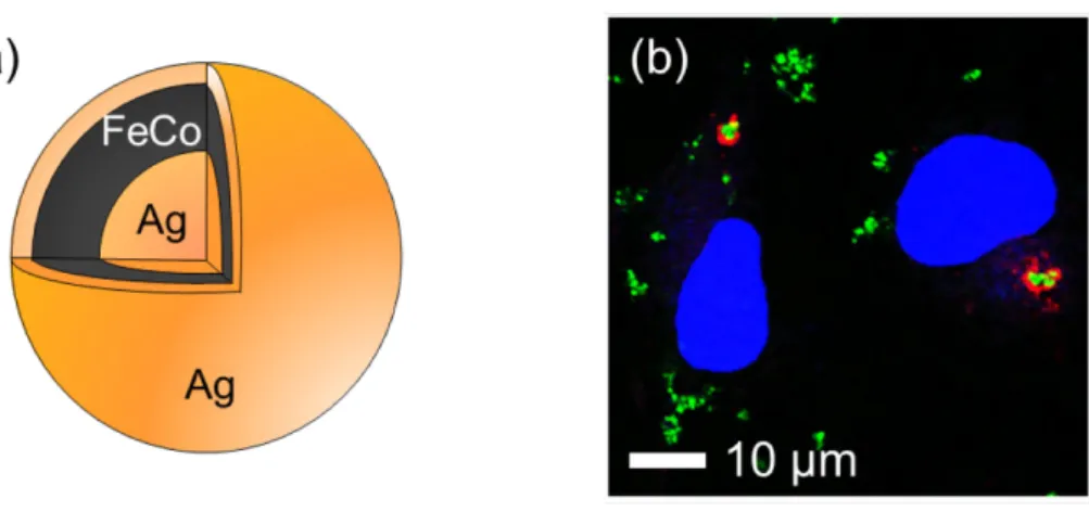
The present research demonstrates the capability of magnetic separation of intracellular organelles using magnetic-plasmonic heterostructured nanoparticles (NPs). Magnetic separation of cellular organelles has been considered to be a powerful analytical technique for cellular organelles which are difficult to be purified by conventional separation techniques. In order to separate these organelles, three aspects are required for the magnetic probe: small size, imaging ability and high magnetic property. In the present research the NP which is composed of Ag@FeCo@Ag core@shell@shell structure (Fig. 1a) is fabricated by a chemical synthesis. The size of the NP is about 15 nm. The formation mechanism and magnetic properties of the NPs are carefully analyzed, then the NPs are made to be water dispersible using hydrophilic polymer. Previously it was reported that a latex bead was transferred to an autophagosome after transfection. In this study we selected the autophagosome as a target organelle with magnetic separation using the current NPs. Incorporation of the NPs into autophagosomes was analyzed by a confocal laser scanning microscope by detecting plasmon scattering from the NPs (Fig. 1b). The results showed the localization of the NPs changed as incubation time increased after transfection of the NP according to the degradation pathway. Finally the autophagosomes which contain the NPs were magnetically separated by an autoMACS Pro Separator. The magnetically separated fraction (MSF) was investigated by western blot analysis. The results indicated the presence of LC3-II which is an autophagosomal membrane protein in the MSF, suggesting the success of magnetic separation of autophagosomes. The advantages of using magnetic-plasmonic heterostructured NPs for magnetic separation include easy organelle tracing due to localization of the probe, and straightforward separation timing. The technique described in this study will provide beneficial knowledge in both fundamental and practical aspects of the medical biology field.
Figure 1. (a) Structure of the Ag@FeCo@Ag core@shell@shell NP. (b) Confocal laser scanning microscope image of COS-1 cells in which the NPs were introduced by transfection. Blue, red and green color represents nucleus, LC3 (autophagosomal marker protein) and plasmon scattering from the NPs respectively.
The framework of this thesis is as follows. Chapter 1 contains the introduction of magnetic separation technique showing some examples and importance of separation of cellular organelles. Then Ag@FeCo@Ag NPs are introduced as a magnetic-plasmonic probe for separation of cellular organelles. Chapter 2 focuses on the formation mechanism of Ag@FeCo@Ag NPs. The NPs are synthesized by combination of a hot injection and a polyol method. The synthesis of the NPs is not just a seed-mediated growth. It contains many important concepts such as size-dependent reducing ability of Ag core, size focusing and surface segregation. In Chapter 3 the magnetic properties of the NPs are discussed. The reduction of saturation magnetization is observed due to surface oxidation. As a result exchange bias at the interface between ferromagnetic FeCo and antiferromagnetic cobalt wüstite is clearly observed. Based on the investigation of the relationship between exchange bias field and structural parameter of the NPs, an analytical tool for estimation of the oxide layer in the NPs from its exchange bias field is produced. Chapter 4 describes the synthesis of hydrophilic polymer and ligand exchange method for water dispersible NPs. Colloidal stability of the NPs is investigated. In addition, the surface of the NPs is modified by specific proteins using biotin-avidin interaction. This demonstrates the potential of surface modification of the NPs by certain proteins. Chapter 5 includes several examples of nanoparticle introduction by endocytosis and/or transfection method. Then the Ag@FeCo@Ag NPs are introduced into cells using a transfection reagent in order to separate autophagosomes. The microscope observation and magnetic separation results are then summarized. Chapter 6 summarizes the conclusion of the present study and significant achievements in each chapter. Finally the ideal magnetic probe for more versatile magnetic separation of cellular organelles is discussed.
Key words
Nanoparticle; Magnetic separation; Plasmon scattering; Cellular organelle; Surface modification 論文審査の結果の要旨
本博士学位論文は、バイオ医療分野での利用が期待される次世代磁気ビーズである磁性
-プラズモンハイブリッドナノ粒子の創製と、その応用の一例としてハイブリッドナノ粒 子を用いたオートファゴソームのイメージングと磁気分離を行ったものである。具体的に は、優れた磁気特性を持つ鉄コバルト合金(FeCo)と高いプラズモン散乱特性を持つ銀(Ag) をナノレベルで複合化したAg@FeCo@Agコア@シェル@シェル型磁性-プラズモンハイブ リッドナノ粒子を化学合成し、このハイブリッドナノ粒子表面を水溶性ポリマー及びタン パク質で修飾し、哺乳類培養細胞へトランスフェクションすることでゼノファジーを誘導 してオートファゴソームへターゲティングさせ、最終的にオートファゴソームを磁気分離 することに成功した。本論文は主に以下の三つの研究成果から構成されている。
第一に、ポリオール法とホットインジェクション法を組み合わせたAg@FeCo@Agコア@ シェル@シェル型磁性-プラズモンハイブリッドナノ粒子の化学合成法を確立し、その粒子 生成機構を解明した。即ち、AgコアがCoイオン及びFeイオンの還元触媒として作用するこ と、AgコアはAg前駆体の注入による粒径集束のため均一化されること、AgシェルはFeCo シェルの形成過程でFeCoシェルに取り込まれたAg原子の表面偏析によって自己組織的に 形成されることなどを見出した。
第二に、FeCoシェルは表面が酸化されてコバルトウスタイト(Co0.5Fe0.5O)相となって いることを明らかにし、強磁性(FM)相であるFeCo層と反強磁性(AFM)相である Co0.5Fe0.5O層の界面で交換バイアスが発現することを見出した。また、交換バイアス磁場 をAFM相とFM相の体積比(vAFM/vFM)に対してプロットしたところ、交換バイアス磁場は vAFM/vFMが小さい時には線形的な応答を示すが、vAFM/vFMが大きくなると振動的な挙動を示 すことを見出した。振動挙動の原因は界面におけるAFMスピンの向きの確率論的ゆらぎに 起因することを、モンテカルロシミュレーションの結果とも比較しながら、明らかにした。
第三に、ハイブリッドナノ粒子をポリリジンで表面修飾しCOS-1細胞へリポフェクショ ンすることで初期エンドソーム→オートファゴソーム→オートリソソームという分解経路 にナノ粒子をターゲティングし、プラズモン散乱によってナノ粒子の細胞内局在を可視化 した。オートファゴソームにナノ粒子が取り込まれた時点で速やかに細胞を破砕して磁気 分離を行い、磁気分画成分をイムノブロッティング法によって解析したところLC3-IIの濃 縮が確認されると同時にトランスフェリン受容体とLAMP2も確認され、GAPDHは検出さ れなかったため、オートファゴソームの分離に成功したことが確認された。
本論文の成果は、バイオ医療分野での応用が大いに期待できる次世代多機能磁気ビーズ の実現に向けて新たな可能性を示しただけでなく、幅広い関連分野において学術的に貢献 するところが大きい。よって博士(マテリアルサイエンス)の学位論文として十分価値あ
るものと認めた。