Japan Advanced Institute of Science and Technology
JAIST Repository
https://dspace.jaist.ac.jp/
Title
熱パルスイオン源を用いた質量分析チップの開発
Author(s)
杉山, 清隆
Citation
Issue Date
2015‑03
Type
Thesis or Dissertation
Text versionETD
URL
http://hdl.handle.net/10119/12769
RightsDescription
Supervisor:高村 禅, マテリアルサイエンス研究科,
博士
Development of on-chip mass spectrometer with pulse-heating ionization source
KIYOTAKA SUGIYAMA
Japan Advanced Institute of Science and Technology
Doctoral Dissertation
Development of on-chip mass spectrometer with pulse-heating ionization source
Kiyotaka Sugiyama
Supervisor: Professor Dr. Yuzuru Takamura
School of Materials Science
Japan Advanced Institute of Science and Technology
March, 2015
i
PREFACE
Mass spectrometry (MS) is one of the highly sensitive and highly selective analytical methods for biochemical samples including DNA, peptides, and proteins. Fundamental technology of the MS relies on the controlling of charged particles such as electrons or ions in a vacuum. MS has been massively developed by the multiple contributions of the ion optics and highly sensitive optical detection in physics, ionization techniques of versatile analyte in chemistry, and high speed data processing in information science. Presently mass spectrometric analysis plays a key role in the health and life sciences research of drug discovery, biomarker analysis for medical diagnosis, genomics, and proteomics.
On the other hand, micro total analysis system (μTAS) and lab-on-a-chip device also contribute to the progress of the life sciences research. The most advantage of the μTAS is the miniaturization of the analytical systems into a desktop or handheld device to apply quick biochemical analysis on the site.
Moreover, remarkable analytical devices which utilize the physical or chemical phenomena in micro to nanoscale were developed such as highly sensitive elemental analysis chip and the highly sensitive immunosensors in a cost effective way.
In this work, the key components for on-chip mass spectrometry including a vacuum pump, an ionization source, and an ion lens for time-of-flight mass analysis were miniaturized toward the highly sensitive detection of versatile
ii
biomarkers. Conventionally, MS analysis system requires a high vacuum.
However, the conceptual step to significantly miniaturize the mass spectrometer into a microchip is to operate it in a low vacuum using microfluidic channel. The pulse-heating ionization source developed herein enables the ionization of a peptide and a protein without laser, high voltage, or ambient gases. On-chip protein mass spectrometry was firstly performed with the integrated device of the ionization source and the ion lens. I hope that developed components for on-chip mass spectrometer in this research can be useful for next generation mass spectrometric devices for highly sensitive analysis of biological materials.
Kiyotaka Sugiyama
iii
CONTENTS
PREFACE --- i CONTENTS --- iii LIST OF ABBREVIATIONS --- vii
CHAPTER 1 GENERAL INTRODUCTION --- Abstract --- 1.1 Recent progress of analytical methods for medical diagnosis --- 1.1.1 Biomarkers in biological samples --- 1.1.2 Detection methods of biomarkers in biological samples --- 1.1.3 Micro total analysis system --- 1.1.4 Proteomic analysis --- 1.2 Miniaturized mass spectrometer --- 1.2.1 General principle of mass spectrometer --- 1.2.2 Miniaturized mass spectrometer --- 1.2.3 Microfabricated vacuum pumps --- 1.2.4 Microfabricated ionization sources --- 1.2.5 Microfabricated mass analyzers --- 1.3 Strategy to miniaturize mass spectrometer on a microchip --- 1.4 Research objectives --- 1.5 Thesis organization --- References ---
CHAPTER2 ON-CHIP VACUUM GENERATION METHOD --- Abstract --- 2.1 Introduction --- 2.2 Objective --- 2.3 Principle ---
1 1 2 2 5 9 10 11 11 13 15 17 21 25 28 29 31
37 37 38 39 39
iv
2.3.1 Principle of vacuum generation on a chip --- 2.3.2 Principle of pressure measurement on a chip --- 2.3.3 Theoretical vacuum --- 2.4 Experiment ---
2.4.1 Design of the chip --- 2.4.2 Chip fabrication --- 2.4.3 Experimental setup for vacuum generation --- 2.4.4 Experimental procedure for vacuum generation --- 2.5 Results and discussion --- 2.5.1 Vacuum measurement on a microchip --- 2.5.2 Vacuum generation with gas-liquid phase transition --- 2.6 Summary --- References ---
CHAPTER 3 ON-CHIP IONIZATION SOURCE FOR PEPTIDE AND PROTEIN ANALYTES --- Abstract --- 3.1 Introduction --- 3.2 Objective --- 3.3 Principle --- 3.4 Experiment --- 3.4.1 Design of ionization chip --- 3.4.2 Chip fabrication --- 3.4.3 Experimental setup for pulse-heating ionization --- 3.4.4 Sample preparation --- 3.4.5 Experimental setup for TOF-MS with pulse-heating ionization - 3.5 Results and discussion --- 3.5.1 Time-of-flight mass spectrometry of inorganic particles --- 3.5.2 Pulse-heating ionization of proteins --- 3.5.3 Time-of-flight mass spectrometry of proteins --- 3.5.4 Time-of-flight mass spectrometry of peptides ---
39 41 42 44 44 45 46 48 49 49 51 55 57
59 59 60 61 62 63 63 70 73 75 76 79 79 80 84 86
v
3.6 Summary --- References ---
CHAPTER 4 EFFECTS OF MATRIX AND SOLVENT FOR SAMPLE FORMATION ON PULSE-HEATING IONIZATION --- Abstract --- 4.1 Introduction --- 4.2 Objective --- 4.3 Principle --- 4.4 Experiment --- 4.4.1 Experimental setup --- 4.4.2 Sample preparation --- 4.5 Results and discussion ---
4.5.1 Effect of the matrix on pulse-heating ionization --- 4.5.2 Effect of the solvent for preparation of the sample layer --- 4.5.3 TOF mass spectrometry with thin layer methods --- 4.6 Summary --- References ---
CHAPTER 5 MINIATURIZED ION LENS AND ON-CHIP MASS SPECTROMETRY OF PROTEIN SAMPLE --- Abstract --- 5.1 Introduction --- 5.2 Objective --- 5.3 Principle --- 5.4 Numerical simulation ---
5.4.1 Design of the chip --- 5.4.2 Numerical simulation of ion optics in a micro channel --- 5.5 Experiment --- 5.5.1 Chip fabrication --- 5.5.2 Experimental setup ---
87 88
90 90 92 93 94 95 95 96 98 98 104 111 116 118
120 120 121 122 122 123 123 124 130 130 132
vi
5.5.3 Sample preparation --- 5.6 Results and discussion ---
5.6.1 Ionization in a micro channel --- 5.6.2 On-chip TOF mass spectrometry --- 5.7 Summary --- References ---
CHAPTER6 CONCLUSIONS ---
ACKNOWLEDGEMENTS --- ACHIEVEMENTS ---
133 134 134 135 139 140
141
143 145
vii
ABBREVIATIONS
AC Alternating Current
BSA Bovine Serum Albumin
CE Capillary Electrophoresis
CHCA α-Cyano-4-HydroxyCinnamic Acid
CNT Carbon NanoTube
Da Dalton
DC Direct Current
DHAP 2,5-DiHydroxyAcetoPhenone
DHB 2,5-DiHyhydroxyBenzoic acid
DI DeIonized
DNA DeoxyriboNucleic Acid
EI Electron Impact
ELISA Enzyme-Linked ImmunoSorbent Assay
ESI ElectroSpray Ionization
FET Field Effect Transistor
GC Gas Chromatography
LEP-AES Liquid Electrode Plasma - Atomic Emission Spectroscopy
M Molar mol/L
m/z Mass to charge ratio
MALDI Matrix Assisted Laser Desorption/Ionization MEMS Micro Electro Mechanical Systems
min Minute
MS Mass Spectrometry
NEMS Nano Electro Mechanical Systems
PDMS Poly(DiMethylSiloxane)
PMMA PolyMethyl MethAcrylate
QOL Quality Of Life
RF Radio Frequency
viii
s Second
S/N Signal to Noise ratio
SA Sinapic Acid or Sinapinic Acid
SPR Surface Plasmon Resonance
TFA TriFluoroacetic Acid
TOF Time-Of-Flight
V Volt
μTAS micro Total Analysis System
1
CHAPTER 1
GENERAL INTRODUCTION
Abstract
In this chapter, recently discovered biomarkers in the biological samples and miniaturized analytical methods used for their analyses for the medical diagnosis are briefly introduced. Immunosorbent assay based analytical methods are widely used for biomarker detection because of highly specific binding affinity of the antibody and the antigen. However, detection of more versatile target molecules on a single analysis is required to utilize the latest research outcomes of the proteomics with mass spectrometry (MS). The preview of the mass spectrometry based miniaturized system such as the integrated devices of a micro vacuum pump, an ionization source, and a mass analyzer was introduced.
The strategy to miniaturize the mass spectrometer on a chip with less bulky instruments was discussed. From the theoretical calculation of the mean free path, it was suggested that ion separation in a micro channel can be operated in a low vacuum at 1-10 Pa.
2
1.1 Recent progress of analytical methods for medical diagnosis
1.1.1 Biomarkers in biological samples
Biological molecules existing in human blood, saliva, urine, and sweat have considerable information for medical diagnosis because of the cause or consequence of many diseases in the body. To understand its nature related to life sciences, lots of analytical methods for biological samples have been developed.
Specifically, many researchers are trying to analyze the functions of proteins which compose cells, organs, and whole human body by these analytical methods.
Whole blood sample contains more than a half million proteins [1] with the different concentration. Figure 1.1 shows the reference intervals for 70 proteins in the plasma reported by Leigh et al [1]. From the literature, proteins seem to be divided into three major classes which are plasma proteins at the high abundance, tissue leakage proteins, and cytokines at the low abundance. Leigh et al [1]
mentioned that “Tissue leakage proteins are important because a serious pathology can be detected in a small volume of tissue by measuring release into plasma of a high abundance tissue protein”. Especially in the low concentration region around ng/ml to pg/ml, significantly meaningful biomarkers for severe diseases such as cancers were discovered [2].
3
Figure 1.1 Normal range abundances of protein analytes in plasma [1] and biomarkers of cancers[2] discovered in blood sample.
Table 1.1 shows biomarkers of severe diseases found in human blood or saliva [3-8]. Some biomarkers of cancers (Interleukin-1β [3], Interleukin-18 [4], and Cathapsin-D [5]) with the concentration in the range of ng/ml to pg/ml were detected from blood and saliva by Enzyme Linked Immunosorbent Assay (ELISA). ELISA is the most famous analytical method of biomarkers by using the highly specific binding of the proteins via the antigen-antibody interactions. Here, blood samples are more frequently used for the medical diagnosis than saliva or urine samples because the blood is directly related to the cellular activities like molecule transportation and biological defence. However, collection of enough blood samples for the analysis is accompanied with the pain and takes time. Saliva
Normal range abundances of protein analytesin plasma
pg
/mlng
/mlμg
/ml
/ml
g
/ml
fg
Classical Plasma Proteins Tissue Leakage Interleukins Hemoglobin
Albumin
e.g.) Cancer markers IL-1β: >10pg/ml IL-18: 53.6-602.5 pg/ml
mg
/ml
Proteins IgG
IgD
4
and urine are expected to be the promising non-invasive sample for cancers diagnosis in the early stage. The issue of saliva in this purpose is that it is usually not enough to evaluate the pathology from a single biomarker due to its lower concentration than the biomarkers in blood [2]. Statistics analysis of peptides and proteins in saliva based on the MS with electrophoresis has been studied [6, 7].
Sugimoto et al reported that 57 principal metabolites [7] are found from saliva sample, which can be used to accurately predict the probability of being affected by oral, breast and pancreatic cancers. Biomarker analysis of volatile chemical compounds as well as proteins was also studied by GC-MS [8] for cancer diagnosis. The detection method of those gaseous biomarkers called “electronic nose” found that organic solvents such as 2-ethyl-1-1hexanol and 3-methyl-1-butanol are related to the lung cancer.
If the prediction of the severe diseases is realized by screening of only saliva even in a small clinic, advanced diagnosis will be provided without a time-consuming thorough checkup in a hospital. Diagnosis in early stage of the diseases will be also realized to discover further biomarkers, leads improvement of Quality of Life (QOL) in the near future.
5
Table 1.1 Lists of biomarkers for serious diseases found in blood or saliva sample, its concentration, and detection method of them.
1.1.2 Detection methods of biomarkers in biological samples
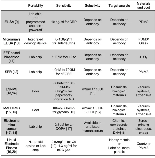
As mentioned in previous section, peptides and proteins in biological samples with low concentration have significant information for the medical diagnosis. To utilize the research outcomes to the clinical application, analytical systems should be considered. Important capabilities of the bio analytical systems are sensitivity, selectivity, target analytes, and cost. Table 1.2 shows the brief
Diseases Sample Concentration range How to detect
IL-1β [3] Cardiovascular disease or Oral
cancer
Blood/
saliva >10pg/ml/ <10pg/ml ELISA
IL-18 [4] Cardiovascular disease or Oral
cancer
Blood/
saliva 34.2-68.2pg/ml/ 53.6-
602.5pg/ml ELISA
Cathapsin-D [5] Breast cancer Blood 45.2pmol/mg ELISA
Statistics analysis of 16
candidate proteins [6]
Lung cancer Saliva - 2-D difference gel
electrophoresis and MS Statistics
analysis of 57 metabolites [7]
Oral, breast and pancreatic
cancers
Saliva - CE-TOF-MS
2-ethyl-1- hexanol, 3-methyl -1-
butanol [8]
Lung cancer Saliva 29 ng/ml ― 90 ng/ml,
3 ng/ml - 400ng/ml GC-MS
6
summarization of developed bio analytical systems. ELISA [9] is the most famous bio analytical method with the specific binding of antigen and antibody. In ELISA, target analyte, usually an antigen is labelled by the antibody with enzyme conjugate, and then unattached antibodies and unfavourable substances in the sample are washed out for high S/N measurement. After that, added substrate and enzyme interaction create color change for the detection. Nie et al reported ELISA in the array of 888 microwells with microspheres [10] for higher S/N measurement than conventional ELISA. Highly sensitive analysis up to a few pg/ml [10] was accomplished to enhance the antigen-antibody interactions labelled with fluorescence by restricting the chamber volume and high surface-to-volume ratio of the microspheres. In those cases, colorimetric changes or fluorescent signal enhancement were used for the detection. In contrast to this, Field Effect Transistor (FET) [11] or Surface Plasmon Resonance (SPR) [12] was also used to detect the target antigen immobilized on the channel or the metal surface via the antibody. FET devices modified by nanomaterials such as silicon nanowires, carbon nanotubes, or graphene [11] were fabricated. These nanomaterials have significant advantage for highly sensitive biosensor with decreasing the non-specific binding of unfavorable substances on the nanomaterials of the channel as well as high electrical conductivity. Tawa et al reported highly sensitive biomarker detection using SPR based lab chip devices [12]. SPR is the collective oscillation of electrons in liquid or solid, usually gold substrate or gold nanoparticles by the specific incident light. They demonstrated that enhanced electric fields by SPR were utilized as the excitation field for
7
fluorescent biomolecules to realize high sensitive detection in the range up to 10-15 M.
Major miniaturized bio analytical devices use antigen–antibody interactions to selectively capture the specific biomolecules in biological samples.
However, it is not easy to simultaneously detect several tens of multiple target analytes using immunochemical reactions because a series of antibodies having the specific binding affinity to each target analyte have to be preliminary equipped on the device. Mass spectrometric analysis like ESI-MS [13, 14] and MALDI-MS [15, 16] is frequently used for discovering biomarkers in life sciences research.
The advantages of MS are high sensitivity and high selectivity, while the equipment is expensive because it is composed of high vacuum systems, high voltage source, and so on. An electrochemical biosensor [17, 18] is one of the applicants for the sensitive detection of multiple target analytes such as chemical compounds, proteins, and nucleic acids [18]. Reported performance of the electrochemical biosensor was not so sensitive in the range of μM for proteins [18]. Another highly sensitive miniaturized elemental analysis system called LEP-AES (Liquid Electrode Plasma Atomic Emission Spectroscopy) was reported by Kitano et al [19]. For the analysis of LEP, generated plasma with applying high voltage excites and atomizes injected samples in the microfluidic channel (width
= 100μm). Atomic emission spectrum is analyzed when the excited atoms return to the ground state. Tung et al reported that LEP-AES can apply to highly sensitive biomolecules analysis as the detection of hCG with the detection limit of 1.3 pg/ml [20]. These studies have been developed for fundamental studies of
8
molecular biology in the cell and biomarker discovering as well as analytical devices for clinical applications. For example, a commercially-available pregnancy test and a glucose sensor for patients of diabetes are widely used.
Table 1.2 Lists of developed bio analytical systems and its characteristics.
Portability Sensitivity Selectivity Target analyte Materials and cost
ELISA [9]
Lab chip, pre- programmed
and self- powered
10 ng/ml for CRP Depends on
antibody Depends on
antibody PDMS
Microarrays
ELISA [10] Integrated
desktop device 6-138pg/ml
for Interleukins Depends on
antibody Depends on
antibody PDMS/
Glass
FET based biosensor
[11] Lab chip 100pM forHER2 Depends on
antibody Depends on
antibody SiO2
SPR [12] Lab chip 10nM to 700fM
for sEGFR Depends on
antibody Depends on
antibody PMMA
ESI-MS
[13,14] Poor
< 50nM for CE- ESI-MS/
50ng/ml for paperspray ionization MS
m/Δm >11000 [13]
Chamicals, biological materials
Vacuum systems, Expensive
MALDI-MS
[15, 16] Poor 10fmol- 50amol
for glycans [15] m/Δm: 40000- 60000 [16]
Chamicals, biological materials
Vacuum systems, Expensive Electroche
mical sensor [17, 18]
Lab chip 2.5μM for L- DOPA [17]
Available in undiluted human serum
Chemical compounds,
proteins , DNA[18]
Scree - printing electrodes,
cheap Liquid
Electrode Plasma
[19,20]
Handheld device or Lab
chip
0.52ng/ml for Cd [19]. 1.3 pg/ml for
hCG [20]
Heavy metals or Labeled metal
particle
Quartz or PMMA
9
1.1.3 Micro total analysis system
The ideas of micro total analysis system (μTAS) and lab on a chip originated from the integration of chemical and biochemical experiments such as chemical handling, reactions, separations, and analyses into a chip to accelerate the research of efficient drug discovery, DNA sequencing, protein analysis, and cell analysis. In 1970s, miniaturized gas chromatography device fabricated on a silicon substrate [21] as a micro analysis system was firstly reported using photolithography and wet etching processes which have been used for semiconductor fabrication. In 1990s, high-throughput capillary electrophoresis in a microchip [22] was studied to accelerate the human genome project to analyze whole human genome. After that, the fabrication method based on inexpensive polymeric materials such as SU-8 [23] and poly (dimethylsiloxane) (PDMS) [24]
were developed and microfluidic controlling elements including a pump and a check valve was also fabricated by soft-lithography techniques reported by Xie et al. The outstanding advantage of μTAS and lab on chip is to fabricate the integrated miniature devices in the mass production, which can be available in the applications of environmental monitoring, food inspection, and medical diagnosis.
Further development of lithography and dry etching process enabled the fabrication of nano structures on a chip having specific functions in nano-scale for the biological analysis of a single cell [25] and a single molecule [26], on the other hand, the development of stable microfluidic pumps and valves on a chip has been still big issue in this field. Integrated and automated systems of immune sensor and electrochemical sensor into desktop device for detection of insulin [27]
10
and cancer markers [10] were developed as successful studies of lab on chip. In near future, the chips for highly sensitive multiple target analysis of several tens of biomarkers, non-invasive diagnosis for cancers with saliva, urine, or sweat, and the diagnosis in very early stage of severe diseases are expected to be developed.
1.1.4 Proteomic analysis
Proteomics is a challenging study of analysis of proteins related to metabolism in the cells to understand its structures and functions. Human genome encodes series of amino acids, and the amino acids compose series of proteins in the body. A human body may contain more than one million different proteins [28], which are having different functions in cells. Understanding the functions of proteins is interesting research for future medical diagnosis and health care.
However, study of the functions of every single protein in cells is difficult because huge proteins existing in the cell interact differently with each other. Proteomics has potential to discover new biomarkers for cancers and the other diseases in very early stage from non-invasive samples such as saliva and urine [7, 29, 30]. In proteomics, cyclopaedic protein expression analysis is required. To accomplish this requirement, massively-developed mass spectrometric analysis of peptides and proteins is one of the desired methods. Characteristics of high sensitivity and high selectivity of the MS supports the analysis of mixtures of peptides and proteins over 10,000 species in proteomic research. As mentioned above, MS is an effective analytical method having key roles in the understanding of the nature of
11
the biological system and in the future diagnosis system. To utilize the analytical methods and findings based on MS more effectively, some works of miniaturization of MS into a desktop device or a chip have been studied for a decade, which are overviewed in the later section.
1.2 Miniaturized mass spectrometer
1.2.1 General principle of mass spectrometer
MS is one of the most powerful and versatile analytical tools to measure gases, metals, chemical compounds and biological substances such as nucleic acids, peptides, and proteins. MS was initially used for precise measurements of atomic weight and isotopes by 1960s. After 1980s, soft ionization methods for biochemical compounds such as ESI [31] and MALDI [32, 33] were developed by Fenn et al, Tanaka et al, and Hillenkamp et al, respectively. These methods become the basis of analytical methods for massively progressed life sciences research.
Figure 1.2 illustrates the typical components of MS. MS is composed of an ionization source, a mass separation filter, and an ion detector in a vacuum chamber. In the initial step of MS, introduced samples are ionized by electron impact, plasma, laser, or high voltage source. The ionization is crucial phase for MS because analyzable substances are restricted by the ionization techniques.
After ionization, generated ions are introduced to the mass analyzer, in which
12
motion of ions is controlled by external electric force and magnetic force. Two major mass analyzers are quadru-pole mass analyzer and a TOF mass analyzer.
For quadru-pole mass analyzer, ions with specific mass-to-charge ratio are filtered to stably pass through the gap of four parallel metal rods applied radio frequency (RF) voltages. For TOF mass analyzer, mass-to-charge ratio is measured via flight time measurement of accelerated ions having the same kinetic energy as any other ion that has the same charge in an electric field. Finally, separated ions are captured and amplified by an electron multiplier. Ions are initially converted to electrons by collisions at the secondary electron emissive surface with high velocity, and then electrons are amplified by consecutive secondary electron emission with collisions to the reactive surface in a vacuum or by electron bombardment in a semiconductor. These components are aligned in a big vacuum chamber evacuated to high vacuum (~10-4 Pa), therefore MS has been expensive and cumbersome in an operation.
Figure 1.2 Schematic of the components of typical mass spectrometer.
Ionization source (by electron, laser, or high voltage etc.)
Sample introduction
Mass separation (by electric or
magnetic)
Ion current amplification and detection
+ +
- - e-
M+ +
Analysis m/z
Relative abundance
13
1.2.2 Miniaturized mass spectrometer
Miniaturization of MS has been studied for the realization of highly sensitive and highly selective analysis on the site, not so much the lab as clinic, field, and environment. For example, miniaturized mass spectrometers were developed in use of real-time in-vivo analysis of the tissues [34] as a surgical tool [35] and on-site detection of chemical warfare agent, illicit drug, and explosives as backpack equipment [36]. Table 1.3 shows the previously reported miniaturized mass spectrometers. Two remarkable desktop mass spectrometers have been developed. Palm portable mass spectrometer developed by Yang et al [37] can analyze organic solvent in the air like dimethyl-methylphosphonate. The important progress in their research is that they created and maintained high vacuum (10-6 Torr ~ 1.3×10⁻⁴ Pa) in a portable device by miniaturized titanium ion getter pump. Mini 12 developed by Li et al [38] can analyze whole blood samples for POCT application. They developed the disposable sample cartridge with the disposable paper ionization source for the miniaturized paper spray ionization source [14]. The method can ionize sample solution containing the analytes under ambient conditions. On the other hand, further challenging studies to miniaturize the MS into microchip have been developed using μTAS techniques.
MEMS or NEMS techniques were used to fabricate a vacuum pump, an ionization source, and a mass analyzer on a chip. Unlike miniaturization of MS to desktop devices, on-chip MS have the significant advantage to be able to separate ions in a low vacuum [39] as well as the miniaturization. Integrated mass spectrometers on a chip [39-41] have been reported by Wapelhorst et al, Chaudhary et al, and
14
Wright et al. They fabricated EI or ESI ionization sources and the mass analyzers for the integration of MS into the chip and succeeded to analyze gaseous compounds [39, 40] and solution containing chemicals [41]. However analysis of peptides and proteins has not been realized by those on-chip MS because of the difficulty to ionize proteins without decomposition. Moreover, the on-chip MS currently works in a chamber maintaining at high vacuum with a conventional ion detector, therefore mass spectrometric analysis in a low vacuum have not been performed.
Table 1.3 Lists of developed miniaturized mass spectrometer into a desktop device, a handheld device, or a chip. Details of its components, ionization source,
mass analyzer, and vacuuming method were shown.
Size Ionization Mass analyzer Vacuuming Palm portable
mass spectrometer [37]
Hand held
(1.48kg) Electron
impact Four-parallel-
disk ion trap Ion getter pump + Roughing pump
Mini 12 [38] Desktop (4kg)
Paper spray ESI
[14]
Rectilinear ion traps
Small turbo pump (1mTorr for MS
scan) A fully integrated
TOF micro mass
spectrometer [39] Lab chip Electron
impact TOF +
Energy filter ―
Ion trap mass spectrometer
arrays [40] Lab chip Electron
impact 25 Cylindrical ion trap arrays
In a vacuum chamer (7.4×10-
7Torr) MEMS-based
nanospray- ionization
mass spectrometer [41]
Lab chip MEMS- based
Nano-ESI Quadru-pole
In a vacuum chamber (1m Torr in and
analysis chamber )
15
1.2.3 Microfabricated vacuum pumps
A vacuum pump is an essential component for MS because the ions are separated in a vacuum with avoiding collisions to any other ions or neutral molecules existing in the chamber. Nowadays, many kinds of vacuum pumps are commercially-available such as a rotary pump, a diaphragm pump, a diffusion pump, a sorption pump, and a turbo-molecular pump. However, there are a few researches of miniaturization of vacuum pump into a microchip [42-44] despite some equipment for handling solution and surface analytical systems like scanning electron microscope (SEM) are utilized by a vacuum. One of the reasons is the difficulty to generate mechanical force in a microfluidic device to evacuate gas molecules from a microchip.
Figure 1.3 shows the fabricated miniaturized vacuum pumps without any moving parts. McNamara et al developed micro-machined Knudsen pump [42]
using MEMS process as shown in Fig 1.3 (a). The Knudsen pump relies on the principle of thermal transpiration which is a net gas flow from the colder chamber to the hotter chamber through the narrow channel. Pressure difference between the two chambers is created through a narrow channel in which the gas is in the free molecular flow regime. They fabricated thermally-isolated chambers connected with micro channels and maintained a vacuum at 46 kPa in a cold chamber on a chip. Doms et al reported a micro-machined vapor jet pump [43] inspired by diffusion pump as shown in the Fig 1.4 (b). Diffusion pump is a vacuum pump to evacuate molecules from a chamber by boiling of the fluid with the high speed jet.
16
Basically, diffusion pump needs roughing vacuum pump and cooling system for capturing boiling fluid. They demonstrated the principle on a chip using the miniaturized device fabricated by MEMS. A vacuum at 87 kPa with the combination of nitrogen gas supply and roughing vacuum pump and a vacuum at 96 kPa with external water vapor and cooling water supply were maintained on a chip. Miniaturized MEMS-type glow-discharge micro pump [44] was reported by Grzebyk et al as shown in Fig 1.3(c). They used gettering of Ti in a miniaturized vacuum chamber where chemically-active gases such as hydrogen, oxygen, nitrogen, and so on are adsorbed on the surface accompanying with sputtered Ti ions. The ion gettering vacuum pump created minimum pressure of 3 Pa while a roughing vacuum pump must be required. High vacuum creation on a chip has not been realized without any roughing vacuum pump. Totally miniaturized vacuum pump can be useful for microfluidic handling and analytical systems if the issues of creating high vacuum, of maintaining a vacuum on a chip, and minimizing the dead volume are solved.
17
Figure 1.3 Images of the miniaturized vacuum pumps. (a) Knudsen pump [42].
(b) Microfabricated vapor jet pump [43]. (c) Titanium ion getter vacuum pump [44].
1.2.4 Microfabricated ionization sources
Ionization is one of significant processes in the mass spectrometric analysis. Biomolecules such as peptides, proteins, and nucleic acids have not been analyzed by MS until 1980s because it was difficult to evaporate without thermal decomposition before the ionization [45]. Fenn et al [31] and Tanaka et al [33]
(a)
(b)
(c)
18
developed the novel ionization methods of ESI and MALDI to ionize biomolecules without decomposition and denaturation via different soft ionization techniques. Such kind of the soft ionization techniques are required to apply biomolecules detection in a miniaturized mass spectrometer.
Figure 1.4 shows the miniaturized ionization sources for gases and solution fabricated by MEMS or NEMS techniques. Microfabricated EI ionization sources with micro tungsten filament [46] and with carbon nano particles [47]
were reported by Yoon et al as shown in Fig. 1.4(a). The electron impact ionization is well-known as a typical ionization method for gases or organic volatile samples. Basically, the analytes are limited to gases to ionize the gaseous molecule with the impact of electron normally accelerated to 70 eV. They demonstrated thermal electron emission from the heated micro filaments fabricated on a chip. Positive ions of organic solvent as acetone were measured by an integrated TOF mass analyzer; however the fabricated miniaturized ionization sources were not used in MS on a chip because of low thermo ionization energy.
Cold cathode electron emitting source using carbon nanotube (CNT) [48] was fabricated by Velasquez-Garcia et al as shown in the Fig. 1.4 (b). Electrons can be generated not only by heated filament (a few thousand K) but also by high electric field (~109 V/m) to remove an electron from metal surface by the tunnelling effect.
CNT is a suitable nano material for electron emissions because of the tiny tip (~ a few dozen nm [48]) in which the electric potential is concentrated.
Perpendicularly-grown CNT forests with its typical diameter of 33 nm were fabricated on the microfabricated structures for the ionization of argon gases.
19
Disadvantage of the both EI sources is that suitable samples are only gases or volatile solvents. Many studies have been studied to evaporate non-volatile samples such as peptides with little fragmentation by rapid heating technique (10
OC/s) and use of Teflon emitter [49] before the EI ionization, but the analysis of the mass spectrum was difficult.
In contrast to the EI ionizations on a chip, nano-ESI was fabricated for the ionization of non-volatile chemical compounds in the solution [50]. Figure 1.4 (c) shows the microfabricated nano-ESI source integrated with miniaturized quadru-pole mass analyzer. ESI is known as a soft ionization method to be able to ionize non-volatile analytes including biomolecules from liquid phase without decomposition of target analytes. Detailed principle of ESI has been still discussed, while aerosol is created at the outlet of nano-capillary in which high voltage (~kV) is applied to sample solution. Initially, positively charged droplets (diameter of μm) were produced after spraying of aerosol if the positive high voltage is applied to the capillary. The charge comes from the ambient excess ions (H+, NH4+, Na+, and K+), mainly protons in acidic solution [51]. The charged droplets undergo rapid solvent evaporation by vacuuming and heating through ambient gases. Finally, nano-size droplets containing the analyte of the interest with multiple charges are analyzed by the mass analyzer. Wright et al reported miniaturized vacuum interfaces to control three different vacuum stages, atmospheric pressure for sample injection, low vacuum for ionization, and high vacuum for MS analysis, as well as nano-capillary for ESI source on a chip. A 150 μg/ml of tetrabutylammonium hydroxide solution was measured by the
20
microfabricated ionization with quadru-pole mass analyzer and the singly-charged target ion was successfully obtained [50]. Drawbacks of the ESIs are that high voltage source (700-850 V for microfabricated nano-ESI) and ambient heated gases are required for the ionization. Moreover, multiply-charged ions are more frequently produced than singly-charged ion, thus deconvolution processing is required for analysis of complicated mass spectrum. On the other hand, MALDI source is also well-known as a soft ionization method. In MALDI, samples containing target analytes and organic matrix having efficient absorption at the specific wavelength are irradiated by a laser, and then are desorbed by rapid thermal desorption. In the plume of desorbed sample gases, the charge transfers to the analyte from the charged matrix. Sometimes MALDI is superior to ESI for protein analysis as following reasons; (i) basically, singly-charged ions are more frequently produced than multiply-charged ions, which give relatively comprehensible mass spectrum. (ii) Integration with TOF mass analyzer provides highly sensitive and highly selective protein analysis up to m/z of 100,000 Da [33].
Currently, miniaturized MALDI source on a chip has not been reported due to the complexity of laser optical components with microfluidic systems.
21
Figure 1.4 SEM images of the miniaturized ionization sources. (a) Electron impact ionization source fabricated by tungsten filament [46]. (b) Carbon nanotube cold emitter [48]. (c) Nano electrospray ionization source [50].
1.2.5 Microfabricated mass analyzers
First ion trapping techniques studied in 1950s such as Penning trap [52]
and Paul trap (Quadru-pole trap) [53] developed by Dehmelt et al and by Paul et al, respectively. An electron or an ion trap is realized by the combination of electric and magnetic fields in a vacuum. For penning trap, charged particles are
(a)
(b)
(c)
22
trapped in a homogeneous static magnetic field and a spatially inhomogeneous static electric field. In contrast to this, charged particles are trapped in oscillating AC electric fields for quadru-pole trap. The quadruple trap system is normally used for MS due to the high scan rate for mass analysis by changing only electric potential. Specific ions circularly travel along the stable trajectory in the both of the trapping systems.
Figure 1.5 shows the fabricated miniaturized mass analyzers using MEMS or NEMS techniques [39-41]. Wapelhorst et al reported on-chip mass analyzer integrated with TOF separation and energy filter [39]. TOF mass analyzer is basically a kind of energy filtering of the ions. Same kinetic energy was applied by the electric potential difference to all the ions with same charge.
After that, ions flight at the different velocity in the free flight region applied no electric and no magnetic fields. Mass to charge ratio m/z of the molecules is analyzed by measuring time of the free flight in a vacuum. Additionally, energy filter placed after TOF region was used to separate the ion having lower or higher initial energy for the highly selective analysis. One of the advantages of TOF mass analyzer is no limitation in the analytical range of m/z in theory if measurement time will be sufficiently long.
Miniaturized ion trap filters, quadru-pole mass analyzer [41] and cylindrical ion trap analyzer [40], were fabricated by Wright et al and Chaundhary et al, respectively. To fabricate miniaturized quadru-pole mass analyzer, electrode rods with a diameter of 650 μm were precisely aligned on the microfabricated
23
devices. Perfluorotributylamine was analyzed by miniaturized quadru-pole mass analyzer with the best resolution of m/Δm ≈ 140 [41]. Cylindrical ion trap chip with 5×5 ion trap arrays [40] were fabricated using MEMS process. Such integrated array of MS components on a chip is challenging issues. Here, theoretical stability of the ions in quadru-pole electrode is discussed to estimate the capability of it in micro systems. The of motion of a charged particle in the x-y cross section of the quadru-pole electrodes applied the superimposed RF and DC voltages (±(U+Vcosωt)) is governed by the following Mathieu equation. [54]
𝑑2𝑥
𝑑𝜏2+ (𝑎𝑥+ 2𝑞𝑥cos 2𝜏)𝑥 = 0 (1.1)
𝑑2𝑦
𝑑𝜏2 + (𝑎𝑦+ 2𝑞𝑦cos 2𝜏)𝑦 = 0 (1.2) where,
𝑎𝑥= −𝑎𝑦 = 4𝑒𝑈
𝑚𝑟02 𝜔2, 𝑞𝑥= −𝑞𝑦 = 2𝑒𝑉
𝑚𝑟02 𝜔2, 𝜏 =𝜔𝑡
2 (1.3)
U is the amplitude of DC voltage, and V is the amplitude of RF voltage applied in the quadru-pole, e is charge of the ion, m is mass of the ion, r0 is the distance to the rod electrode from the center, and ω=2πf is angular frequency. Ions having the specific mass-to-charge ratio pass through the quadru-pole with stable oscillation in the x-y plane. Basically, amplitude of RF voltage is scanning for mass separation with holding a constant value of 2U/V, thus the trapped ions satisfy the following equation.
24
𝑚
𝑒 = 𝐾 𝑉
𝑟02 𝜔2 (1.4)
where, K is a constant depending on the geometry of quadru-pole electrodes.
From the relation, high voltage is required to stabilize heavier ions such as proteins with the constant RF frequency. For the miniaturized quadru-pole, relatively high voltage V = 770V and f ≈ 6.5MHz were used to trap the ion with m/z of 1,200 [41] despite the value of r0 decreases.
25
Figure 1.5 Images of the miniaturized mass analyzers. (a) TOF mass analyzer with an energy filter [39]. (b) Quadru-pole mass analyzer [41]. (c) Cylindrical
ion trap mass analyzer [40].
1.3 Strategy to miniaturize mass spectrometer on a microchip
Miniaturized mass spectrometers composed of various MS components were developed using the fabrication process, MEMS and NEMS due to its high sensitivity and high selectivity for multiple target analysis. Interestingly, Siebert et
(a)
(b)
(c)
26
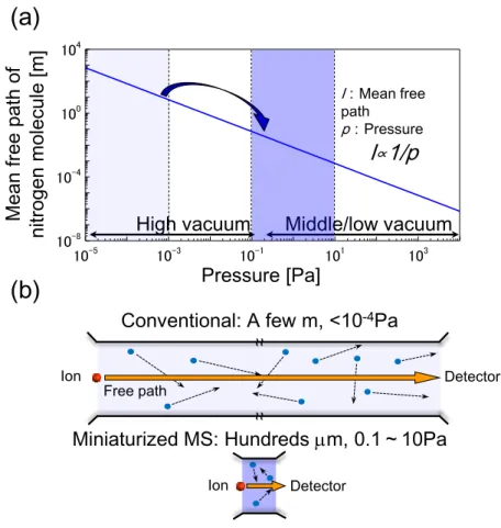
al pointed that mass separation in microsystems can be employed at the pressure of 1-10 Pa [55] and calculated ion trajectory in the microstructure by numerical simulation. To discuss the ion separation, mean free path, the average distance travelled by gas molecules between collisions, is estimated by the following equation [56].
𝑚𝑒𝑎𝑛 𝑓𝑟𝑒𝑒 𝑝𝑎𝑡ℎ =7×10⁻³
𝑝 [𝑃𝑎] [𝑚] (1.5)
where, p is the ambient pressure. The relation of mean free path versus pressure was shown in the Figure 1.6 (a). In conventional high vacuum system at the pressure of 10-3 Pa, mean free path of the remaining gases is about 7 m which is greater than the dimension of usual laboratory vacuum chamber. Figure 1.6 (b) shows the illustration of the concept of MS in the microsystem. Presently available mass spectrometer is used in a high vacuum (below 10-3 Pa) to prevent ion disruption and disturbance by collisions with the other ions or molecules as well as in a large chamber (~ 1 m) for highly selective analysis. If the ion generation and handling in the miniaturized chamber with the size of mm or μm are realized, required mean free path for MS to prevent the ion interactions in the miniaturized chamber will decrease to mm or μm scale. From the calculated mean free path, mass separation in a microchip can be operated even in a low vacuum approximately 10 Pa. Thus, bulky high vacuum system like a turbo-molecular pump will be not employed, if the required vacuum is down to 1-10 Pa. These
27
concepts have not been realized on a chip even the miniaturized ionization sources and mass analyzers were developed. The reasons of this are follows; (i) Development of the miniaturized vacuum pump having enough performance for the on-chip MS (1-10 Pa) without any external vacuum pump has not been realized. (ii) Currently developed miniaturized ionization source for biological samples i.e. on-chip nano-ESI was employed with high vacuum system at high exhaust velocity and with high voltage (~0.85 kV) which may cause problems of discharge in a low vacuum. (iii) Integration of whole components of MS, an ionization source, the interface for sample introduction, a mass analyzer, and a detector into a single device have not been realized.
28
Figure 1.6 Schematic of the advantage of miniaturized mass spectrometry on a chip. (a) Dependence of mean free path on ambient pressure. (b) Comparison
between conventional MS and on-chip MS.
1.4 Research objectives
In this thesis, the components of MS, the vacuum pump, the ionization source, and the TOF mass analyzer, are miniaturized using MEMS fabrication techniques for the development of on-chip mass spectrometer toward highly sensitive analysis of biological samples. A vacuum generation method on a chip with gas-liquid phase transition is studied for realization of a low vacuum toward
10-5 10-3 10-1 101 103
10-8 10-4 100 104
Mean free path of nitrogen molecule [m]
Pressure [Pa]
High vacuum Middle/low vacuum
l:Mean free pathp:Pressure
l∝1/p (a)
Ion Free path Detector
~ ~
~ ~
Conventional: A few m, <10-4Pa
Detector
Miniaturized MS: Hundreds m, 0.1~10Pa
Ion
(b)
29
MS analysis without any external pumps. Second, an ionization source for biological samples such as peptides and proteins is developed on a chip without laser, high voltage, and ambient heated gases. The novel ionization source utilizes nano-second Joule heating from the micro heater to the solid-phase samples on a chip, which easily combines to relatively small homebuilt TOF mass analyzer for protein mass spectrometry. Finally, the developed ionization source is integrated with the miniaturized ion lens and TOF path fabricated on a chip to realize on-chip MS analysis. Protein MS is performed in the chip-based TOF mass analyzer.
1.5 Thesis organization
Chapter 1 explains recent progress of analytical methods for medical diagnosis including desktop or microfabricated lab on chip devices and miniaturized mass spectrometers and the strategy to effectively miniaturize the mass spectrometer on a chip.
Finally research objectives are presented.
Chapter 2 shows the study of vacuum generation on a chip. Pressure measurement in a chamber and the vacuum generation method using gas-liquid phase transition on a chip are shown.
Chapter 3 presents pulse-heating ionization for peptides and proteins without laser, high voltage, and ambient gases. Numerical simulation, fabrication process of the chip, experimental setup for the ionization, and TOF mass analysis
30
are explained. And then protein mass spectrometry with the pulse-heating ionization chip and relatively small TOF mass analyzer is performed.
Chapter 4 discusses the effects of matrix and solvent for sample formation on the pulse-heating ionization. To minimize the generation of fragment ions in the ionization, effective matrix and sample preparation method are studied.
Finally, the sensitivity of the pulse-heating ionization is estimated.
Chapter 5 explains the miniaturized ion lens and the TOF mass analyzer on a chip. Miniaturized TOF mass analyzer is studied by numerical simulation of the ion trajectory controlled by the electric fields with micro electrodes. On-chip mass spectrometry of protein sample is realized by the integration of the pulse-heating ionization source and the miniaturized TOF mass analyzer of 5 mm length.
Chapter 6 summarizes the works in this thesis.
31
References
[1] Anderson, N. Leigh, and Norman G. Anderson. "The human plasma proteome history, character, and diagnostic prospects." Molecular & cellular proteomics 1.11 (2002): 845-867.
[2] Pfaffe, Tina, et al. "Diagnostic potential of saliva: current state and future applications." Clinical chemistry 57.5 (2011): 675-687.
[3] Morris, Andrew Conway, et al. "Diagnostic importance of pulmonary interleukin-1β and interleukin-8 in ventilator-associated pneumonia." Thorax 65.3 (2010): 201-207.
[4] Mallat, Z., et al. "Increased plasma concentrations of interleukin-18 in acute coronary syndromes." Heart 88.5 (2002): 467-469.
[5] Foekens, J. A., et al. "Cathepsin-D in primary breast cancer: prognostic evaluation involving 2810 patients." British journal of cancer 79.2 (1999):
300.
[6] Xiao, Hua, et al. "Proteomic analysis of human saliva from lung cancer patients using two-dimensional difference gel electrophoresis and mass spectrometry." Molecular & Cellular Proteomics 11.2 (2012): M111-012112.
[7] Sugimoto, Masahiro, et al. "Capillary electrophoresis mass spectrometry-based saliva metabolomics identified oral, breast and pancreatic cancer-specific profiles." Metabolomics 6.1 (2010): 78-95.
[8] del Nogal Sánchez, Miguel, et al. "Sensitivity Enhancement in the Determination of Volatile Biomarkers in Saliva Using a Mass Spectrometry-Based Electronic Nose with a Programmed Temperature Vaporizer." Analytical chemistry 86.15 (2014): 7890-7898.
[9] Safavieh, Roozbeh, and David Juncker. "Capillarics: pre-programmed, self-powered microfluidic circuits built from capillary elements." Lab on a Chip 13.21 (2013): 4180-4189.
[10] Nie, Shuai, et al. "An automated integrated platform for rapid and sensitive multiplexed protein profiling using human saliva samples." Lab on a Chip 14.6 (2014): 1087-1098.
32
[11] Myung, Sung, et al. "Graphene-encapsulated nanoparticle-based biosensor for the selective detection of cancer biomarkers." Advanced Materials 23.19 (2011): 2221-2225.
[12] Tawa, Keiko, et al. "Application of 300× Enhanced Fluorescence on a Plasmonic Chip Modified with a Bispecific Antibody to a Sensitive Immunosensor." ACS applied materials & interfaces 5.17 (2013): 8628-8632.
[13] Lapainis, Theodore, Stanislav S. Rubakhin, and Jonathan V. Sweedler.
"Capillary electrophoresis with electrospray ionization mass spectrometric detection for single-cell metabolomics." Analytical chemistry 81.14 (2009):
5858-5864.
[14] Liu, Jiangjiang, et al. "Development, characterization, and application of paper spray ionization." Analytical chemistry 82.6 (2010): 2463-2471.
[15] Kaneshiro, Kaoru, et al. "Highly sensitive MALDI analyses of glycans by a
new aminoquinoline-labeling method using
3-aminoquinoline/α-cyano-4-hydroxycinnamic acid liquid matrix." Analytical chemistry 83.10 (2011): 3663-3667.
[16] Sato, Hiroaki, et al. "Application of high-resolution MALDI-TOFMS with a spiral ion trajectory for the structural characterization of free radical polymerized methacrylate ester copolymers." Mass Spectrometry 2.1 (2013).
[17] Brunetti, Barbara, et al. "A disposable electrochemical biosensor for L-DOPA determination in undiluted human serum." Electrochemistry Communications (2014).
[18] Vashist, Sandeep Kumar, et al. "Advances in carbon nanotube based electrochemical sensors for bioanalytical applications." Biotechnology advances 29.2 (2011): 169-188.
[19] Kitano, Atsushi, et al. "Highly sensitive elemental analysis for Cd and Pb by liquid electrode plasma atomic emission spectrometry with quartz glass chip and sample flow." Analytical chemistry 83.24 (2011): 9424-9430.
[20] Tung, Nguyen Hoang, et al. "Sensing technique of silver nanoparticles as labels for immunoassay using liquid electrode plasma atomic emission spectrometry." Analytical chemistry 84.3 (2012): 1210-1213.
33
[21] Terry, Stephen C., John H. Jerman, and James B. Angell. "A gas chromatographic air analyzer fabricated on a silicon wafer." Electron Devices, IEEE Transactions on 26.12 (1979): 1880-1886.
[22] Woolley, Adam T., and Richard A. Mathies. "Ultra-high-speed DNA sequencing using capillary electrophoresis chips." Analytical chemistry 67.20 (1995): 3676-3680.
[23] Lorenz, Hubert, et al. "Fabrication of photoplastic high-aspect ratio microparts and micromolds using SU-8 UV resist." Microsystem Technologies 4.3 (1998): 143-146.
[24] Anderson, Janelle R., et al. "Fabrication of microfluidic systems in poly (dimethylsiloxane)." Electrophoresis 21 (2000): 27-40.
[25] Sims, Christopher E., and Nancy L. Allbritton. "Analysis of single mammalian cells on-chip." Lab on a Chip 7.4 (2007): 423-440.
[26] Craighead, Harold. "Future lab-on-a-chip technologies for interrogating individual molecules." Nature 442.7101 (2006): 387-393.
[27] Shiohara, Suguru, et al. "Development of the automated gold-linked electrochemical immunoassay system for blood monitoring." Microsystem Technologies 20.2 (2014): 273-279.
[28] Lund, Ole, ed. Immunological bioinformatics. MIT press, 2005.
[29] Soga, Tomoyoshi, et al. "Metabolomic profiling of anionic metabolites by capillary electrophoresis mass spectrometry." Analytical chemistry 81.15 (2009): 6165-6174.
[30] Hirayama, Akiyoshi, et al. "Quantitative metabolome profiling of colon and stomach cancer microenvironment by capillary electrophoresis time-of-flight mass spectrometry." Cancer Research 69.11 (2009): 4918-4925.
[31] Fenn, John B., et al. "Electrospray ionization for mass spectrometry of large biomolecules." Science 246.4926 (1989): 64-71.
[32] Hillenkamp, Franz, et al. "Matrix-assisted laser desorption/ionization mass spectrometry of biopolymers." Analytical chemistry 63.24 (1991):
1193A-1203A.
[33] Tanaka, Koichi, et al. "Protein and polymer analyses up to m/z 100 000 by
34
laser ionization time of flight mass spectrometry." Rapid communications in mass spectrometry 2.8 (1988): 151-153.
[34] Schäfer, Karl-Christian, et al. "Real time analysis of brain tissue by direct combination of ultrasonic surgical aspiration and sonic spray mass spectrometry." Analytical chemistry 83.20 (2011): 7729-7735.
[35] Balog, Julia, et al. "Identification of biological tissues by rapid evaporative ionization mass spectrometry." Analytical chemistry 82.17 (2010):
7343-7350.
[36] Hendricks, Paul I., et al. "Autonomous in Situ Analysis and Real-Time Chemical Detection Using a Backpack Miniature Mass Spectrometer:
Concept, Instrumentation Development, and Performance." Analytical chemistry 86.6 (2014): 2900-2908.
[37] Yang, Mo, et al. "Development of a palm portable mass spectrometer."
Journal of the American Society for Mass Spectrometry 19.10 (2008):
1442-1448.
[38] Li, Linfan, et al. "Mini 12, Miniature Mass Spectrometer for Clinical and Other Applications Introduction and Characterization." Analytical chemistry 86.6 (2014): 2909-2916.
[39] Wapelhorst, Eric, Jan-Peter Hauschild, and Jörg Müller. "Complex MEMS: a fully integrated TOF micro mass spectrometer." Sensors and Actuators A:
Physical 138.1 (2007): 22-27.
[40] Chaudhary, Ashish, F. Van Amerom, and R. Timothy Short. "Development of microfabricated cylindrical ion trap mass spectrometer arrays."
Microelectromechanical Systems, Journal of 18.2 (2009): 442-448.
[41] Wright, Steven, et al. "Microfabricated quadrupole mass spectrometer with a Brubaker prefilter." Microelectromechanical Systems, Journal of 19.2 (2010):
325-337.
[42] McNamara, Shamus, and Yogesh B. Gianchandani. "On-chip vacuum generated by a micromachined Knudsen pump." Microelectromechanical Systems, Journal of 14.4 (2005): 741-746.
[43] Doms, Marco, and J. Mueller. "A micromachined vapor jet pump." Sensors
35
and Actuators A: Physical 119.2 (2005): 462-467.
[44] Grzebyk, Tomasz, et al. "Integration of a MEMS-type vacuum pump with a MEMS-type Pirani pressure gauge." Vacuum Nanoelectronics Conference (IVNC), 2014 27th International. IEEE, 2014.
[45] Cotter, Robert J. "Mass spectrometry of nonvolatile compounds by desorption from extended probes." Analytical Chemistry 52.14 (1980): 1589-1606.
[46] Yoon, Hyeun Joong, et al. "Fabrication of a novel micro time-of-flight mass spectrometer." Sensors and Actuators A: Physical 97 (2002): 441-447.
[47] Yoon, Hyeun Joong, et al. "Fabrication of two types of micro ion sources for a micro time-of-flight mass spectrometer." Journal of Micromechanics and Microengineering 17.8 (2007): 1542.
[48] Velasquez-Garcia, Luis Fernando, Blaise Laurent Patrick Gassend, and Akintunde Ibitayo Akinwande. "CNT-based MEMS/NEMS gas ionizers for portable mass spectrometry applications." Microelectromechanical Systems, Journal of 19.3 (2010): 484-493.
[49] Wright, Steven, et al. "MEMS-based nanospray-ionization mass spectrometer." Microelectromechanical Systems, Journal of 19.6 (2010):
1430-1443.
[50] Beuhler, R. J., et al. "Proton transfer mass spectrometry of peptides. Rapid heating technique for underivatized peptides containing arginine." Journal of the American Chemical Society 96.12 (1974): 3990-3999.
[51] Konermann, Lars, et al. "Unraveling the mechanism of electrospray ionization." Analytical chemistry 85.1 (2012): 2-9.
[52] Dehmelt, H. G., and F. L. Walls. "" Bolometric" Technique for the rf Spectroscopy of Stored Ions." Physical Review Letters 21.3 (1968): 127.
[53] Paul, Wolfgang. "Electromagnetic traps for charged and neutral particles."
Reviews of Modern Physics, 62.3 (1990): 531-540.
[54] Gross, Jürgen H. Mass spectrometry Springer, 2004.
[55] Siebert, Peter, et al. "Surface microstructure/miniature mass spectrometer:
processing and applications." Applied Physics A: Materials Science &
Processing 67.2 (1998): 155-160.
36
[56] Chambers, Austin. Basic vacuum technology 2nd edition. IOP Publishing, 1998.
37
CHAPTER 2
ON-CHIP VACUUM GENERATION METHOD
Abstract
Development of an on-chip vacuum generation technique employing the gas-liquid phase transition was studied toward the use for miniaturized mass spectrometer on a chip. A vacuum-tight quartz chip was fabricated using conventional MEMS process. The fabricated chip was composed of a simple structural design of vacuum chamber and a diaphragm for pressure measurement with laser displacement meter. Efficient reduction of pressure in the chamber was realized by phase transition from the gas phase to liquid phase on a chip. The lowest pressure attained to 8.5 kPa from atmospheric pressure using degassed deionized (DI) water without any external vacuum pump. The highest performance was achieved among the previously reported on-chip vacuum pumps of Knudsen pump and micro vapor jet pump without any external vacuum pump.
![Figure 1.1 Normal range abundances of protein analytes in plasma [1] and biomarkers of cancers[2] discovered in blood sample](https://thumb-ap.123doks.com/thumbv2/123deta/6185182.1086256/14.892.205.678.175.558/figure-normal-abundances-protein-analytes-biomarkers-cancers-discovered.webp)