Characterization of Trichoderma Species Isolated in Ecuador and Their Potential as a Biocontrol Agent Against
Phytopathogenic Fungi from Ecuador and Japan
(エクアドルにおいて分離された Trichoderma属菌の同定・機能解析と エクアドルおよび日本産植物病原菌に対する生物防除剤としての可能性)
Galarza Romero Luis Lenin
2015
i
CONTENST
CONTENTS
LIST OF TABLES
LIST OF FIGURES
Chapter 1 General Introduction
1.1 Trichoderma morphology
1.2 Identification of Trichoderma species 1.3 Ecology
1.4 Trichoderma species as biocontrol agent
1.5 Mechanism of biocontrol of Trichoderma species 1.6 Lytic enzymes
1.7 Genes involved in the mycoparasitism
1.8 Goals of this study
Chapter 2 Identification of Trichoderma strains to species level
2.1 Introduction
2.2 Materials and Method
2.2.1. Isolation and identification of Trichoderma species
2.2.2. Pathogens
2.2.3. DNA sequencing and phylogenetic analysis of Trichoderma species 2.2.4. In vitro mycoparasitism assay
i v vi
1 1 2 3 5 6 9 10 14
16 16 19 19 22 24 25
ii 2.3 Result
2.3.1 Molecular identification of Trichoderma species 2.3.2 Phylogenetic analysis of Trichoderma species
2.3.3 Growth inhibition
2.3.4 Mycoparasitism 2.4 Discussion
Chapter 3 Microscopy interaction of Trichoderma harzianum T36 using Ds-red and green fluorescent protein reporter systems
3.1 Introduction
3.2 Materials and Methods
3.2.1. Fungal samples
3.2.2. Plasmid and Fungal protoplast preparation and transformation 3.2.3. In vitro mycoparasitism interactions assay
3.3 Result
3.3.1. Ds-red and GFP expression and stability in transformants strains 3.3.2. Morphology of T. harzianum T36 (ThDsred) and F. oxysporum
f. sp. cubense Fo-01 (FocGFP)
3.3.3. Interactions between T. harzianum T36 (ThDsred) and F.
oxysporum f. sp. cubense Fo-01 (FocGFP)
3.3 Discussion
27 27 34 40 44 49
55
55 57 57 57 58 59 59
61 63 70
iii
Chapter 4 Involvement of ThSNF1 in development and virulence of a biocontrol
agent Trichoderma harzianum
4.1 Introduction
4.2 Materials and Method
4.2.1 Fungal strains and culture conditions 4.2.2 Isolation and gene targeting of ThSNF1
4.2.3 Gene expression analysis
4.2.4 Morphology and colony growth
4.2.5 In vitro mycoparasitism assay
4.3 Result
4.3.1. Cloning and targeted disruption of ThSNF1 in T. harzianum 4.3.2. Phenotypic characterization of the ThSNF1-targeted strain 4.3.3. The expression of the genes encoding wall-degrading enzymes in
the ThSNF1-targeted strain
4.3.4. Mycoparasitism ability of the ThSNF1-targeted strain
4.4 Discussion
Chapter 5 Compressive Discussion
5.1 Identification of Trichoderma isolates 5.2 Trichoderma genus as biocontrol agent 5.3 Genes involved in the mycoparasitism
73
73 75 75 75 77 77 78 80 80 84 87
88 91
95 95 97 99
iv
ACKNOWLEDGMENTS
REFERENCE
APPENDIX
SUMMARY
和文摘要
List of Publications
102 103
127 137
145 141
v
LIST OF TABLES
Table 2.1. Morphological classification of Ecuadorian Isolated
Table 2.2 List of pathogenic fungi used in this study
Table 2.3. Molecular classification of the Ecuadorian isolated.
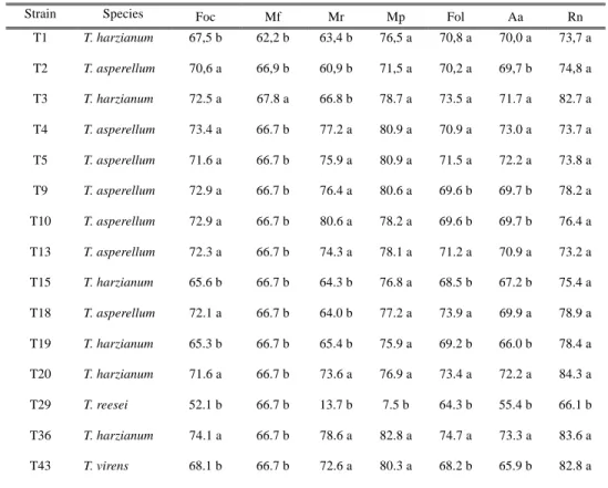
Table 2.4. Inhibitory effects of Trichoderma sp. against pathogenic fungi
Table 4.1. Primers used in this study
Table S1. List of media and buffer
Table S2. Primers used in this study
Table S3. Mix PCR using in this study
Table S4. PCR conditions using in this study
Table S5. Ecuadorian Trichoderma isolates, morphological and molecular information
Table S6. Inhibition activity of Trichoderma strains (+) indicted more that 70% of inhibition (-) indicated less than 70% of inhibition.
21
23
28
42
79
127
130
131 132
133
134
vi
LIST OF FIGURES
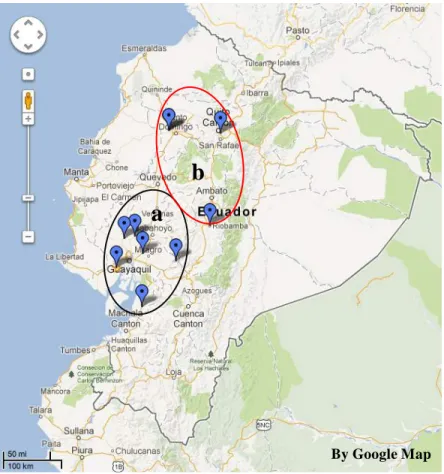
Fig. 2.1. Ecuador sampling map for Trichoderma isolates (a-coastal region, b- highland region)
Fig. 2.2. Colonies of pathogenic fungi on PDA, (Foc) Fusarium oxysporum f. sp.
cubense (Fo-01), (Mf) Mycosphaerella fijiensis (Ec-01), (Mr) Moniliophthora roreri (Cp-01), (Mp) M. perniciosa (MrEO-1), (Fol) F. oxysporum f. sp. lycopersici (Chz1-A), (Aa) Alternaria alternata tomato pathotype (As-27), (Rn) Rosellinia necatrix (ES-0601).
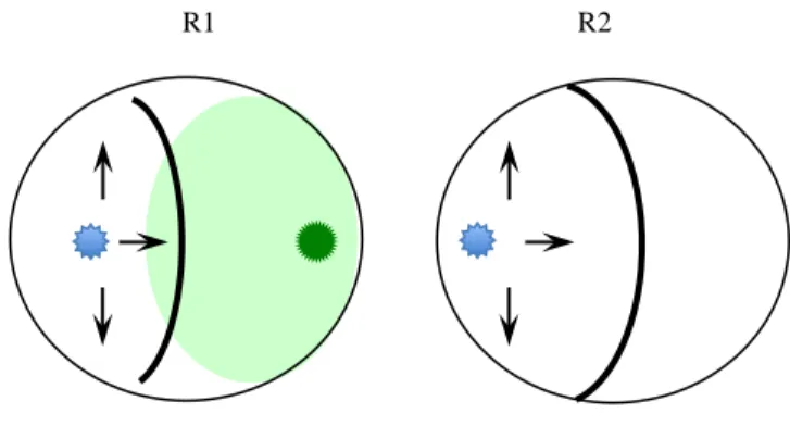
Fig. 2.3. Graphic illustration of antagonistic test, pathogen in contrast with Trichoderma strains (R1) and growth of the pathogen in control dishes (R2). Based in the formula PIRGP = (R1 – R2)/R1 x 100.
Fig 2.4. T. harzianum strains isolated in different region of Ecuador. T1, T3 and T36 Coast Region and T15, T19 and T20 Highland Region. 30
Fig 2.5. T. asperellum strains from different region of Ecuador. T2, T4, T9 and T10 Coast Region. T5, T13 and T18 Highland Region. 32
Fig 2.6. T. reesei (T29) and T. virens (T43) isolated from different region of Ecuador 20
23
26
30
32
33
vii
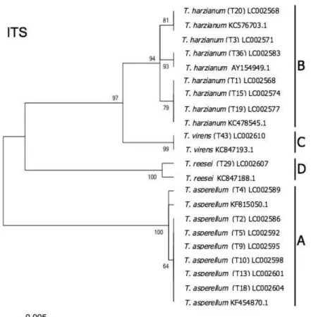
Fig. 2.7. Phylogenetic relations of Trichoderma taxa based on neighbor-joining analysis of ITS sequence data. The evolutionary history was inferred using the Neighbor-Joining method [1]. The optimal tree with the sum of branch length = 0.11417193 is shown. The percentage of replicate trees in which the associated taxa are clustered together in the bootstrap test (2000 replicates) are shown next to the branches [2]. The tree is drawn to scale, with branch lengths in the same units as those of the evolutionary distances used to infer the phylogenetic tree. The evolutionary distances were computed using the Kimura 2-parameter method [3]
and are in the units of the number of base substitutions per site. The analysis involved 25 nucleotide sequences. Codon positions included were 1st+2nd+3rd+Noncoding. All positions containing gaps and missing data were eliminated. There were a total of 500 positions in the final dataset. Evolutionary analyses were conducted in MEGA 5.1 [4].
Fig. 2.8. Phylogenetic relations of Trichoderma taxa based on neighbor-joining analysis of EF-1α sequence data. The evolutionary history was inferred using the Neighbor-Joining method [1]. The optimal tree with the sum of branch length = 1.18257330 is shown. The percentage of replicate trees in which the associated taxa are clustered together in the bootstrap test (2000 replicates) are shown next to the branches [2]. The evolutionary distances were computed using the Kimura 2- parameter method [3] and are in the units of the number of base substitutions per site. The analysis involved 25 nucleotide sequences. Codon positions included were 1st+2nd+3rd+Noncoding. All positions containing gaps and missing data were eliminated. There were a total of 60 positions in the final dataset.
Evolutionary analyses were conducted in MEGA 5.1 [4].
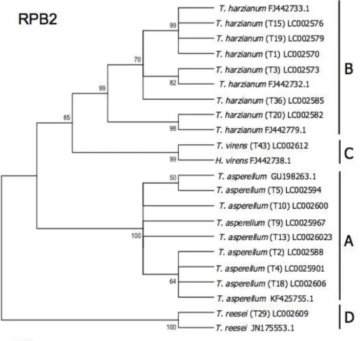
Fig. 2.9. Phylogenetic relations of Trichoderma taxa based on neighbor-joining analysis of RPB2 sequence data. The evolutionary history was inferred using the Neighbor-Joining method [1]. The optimal tree with the sum of branch length = 0.39840624 is shown. The percentage of replicate trees in which the associated taxa are clustered together in the bootstrap test (2000 replicates) are shown next to the branches [2]. The evolutionary distances were computed using the Kimura 2- parameter method [3] and are in the units of the number of base substitutions per site. The analysis involved 23 nucleotide sequences. Codon positions included were 1st+2nd+3rd+Noncoding. All positions containing gaps and missing data were eliminated. There were a total of 317 positions in the final dataset.
Evolutionary analyses were conducted in MEGA 5.1 [4].
35
37
39
viii
Fig. 2.10. Percentage of inhibition of radial growth of pathogens, Trichoderma strains against several pathogen fungi (a) T. harzianum strains, (b) T. asperellum strains (c) T. reesei and T. virens.
Fig. 2.11. Mycoparasitism index of Trichoderma strains against several pathogens fungi (Foc) Fusarium oxysporum f. sp. cubense (Fo-01), (Mf) Mycosphaerella fijiensis (Ec-01), (Mr) Moniliophthora roreri (Cp-01), (Mp) Mo. perniciosa (MrEO-1), (Fol) F. oxysporum f. sp. lycopersici (Chz1-A), (Aa) Alternaria alternata tomato pathotype (As-27), (Rn) Rosellinia necatrix (ES-0601).
Fig. 2.12. Antagonism test of T. harzianum strains (T1, T3, T15, T19, T20, T36).
Photo taken after ten days of incubation. (Foc) F. oxysporum f. sp. cubense Fo-01, (Mf) M. fijiensis Ec-01, (Mr) M. roreri CP-01, (Mp) M. perniciosa MrEO-1, (Fol) F. oxysporum f. sp. lycopersici Chz1-A, (Aa) A. alternate As-27, (Rn) R. necatrix ES-0601.
Fig. 2.13. Antagonism test of T. asperellum strains (T2, T4, T5, T9, T10, T13, T18). Photo taken after ten days of incubation. (Foc) F. oxysporum f. sp. cubense Fo-01, (Mf) M. fijiensis Ec-01, (Mr) M. roreri CP-01, (Mp) M. perniciosa MrEO- 1, (Fol) F. oxysporum f. sp. lycopersici Chz1-A, (Aa) A. alternate As-27, (Rn) R.
necatrix ES-0601.
Fig 2.14. Antagonism test of T. reesei strain (T29) and T. virens strain (T43). Photo taken after ten days of incubation. (Foc) F. oxysporum f. sp. cubense Fo-01, (Mf) M. fijiensis Ec-01, (Mr) M. roreri CP-01, (Mp) M. perniciosa MrEO-1, (Fol) F.
oxysporum f. sp. lycopersici Chz1-A, (Aa) A. alternate As-27, (Rn) R. necatrix ES-0601.
Fig. 3.1. Graphic representation of fungi interaction on petri dish.
43
45
46
47
48
58
ix
Fig. 3.2. Hyphae from (a) T. harzianum T36 (ThDsred) and (b) F. oxysporum f. sp.
cubense Fo-01 (FocGFP) using fluorescent microscopy 40x.
Fig. 3.3. T. harzianum T36 (ThDsred) morphology, (a) conidiophores, (b) phyliades and (c) conidia.
Fig. 3.4. F. oxysporum f. sp. cubense Fo-01 (FocGFP) morphology, (a) hypha, (b) conidia.
Fig. 3.5. T. harzianum T36 (ThDsred) (a) mycelia growth alongside F. oxysporum f. sp. cubense Fo-01 (FocGFP) after 24h of co-cultivation. (b) Arrows indicate T.
harzianum T36 (ThDsred) make damage to F. oxysporum f. sp. cubense Fo-01 (FocGFP).
Fig. 3.6. Different light stage showed T. harzianum T36 (ThDsred) coiling and growth alongside and between F. oxysporum f. sp. cubense Fo-01 (FocGFP).
Fig. 3.7. T. harzianum T36 (ThDsred) (a) mycelia growth alongside F. oxysporum f. sp. cubense Fo-01 (FocGFP) after 48h of co-cultivation. (b) Arrows indicate T.
harzianum T36 (ThDsred) make damage to F. oxysporum f. sp. cubense Fo-01 (FocGFP) interaction.
Fig. 3.8. T. harzianum T36 (ThDsred) (a) mycelia growth alongside F. oxysporum f. sp. cubense Fo-01 (FocGFP) after 72h of co-cultivation. (b) Arrows indicate T.
harzianum T36 (ThDsred) make damage to F. oxysporum f. sp. cubense Fo-01 (FocGFP) interaction.
Fig. 3.9. Micoparasitic activity of T. harzianum T36 (ThDsred) against F.
oxysporum f. sp. cubense Fo-01 (FocGFP) with formation of papilla-like structures (arrows).
60
62
62
64
65
66
66
67
x
Fig. 3.10. Hypha of F. oxysporum f. sp. cubense Fo-01 (FocGFP) degradeted by T.
harzianum T36 (ThDsred) arrows micoparasitic activity. (a) T. harzianum T36 (ThDsred) hypha coil host hypha, (b, c, d) host hypha degradation, (e) fungal conidia after 4 days of growth 40x.
Fig. 4.1. Alignment of the deduced amino acid sequence of ThSNF1 and the SNF1 orthologs in other ascomycetes fungi. The amino acid sequences were aligned by the program Clustalw2 (http://www.ebi.ac.uk/Tools/msa/clustalw2/). Protein domain prediction of ThSNF1 was performed by InterProScan 5 (http://www.ebi.ac.uk/Tools/pfa/iprscan5/). CcSNF1 and FoSNF1 are SNF1 orthologs in Cochliobolus carbonum (AF159253) and Fusarium oxysporum (AF420488), respectively. The underlines show serine/threonine protein kinases, catalytic domain.
Fig. 4.2. Deletion strategies for the ThSNF1 in the genome of T. harzianum T36 strain. (a) A fusion PCR method was used to construct the ThSNF1 replacement vector. All PCR primers used in this figure are listed in Table 1. (b) PCR analysis of gene replacement events in ThSNF1 in the wild-type strain (WT) and the ΔThSNF1 mutant using primer pairs HphF/HphR (b)-Hph, Thsnf1inF/Thsnf1inR (b)-IN, Thsnf1homoF/ HphhomoR (b)-Homo 1, and HphhomoF/Thsnf1homoR (b)-Homo 2. (c) Expression of ThSNF1 in the wild type (T36) and the ΔThSNF1 strains of T. harzianum. For RT-PCR the primer sets listed in Table 1 were used for detection of ThSNF1 and β-tubulin gene of T. harzianum.
Fig. 4.5. Growth of the T. harzianum strains WT and ΔThSNF1 on different media after seven days of incubated at 25°C. Arrows indicated slow growth.
69
81
83
85
xi
Fig. 4.4. Comparison of the growth rates between the wild type (T36) and the ΔThSNF1 strains of T. harzianum on minimal media supplemented with different carbon sources. The plates were observed every day until 7 days of growth.
Fig. 4.5. Conidia production of the wild type (T36) and the ΔThSNF1 strains of T.
harzianum. Conidia produced on the minimal media supplemented with glucose were harvested 7 days after culture and the number of conidia was counted.
Fig. 4.6. Expression of genes encoding cell wall-degrading enzymes in the wild type T36 (WT) and ΔThSNF1 strains of T. harzianum. Total RNA was extracted from fungal mycelia grown in minimal media supplemented with glucose as the only carbon source. RT-PCR primer sets listed in Table 1 were used for detection of chitinase gene Chi18-17 (lane 1), polygalacturonase gene PGX1 (lane 2) and the β-tubulin gene (lane 3) of T. harzianum.
Fig. 4.7. Mycoparasitism test of the wild type (WT) and ΔThSNF1 strains of T.
harzianum using the dual culture method. (a) The antagonism test was performed on PDA by placing a mycelium disc (5 mm in diameter) of each pathogenic fungus (F. oxysporum f. sp. cubense (Foc) or F. graminearum (Fg)) on one side of a Petri dish; the opposite side of each dish was inoculated with the Trichoderma strains.
The plates were incubated at 25°C for 10 days. (b) The mycoparasitism of the Trichoderma strains against the pathogens F. oxysporum f. sp. cubense and F.
graminearum was determined in triplicate using the scale described in the Materials and methods section. Each value is the average of experiment with three replicates per treatment. Error bars represent the standard deviation.
Fig. 4.8. Control strains of T36 wild type, T36 ΔThSNF1, F. oxysporum f. sp.
cubense (Foc) and F. graminearum (Fg).
86
86
87
89
90
xii
Fig. S. 1. Growth of the T. harzianum wild type and mutant strain ΔThSNF1 in YPG liquid media after 24h.
Fig. S. 2. Growth of the T. harzianum wild type and mutant strain ΔThSNF1 in MM supplemented with colloidal chitin, photo was take after four days of growth.
Fig. S. 3. Conidial morphology and the germination rate of the T. harzianum strains T36 (WT) and ΔThSNF1 (mutant) in different media photo was taken after 10 hours of growth.
135
135
136
1 CHACTER 1 General Introduction
1.1 Trichoderma morphology
The genus Trichoderma is one of the most common genera isolated from soil and can be found widely in agricultural, prairie, forest, salt marsh and desert soil in all climates. These general saprophytes are highly interactive in root, soil and foliar environments. They are fast-growing filamentous deuteromycetes, which can sporulate abundantly, and have been described to constitute up 3% of the total fungus propagules in forest soil (Harman et al. 2004; Klein and Eveleigh 1998; Pinto et al. 2006).
Persoon first described the genus Trichoderma in 1794 but its classification remained unclear and contradictory until Rifai (1969) made the first real attempt to produce a workable classification system of the genus. The system was based on species morphology and on the concept of species aggregates. Bissett (1991) proposed a revised classification of the genus Trichoderma based on key morphological characteristics and four sections were established (Sect. Trichoderma, Sect. Pachybasium, Sect.
Longibrachiatum, and Sect. Hypocreanum) (Bissett 1991a; b; c).
The genus Trichoderma possesses key morphological characteristics that are still used to identify Trichoderma species. It is a septate fungus and produces highly branched conidiophores with a conical or pyramidal outline (Rifai 1969). Flask-shaped structures called phialides are found at the tip of the conidiophores. Phialospores, also known as conidia, are produced at the end of the phialides where they accumulate to form a conidial head (Gams and Bissett 1998). Under certain nutritional or against drying conditions, resistance structures called chlamydospores are produced. These are very
2
important for the survival of the fungus (Lewis and Papavizas 1984). Like all deuteromycetes, Trichoderma species can only reproduce asexually through intense sporulation or clonal growth from hyphal fragments (Gams and Bissett 1998). However, the genus Trichoderma also has a sexual stage (teleomorph) known as Hypocrea, which is in the ascomycete order Hypocreales (Samuels 1996). Trichoderma teleomorphs possess the entire key characteristics of Trichoderma anamorphs; they can reproduce sexually to form ascospores. Trichoderma species form floccose or tufted colonies of various colors (white, yellow, green), which in the past were used to identify species (Rifai 1969). Most of the Trichoderma species have a rapid growth over different substrates with a large number of green conidia, unusually white, formed from enteroblastic phialidic conidiogenous cells (Kirk et al. 2001). Today, the use of morphological characteristics for identifying Trichoderma species is being progressively replaced by molecular tools, which provide a more robust and reliable form of species identification (Lieckfeldt et al. 1998).
1.2 Identification of Trichoderma species
Molecular methodologies have greatly improved our understanding of the genus especially at species level. Today around 104 species of Trichoderma have been identified (http://www.isth.info/biodiversity/index.php), many of which are important biological control agents, such as T. harzianum and T. polysporum (Rifai 1969), T.
hamatum (Bainier 1907), T. koningii (Samuels et al. 2006), T. asperellum and T. virens (Samuels 1999).
Trichoderma species were allocated into four sections, sect. Trichoderma, sect.
3
Longibrachiatum, sect. Pachybasium, and sect. Hypocreanum (Bissett 1991 a; b; c) and redefined by Samuels (1996). Trichoderma is a septate genus that produces highly branched conidiophores that release numerous conidia. This genus is less frequently found in sexual state (teleomorph) belonging to the Hypocreales. Ascospores are formed in sexual reproduction (Kubicek and Harman 1998).
The first studies that involved characterization of the genus Trichoderma were with molecular tools like RFLP, RAPD-PCR, and sequences from different areas of the genome or markers SCAR (Muthumeenakshi et al. 1994; Arisan-Atac et al. 1995;
Hermosa et al. 2000, 2001; Rubio et al. 2005), are useful for the identification and for the systematic of the genus. Especially useful are the sequence data obtained from regions internal transcribed spacer (ITS) and encoding gene as translation elongation factor 1 (EF-1α) consisted in several relative large and variable introns and exons. Although the coding portion of endochitinase (ech42) and RNA polymerase subunit 2 (RBP2), have displayed significant intra and inter specific variability while other genes (Chaverri et al.
2003).
Safe identification of new species was significantly facilitated by development of an oligonucleotide barcode (TrichOKEY) and a customized similarity search tool (TrichoBLAST), both available online at www.isth.info (Druzhinina et al. 2005;
Kopchinskiy et al. 2005).
1.3 Ecology
The genus Trichoderma, which comprises of a group of fast-growing fungi, is common in forest and agricultural soils. Trichoderma species adapt to all climates but
4
occur abundantly on plant debris in wetlands (Klein and Eveleigh 1998). The composition, biomass and biological activity of microbe communities in the soil depend on important physical and chemical factors (Garbeva et al. 2004; Killham 1994; Lavelle and Spain 2001), fungi and more specifically Trichoderma species are no exception.
Environmental parameters such as soil temperature, moisture, atmosphere, pH, organic matter (OM); nutrient content and plant types are key factors that influence soil colonization by Trichoderma species (Carreiro and Koske 1992; Danielson and Davey 1973b; Domsch et al. 1980; Eastburn and Butler 1988; 1991; Klein and Eveleigh 1998;
Widden and Abitbol 1980). The substrates to be used by Trichoderma growth are varied but show preference for acidic soils rich in organic matter (Hubbard et al. 1983; Klein and Eveleigh 1998). Concerning the ambient parameters needed for growth, Trichoderma can grow within a wide temperature range, also can be influenced by soil temperature in terms of their world distribution. Species like T. harzianum are generally isolated from warm tropical soils whereas T. polysporum and T. viride are mostly found in cool temperate regions (Danielson and Davey 1973a; Klein and Eveleigh 1998). The range of temperatures at which Trichoderma species can grow is fairly wide, it can be as low as 0°C for T. polysporum and as high as 40°C for T. koningii (Domsch et al. 1980; Tronsmo and Dennis 1978). Temperature does not only affect the growth of Trichoderma species, it also affects their metabolic activity especially the production of volatile antibiotics and enzymes (Tronsmo and Dennis 1978).
Another aspect is the relative resistance of various species of Trichoderma numerous chemicals used in agriculture as fungicides; organochlorine, endosulfan types (Katayama and Matsumura 1993; Shaban and El-Komy 2001) or Benzimidazoles
5
(Mukherjee et al. 2003). These features, along with its growth rate and its easy adaptation to different climatic and soil conditions (López-Errasquin and Vázquez 2003), give Trichoderma advantage over many other filamentous fungi in the soil colonization after application of these treatments.
1.4 Trichoderma species as biocontrol agent
Trichoderma species have been associated as biocontrol agent in agriculture over several pathogenic fungi (Monte 2001; Benítez et al. 2004; Harman et al. 2004). T.
harzianum, T. viride, T. asperellum and T. virens are the most useful as biocontrol agents (Papavizas 1985; Chet 1987). Actually, T. harzianum alone or combination with other Trichoderma specie is currently being used in commercial preparations for the control of numerous plant diseases caused by fungi (Cook 1997; Grondona et al. 2001), like Rhizoctonia solani and Fusarium oxysporum (Faull and Scarseletti 1994), Colletotrichum species (Tronsmo and Hjeljord 1995), Sclerotinia sclerotiorum and Sclerotium rolfsii (Knudsen and Eschen 1991), Phytophthora parasitica and Pythium ultimum (Lumsden and Locke 1989), Verticillium species (Santamarina and Roselló 2006), Moniliophthora perniciosa (De Marco et al. 2003).
Numerous other Trichoderma species have antagonistic capacity through different mechanisms of biocontrol such as T. hamatum against Botryris cinerea (Nelson and Powelson 1988), T. lignorum and T. pseudokoningii against R. solani (Aziz et al. 1997;
Askew and Laing 1994), T. auroviride against Fusarium species, Phythium species and R.
solani (Kredics et al. 2001), T. koningii against Sclerotinia sclerotiorum, Sclerotium cepivorum (Metcalf and Wilson 2001), T. asperellum against F. oxysporum (Cotxarrera et
6
al. 2002), T. koningii is also useful in increasing of wheat seed (Duffy et al. 1996).
Additionally, new Trichoderma isolate showed biocontrol capacity as T. martiale against Phytophthora palmivora (black-pod disease) in cacao (Hanada et al. 2009).
In addition, some Trichoderma species are useful in bioremediation, T. atroviride strain F6 applied to soils Brassica juncea plants, detoxifying heavy metals such as cadmium, nickel or a combination of both (Cao et al. 2008).
On the other hand, species like T. reesei have industrial application because they can produce cellulolytic enzymes able to degraded cellulolytic material (Reese and Mandels 1989; Kubicek et al. 1990). Furthermore, the Trichoderma species has the capacity to secreteproteins, and is one of the organisms used today as a “cell factory” for the production of proteins for pharmaceutical purposes (Rey et al. 2004). Similarly, the chitinases produced by this species are used in the degradation of chitin remains in various industries, such as working with crustaceans, to obtain N-acetyl-glucosamine, which is used as a food supplement (Cosio et al. 1982).
1.5 Mechanism of biocontrol of Trichoderma species.
Over 80 years ago Weindling (1932) described the antagonistic capacity of Trichoderma lignorum. Since then there have been various mechanisms by which Trichoderma prevents and protects plants against attack by other fungi. Currently, the mechanisms of action of Trichoderma are described in five categories: (i) mycoparasitism, (ii) antibiosis, (iii) competition, (iv) promoting growth and/or (v) induction of resistance in the host plant (Hjeljord and Tronsmo 1998; Howell 2003;
Harman et al. 2004). These mechanisms are not mutually exclusive, and the relative
7
importance of each is not well established, although it seems to depend of the Trichoderma strain, the target pathogen and soil and environmental conditions.
Mycoparasitism is regarded as a direct attack of one fungus on another and can be subdivided into four stages of interaction: Chemotrophic growth includes the direct growth of the mycoparasite towards the host fungus. Trichoderma species are attracted by chemical stimuli released by the target fungus, which to some extent is due to expression of cell wall degrading enzymes. Specific recognition arises by the binding of carbohydrates on the Trichoderma cell wall to lectins on the host surface. When both funguses come into contact, Trichoderma hyphae attach and coil around the host fungus hyphae through formation of hook-like structures and appresorium-like bodies. The breakdown of the host cell wall begins with the secretion of lytic enzymes, the subsequent penetration into the lumen of the target fungus together with the production of specific antibiotics causes the death of the host fungus (Chet et al. 1998; Harman 2000;
Harman et al. 2004). In mycoparatism some of the lytic enzymes produced by this genus are thought to play a fundamental role, due to their function in direct physical interactions (Brunner et al. 2003).
The antibiosis consists in the production of a large number of volatile and no- volatile secondary metabolites by Trichoderma sp., the most common anti-fungal metabolites are viridian (steroid group), gliotoxin and gliovirin (diketopiperazine group) (Howell 1998). The capacity to synthesize antibiotics differs considerably between and within species, both quality and quantitatively can be affected by environment conditions.
Competition happens when two or more microorganisms demand the same limited resource, such as nutrients or space. Competition between Trichoderma and plant
8
pathogen evidently leads to disease control if Trichoderma shows better saprophytic abilities and as result reduces the pathogen growth or propagule production (Hjeljord and Tronsmo 1998). Competition can be classified into “interference competition”, indicated behavioral or chemical mechanism by which one organism limits another organism’s access to the resource due to both inter and intraspecific mycelial interactions and indirect inhibition and “exploitation competition”. suggested the antagonist directly competes with the opponent for a resource (Tuininga 2005).
Trichoderma species, colonize the substrate rapidly, conidiate prolifically and utilize a wide range of substrates as aggressive soil saprophytic. Through, their ability to produce antibiotics and directly parasite other organisms, they can affect their competitors by successfully conquering the resources. The most efficient antagonistic mechanism to prevent pathogenic infection is related to nutrient competition and is likely to be a key component in biological control systems (Hjeljord and Tronsmo 1998).
The genus Trichoderma has the capacity to protect plants against root pathogens;
this was attributed for a long time to a direct effect against the pathogens (Chet et al.
1998). However, Trichoderma species in direct association with plant roots has been found, stimulating the defense mechanism of the plants (Yedidia et al. 1999), leading to a resistance against a variety of phytopathogenic microorganism and even nematodes (Harman et al. 2004). Parallel to the production of antibiotics metabolites some Trichoderma strains can strongly stimulate plants to synthesise their own antimicrobial compounds. Trichoderma species can induce both locally and systemically defensive response in the plant by colonizing the root surface and penetrating the superficial root cell. A zone of interaction with Trichoderma isolated is established and releases elicitors
9
of resistance including peptides, proteins and low-molecular-weight compounds, as well as, plant resistance response increases by the release of the cell wall fragments (Harman et al. 2004). The modification induced in the metabolic machinery of the plant results in increased level of pathogenesis-related proteins, accumulation of phytoalexins and deposition of structural polymers (Yedidia et al. 2000).
1.6 Lytic enzymes of Trichoderma
The genus Trichoderma has an astonishingly remarkable fast and efficient exploitation of almost any compound and therefore nutrients, by their enormous equipment of lytic enzymes. Organic matter consists of a wide range of sugars, homo and heteropolysaccharides (Dix and Webster 1995; Klein and Eveleigh 1998).
In order to absorb nutrients, Trichoderma species like other fungi need to breakdown large insoluble compounds such as polysaccharides and proteins exocellularly (Griffin 1994; Lavelle and Spain 2001). Trichoderma species grow on different carbon and nitrogen substrates and molecules like cellulose, chitin, xylose or large peptide chains represent a challenge to enzymatic hydrolysis because of the heterogeneity of their constituents (Griffin 1994; Koivula et al. 1998). Most Trichoderma species overcome this problem through large secretion of complex mixtures of specific exocellular enzymes (Dix and Webster 1995; Griffin 1994; Klein and Eveleigh 1998; Koivula et al. 1998;
Kubicek-Pranz 1998). The ability of some species, such as T. viride and T. reesei to produce huge quantities of hydrolytic enzymes such as cellulase, is applied commercially in diverse areas as cloth-washing detergents and the paper industry (Griffin 1994;
Kubicek-Pranz 1998). Some of the other important hydrolytic enzymes produced by
10
Trichoderma species include hemicellulase (xylanase, mannanase, galactanase), chitinase, glucanase, amylase (Griffin 1994; Worasatit et al. 1994) and cell wall lytic enzymes (Lorito et al. 1996; Steyaert et al. 2003).
With the objective of degrading proteins and peptide chains, Trichoderma species also produce a wide range of exocellular proteolytic enzymes with low substrate specificity, enabling then to absorb small amino acid units and use them as nitrogen and sulphur sources. In the middle of those enzymes, multiple forms of serine and aspartic proteinases are very common in most saprophytic fungi (Dix and Webster 1995; Griffin 1994). Most Trichoderma species also produce high levels of key enzymes (glutamine synthetase and NADPH-glutamate dehydrogenase) for ammonia assimilation in the presence of ammonium ions (Ahmad et al. 1995).
1.7 Genes involved in the mycoparasitism
Complete sequencing of T. reesei/Hypocrea jecorina genome provided finally the data for a detailed study on the genes encoding for those enzymes and how they are regulated. Additionally, H. atroviridis genome is almost completely sequenced, creating a broad basis for further studies on genes involved in biocontrol mechanism (Lorito et al.
2010; Seidl et al. 2009).
Chitin is one of the most abundant polymers in the biosphere and its degradation is involved many biological process. Chitinolytic enzymes can be divided into exo and endo-acting enzymes based on their reactions and products and catalytic mechanisms. β- N-acetylglucosaminidases (NAGases) catalyse the hydrolysis of terminal non-reducing N-acetyl-D-glucosamine (GlcNAc) residues and transglycosilation reactions (Sahai and
11 Manocha 1993).
Chitinases have been found in many Trichoderma species, inclusive explored their potential presence in the already sequenced genomes of Trichoderma (Seidl et al. 2005).
Genes encoding some chitinases have been cloned in different strains that encoding chitinases like CHIT37, CHIT42 and CHIT33 of T. harzianum (García et al. 1994; Limón et al. 1995; Viterbo et al. 2001) and CHIT36 from T. asperellum (Viterbo et al. 2002).
The action of chitinases was studied in in vitro tests on germination of spores of various pathogens (Lorito et al. 1993). Gene over-expression of various Trichoderma strains confirmed the antifungal effect of these enzymes (Limón et al. 1999; Viterbo et al.
2001; Limón et al. 2004).
During mycoparasitism was observed sequential transcription of genes encoding chitinases, sometimes the loss of expression of these genes leads to deregulation of the chitinolytic system (Inbar and Chet 1995; Zeilinger et al. 1999; Brunner et al. 2003).
Glucanases, chitin and β-1,3-glucan are the principal modules fungal cell walls are made by Trichoderma, posses a range of chitinases and β-1,3-glucanases, which do the main work in the degradation of the latter (Mahadevan and Tatum 1967).
Benítez et al. (2004) reported that after breaking down the obstacle to the pathogen cell wall, Trichoderma species are able to prevent the pathogen from further growth by release of β-1,3-glucanases that collaborate with chitinases and antibiotics. Some of the β-1,3-glucanases have been isolated and cloned so far, as bgn13.1 (De la Cruz et al. 1993) and lam1.3 (Cohen-Kupiec et al. 1999) from T. harzianum/H. lixii, glu78 from T.
atroviride (Donzelli and Harman 2001) and Tv-bgn1 and Tv-bgn2 from T. virens (Kim et al. 2002). Moreover, an effective antagonist needs further glucanases to degrade other
12
structurally important cell wall components like β-1,6-glucan, α-1,3-glucan or faced with chitin as only carbon sources. In addition, β-1,6-glucanases from T. harzianum/H. lixii (BGN16.1, BGN16.2, BGN16.3) have been purified and could be essential in the fungal cell wall degradation , rich in chitin (Montero el al. 2005; De la Cruz and Llobell 1999;
Delgado-Jarana et al. 2001). The α-1,3-glucanases can be divided into endo and exo- glucanases corresponding to the final degradation product is a small glucose.
Proteases are described as another important set of enzymes enabling a well- organised biocontrol reaction. Different extracellular proteases have been isolated from the genus Trichoderma (Antal et al. 2001; Delgado-Jarana et al. 2002; Williams et al.
2003; Suárez et al. 2004). Some proteases of T. harzianum/H. lixii could neutralize certain hydrolytic enzymes released from pathogens as Botrytis cinerea, with subsequent reduced germination activity by the pathogen (Kapat et al. 1998; Elad and Kapat 1999).
Proteases may be important for the mycoparisitism activity by degradation of protein components of the host cell wall, through the deactivation of plant pathogen enzymes.
The reaction and involvement of protease in mycoparasitic interaction protease gene were cloned, in order to understand this way biocontrol of Trichoderma species on Prb1 protease of T. atroviride, like it’s orthologues from T. virens and T. hamatum (Pozo et al.
2004; Steyaert et al. 2004), is a subtilisin-like serine protease and was shown to play its part in mycoparasitism of R. solani (Geremia et al. 1993; Flores et al. 1997; Cortes et al.
1998; Olmedo-Monfil et al. 2002). Expression analyses based on mycoparasitic activity with fungal cell walls, confrontation assays with pathogens, carbon or nitrogen as limiting factors showed the upregulation of pra1 (trypsin-like serine protease), papA (aspartic protease), papB (vacuolar aspartic protease), and an extracellular aspartic
13
protease (Suárez et al. 2004, 2005; Delgado-Jarana et al. 2002; Viterbo et al. 2004).
Serine threonine protein kinase is probable to be important mediator of fungal proliferation and development as well as signal transduction and infection-related morphogenesis. Protein kinase-mediated phosphorylation regulates protein functions (directly or via transducing the relevant signal) implicated in the entire spectrum of cellular process (Dickman and Yarden 1999). Carbon catabolite repression is involved in the energy mechanism by the utilization of complex sugar like glucose. Mig1p mediated carbon repression in yeast, in absence of glucose is phosphorylated and hence inactivated by Snf1-kinase (Johnston et al. 1994; Lutfiyya et al. 1998; Treitel and Carlson 1995).
In filamentous fungus, as Aspergillus nidulans (Arst and MacDonald 1975), Aspergillus niger (Drysdale et al. 1993), Metarhizium anisopliae (Screen et al. 1997) and Cochliobolus carbonum (Tonukari et al. 2000), CreA have been identified to be involved in carbon catabolite repression and is homolog of yeast MIG1 gene.
Disruption of SNF1 homologue in C. carbonum produces a substantial reduction of cell wall-degrading enzyme action and their transcript under derepressive. conditions and also caused reduced growth in different carbon sources (Tonukari et al. 2000). Identical result with Fusarium oxysporum showed a diminished transcription of genes encoding cell wall-degrading enzymes (Ospina-Giraldo et al. 2003).
14 1.8 Goal of this study
This study involves the use of a fungal collection maintained by CIBE-ESPOL, consisting of several Trichoderma species isolates, in order to identify and select beneficial isolates to be used as biocontrol agents against important diseases of the principal cultivars in Ecuador.
The population dynamics of Trichoderma isolates in different environments and cultivars is of special interest to our working group, in addition to the identification to species level, as well as, the biocontrol activity of Trichoderma isolates against several pathogenic fungi, by different mechanisms/modes of action. The mycoparasitism involve the production of several enzymes like proteases, glucanases and chitinases by activation of many genes during the interaction with both fungi.
Trichoderma isolates have been identified as being able to act as endophytic plant symbionts. The strains become endophytic in roots, but the greatest changes in gene expression occur in shoots. These changes alter plant physiology and may result in the improvement of abiotic stress resistance, nitrogen fertilizer uptake, and resistance to pathogens and photosynthetic efficiency. Typically, the net result of these effects is an increase in plant growth and productivity (Hermosa et al., 2000).
The goals of the present study are: molecular identification of Trichoderma isolates from different regions of Ecuador at species level. Re-description of T. viride strains, antagonistic activity of Trichoderma strains against important pathogenic fungi from Ecuador and Japan. The mycoparasitism one of the most important mechanisms of action used by this genus was analyzed by Ds-Red T. harzianum T36 and GFP Fusarium
15
oxysporum f. sp. cubense. During the mycoparasitism several genes are activated. In order to determine those involved of Serine/threonine protein kinase in the virulence and morphology of T. harzianum T36, a disruption of ThSNF1 was performed.
16
CHAPTER 2. Identification of Trichoderma strains at species level
2.1 Introduction
Trichoderma is a cosmopolitan soil-borne fungus that interacts with root systems, soil and the foliar environment (Hjeljord and Tronsmo 1998) and is an important biological agent for controlling plant pathogens. Trichoderma species have been reported to control several phytopathogens of diverse crops based on various mechanisms, such as the production of antifungal metabolites, competition for nutrients and space, mycoparasitism and efficiency in promoting defense mechanisms (Hoyos-Carvajal et al.
2008; Woo and Lorito 2007).
Morphological characters are reported to be variable to a certain degree in their color, shape of conidia, conidiophore, pustules and phialides. These characteristics allow a comparatively easy means of identification of Trichoderma as a genus, but the species concept is difficult to deduce and there is considerable confusion over the application of specific names. To identify and characterize Trichoderma species morphological characteristics should be considered along with molecular data from DNA sequencing (Samuels 2006). Additionally, a multi-gene approach using at least two unlinked loci is desirable for the molecular identification of closely related Trichoderma spp. The internal transcribed spacer (ITS) region of the ribosomal DNA (rDNA) is one of the most reliable targets in identifying a strain at the species level (Kullnig-Gradinger et al. 2002). EF-1α gene encode the translation elongation factor 1-α and RPB2 gene encode the subunit of RNA polymerase II, which are more variable and can be used to reflect differences within and between groups of closely related species (Liu and Hall 2004; Matheny 2005; Zhang
17
et al. 2005). The grouping of these three genes enables the identification of most Trichoderma at species level (Samuels 2006). Together with TrichOKEY and TrichoBLAST (www.isth.info) a convenient online tool for the molecular identification of Trichoderma isolates, are based on the sequence comparisons of these genes (Druzhinina et al. 2005; Kopchinskiy et al. 2005). However, molecular identification has also shown a high level of heterogeneity at species level in, for instance e.g., T.
harzianum (Chaverri et al. 2003; Hermosa et al. 2000; Samuels et al.1999).
Trichoderma species have been used as biological control agents of a wide range of foliage diseases (Nelson 1991). The most widespread species used as biocontrol agents are T. harzianum (Sharma et al. 2009), T. viride (Karthikenyan et al. 2006), T. hamatum (Harman et al. 1981), T. atroviride (Hjeljord and Tronsmo 2003), T. asperellum (Watanabe et al. 2005) and T. virens (Mukherjee et al. 2006). The control efficiency for each disease differs between Trichoderma strains and depends on the target disease(s) (Harman et al. 2004).
Banana, including plantain Musa species is one of the most important crops in the world. Nevertheless, banana production in tropical areas has recently faced a crisis due to outbreak of several diseases, such as black Sigatoka or black leaf streak disease, which is caused by Mycosphaerella fijiensis, as well as Panama disease (Fusarium wilt), which is caused by Fusarium oxysporum f. sp. cubense (Ploetz 2006; Stover 1962). On the other hand, Cacao (Theobroma cacao) is grown in many tropical environments (Wood and Lass 2001), and is one of the most economically important crops in Ecuador. The well known diseases of cultivated cacao include Moniliophthora roreri and M. perniciosa, which cause frosty pod rot (Bowers el al. 2001; Wood and Lass 2001) and witches' broom
18
disease (Aime and Phillips-Mora 2005), respectively. These pathogens can cause complete yield loss. Therefore, control of these diseases is the most important agricultural issue in many counties in South America, Southeast Asia and Africa.
The employment of endogenous and domestic microorganisms as biocontrol agents is the most important factor in biosafety, environmental conservation and sustainability.
In this study we characterized native Trichoderma isolates using both morphological observation and molecular identification. The antagonistic activity and mycoparasitism of the isolates were evaluated against banana and cacao disease pathogens common in Ecuador. The antagonistic activity of these isolates against the important foliar disease pathogen Alternaria alternata, as well as soil-borne disease pathogens Rosellinia necatrix and F. oxysporum f. sp. lycopersici (from Japan), were also examined for comparison.
Molecular markers are important to develop tools that monitor the genetic and environmental fate of biocontrol agents Trichoderma as more fungal biocontrol agents are registered as alternatives to chemical pesticides. Molecular characterization provides an immense source of data that can assist scientists in the study of identity, relatedness, diversity and selection of proper candidates for biological control.
19 2.2 Materials and Methods
2.2.1 Isolation and identification of Trichoderma species
Trichoderma species were isolated from soils of different fields, cacao bark and substrate the Pleurotus species in various regions of Ecuador (Fig. 2.1) (Table 2.1). The isolation of Trichoderma strains from these samples was performed as described previously (Elad et al. 1981). Single spore strains were grown on potato dextrose agar (PDA) (Difco, Detroit, MI, USA) at 28°C and stored at -80°C in 20% (v/v) glycerol.
Trichoderma species were identified via microscopic observation of the morphology of conidia, conidiophore, phialides and chlamydospore using taxonomic keys (http://nt.ars- grin.gov/taxadescriptions/keys/FrameKey.cfm?gen=Trichoderma) (Samuels et al. 2014).
20
Fig. 2.1. Ecuador sampling map for Trichoderma isolates (a-coastal region, b- highland region)
By Google Map
a
b
21
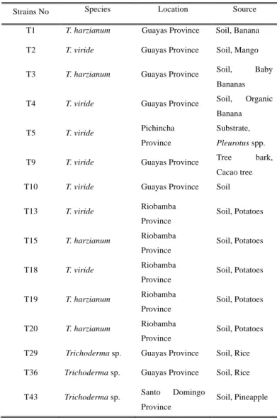
Table 2.1. Morphological classification of Ecuadorian Isolates.
Strains No Species Location Source
T1 T. harzianum Guayas Province Soil, Banana T2 T. viride Guayas Province Soil, Mango T3 T. harzianum Guayas Province Soil, Baby
Bananas T4 T. viride Guayas Province Soil, Organic
Banana
T5 T. viride Pichincha
Province
Substrate, Pleurotus spp.
T9 T. viride Guayas Province Tree bark,
Cacao tree
T10 T. viride Guayas Province Soil
T13 T. viride Riobamba
Province
Soil, Potatoes
T15 T. harzianum Riobamba
Province
Soil, Potatoes
T18 T. viride Riobamba
Province
Soil, Potatoes
T19 T. harzianum Riobamba
Province
Soil, Potatoes
T20 T. harzianum Riobamba
Province
Soil, Potatoes T29 Trichoderma sp. Guayas Province Soil, Rice T36 Trichoderma sp. Guayas Province Soil, Rice T43 Trichoderma sp. Santo Domingo
Province
Soil, Pineapple
22 2.2.2 Pathogens
Pathogenic fungi used in this study are listed in Table 2.2. Four banana and cacao pathogens originating from Ecuador, M. fijiensis (Ec-01), F. oxysporum f. sp. cubense (Fo-01), M. roreri (Cp-01) and M. perniciosa (MrEO-1), were obtained from stock collections at the Biotechnology Research Center of Ecuador (CIBE-ESPOL). F.
oxysporum f. sp. lycopersici (Chz1-A), Rosellinia necatrix (ES-0601) and the tomato pathotype of A. alternata (As-27) (Fig. 2.2) were kindly provided by Dr. Arie (Tokyo University of Agriculture and Technology), Dr. Yasuda (Tottori Prefectural Agriculture and Forest Research Institute) and Dr. D. G. Gilchrist (University of California, Davis), respectively. The Fungus/Mushroom Resource and Research Center, Tottori University (FMRC) provided standard Trichoderma strains (T. harzianum TUFC 60692, T. atroviride TUFC 60732, T. koningiopsis TUFC 60205, T. citroviride TUFC 61231 and T.
longibrachiatum TUFC 60819) as references. All isolates were maintained on PDA (Difco) slants or in 20% (v/v) glycerol at -80°C.
23
Table 2.2 List of pathogenic fungi used in this study
Fig. 2.2. Colonies of pathogenic fungi on PDA, (Foc) Fusarium oxysporum f. sp. cubense (Fo-01), (Mf) Mycosphaerella fijiensis (Ec-01), (Mr) Moniliophthora roreri (Cp-01), (Mp) M. perniciosa (MrEO-1), (Fol) F. oxysporum f. sp. lycopersici (Chz1-A), (Aa) Alternaria alternata tomato pathotype (As-27), (Rn) Rosellinia necatrix (ES-0601).
Pathogen Disease Origen
(Foc) Fusarium oxysporum f. sp. cubense Panama disease Ecuador (Mf) Mycosphaerella fijiensis Black sigatoka Ecuador
(Mr) Moniliophthora roreri Frosty pod rot Ecuador
(Mp) Moniliophthora perniciosa Witches’ broom disease Ecuador (Fol) Fusarium oxysporum f. sp. lycopersici Vascular wilt Japan (Aa) Alternaria alternata Stem cancer of tomatoes Japan
(Rn) Rosellinia necatrix White root-rot Japan
Foc Mf Mr Mp
Fol Aa Rn
24
2.2.3 DNA sequencing and phylogenetic analysis of Trichoderma species
For the extraction of DNA, fungi were grown in 50 ml of potato dextrose broth (PDB) in 100-ml Erlenmeyer flasks at 25°C for 2 days on an orbital shaker (120 rpm).
The resulting mycelia were ground in liquid nitrogen using a mortar and pestle. Total genomic DNA was extracted from the mycelia as described previously (Garber and Yoder 1983).
PCR amplification of the Internal translation spacer of ribosomal DNA (ITS), translation elongation factor 1-α (EF-1α) gene and RNA polymerase II (RPB2) gene was achieved using three sets of primers: ITS1/ITS2 (White et al. 1990), EF1-728F/EF-986R (Samuels 2006), and fRBP2-5F/fRPB2-7cR (Lieckfeldt et al. 1999), respectively. PCR reactions were performed using a Thermal Cycler Dice TP650 (Takara Bio, Ohtsu, Japan) or a MyCycler 170-9703JA (Bio-Rad Laboratories, Hercules, CA, USA) with an initial step of 2 min at 95°C, followed by 30 cycles of 20 s at 94°C, 20 s at 55°C, and 30 s at 72°C and a final step of 5 min at 72°C. For the molecular identification of Trichoderma species, several online tools were employed: the International Subcommission on Trichoderma and Hypocrea (ISTH, www.isth.info), TrichOKEY v. 2.0 based on an oligonucleotide barcode within the ITS1 and ITS2 sequences, TrichoMARK and TrichoBLAST (Druzhinina et al. 2005; Kopchinskiy et al. 2005). A phylogenetic analysis was carried out using the MEGA 5.1 program (Tamura et al. 2011), and a neighbor- joining tree was constructed using the Kimura 2-parameter distance model. Confidence values were assessed using 2,000 bootstrap replicates of the original data.
25 2.2.4 In vitro mycoparasitism assay
In total, 15 Trichoderma isolates were used for further screening for their growth inhibition and mycoparasitism abilities against pathogenic fungi from Ecuador and Japan using the dual culture method, based on grown rate and sporulation. The growth inhibition/antagonism test was performed in triplicate on PDA by placing a mycelium disc (5 mm in diameter) of each pathogenic fungus at one side of a petri dish; the opposite side of each dish was inoculated with Trichoderma species.
The plates were incubated at 25°C for 10 days, and measurements were performed every 24 h to measure the radial growth of each fungus. The percentage inhibition of radial growth of pathogens (PIRGP) was determined with the formula used by Ezziyyani et al. (2004): PIRGP = (R1 – R2)/R1 x 100, where R1 is the colony radius (distance from the inoculation site to the edge of colony) of the control pathogens without Trichoderma species. R2 is the colony radius of the pathogens with Trichoderma species The following scale (Ezziyyani et al. 2004) was used to evaluate the 10-day mycoparasitism against Trichoderma species in these dual culture plates (Fig. 2.3).
0: No invasion of Trichoderma on the surface of the pathogenic fungus 1: 25% invasion on the surface of the pathogenic fungus colony
2: 50% invasion on the surface of the pathogenic fungus colony
3: 100% invasion on the colony surface of the pathogenic fungus colony
4: 100% invasion on the colony surface of the pathogenic fungus colony and sporulation on it
26
R2
Fig. 2.3. Graphic illustration of antagonistic test, pathogen in contrast with Trichoderma strains (R1) and growth of the pathogen in control dishes (R2). Based in the formula PIRGP = (R1 – R2)/R1 x 100.
R1
27 2.3 Results
2.3.1 Molecular identification of Trichoderma species
Trichoderma species isolated in Ecuador were identified preliminarily via morphological observation in CIBE-ESPOL (Ecuador) as T. harzianum, T. viride and Trichoderma sp., following the taxonomic key from Trichoderma Home provided by (Samuels et al. 2014), with reference of conidiosphores, conidia, phialides and clamydospores. Growth features in different media were including in the analysis.
Further molecular identification of these Trichoderma strains was performed using sequence analyses of three unlinked loci: the ribosomal internal transcribed spacer (ITS) region, translation elongation factor 1-α gene (EF-1α) and the second largest subunit of RNA polymerase II gene (RPB2). The identification of the strains was performed via Blast search on GenBank, DDBJ and the International Subcommission on Trichoderma and Hypocrea Taxonomy (ISTH), as well as using the TrichOKEY and TrichoBLAST programs. The identification, origin, and GenBank accession numbers of all of the isolates are provided in Table 2.3.
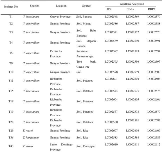
28
Table 2.3. Molecular classification of the Ecuadorian Trichoderma isolates.
Isolates No Species Location Source
ITS EF-1α RBP2
T1 T. harzianum Guayas Province Soil, Banana LC002568 LC002569 LC002570
T2 T. asperellum Guayas Province Soil, Mango LC002586 LC002587 LC002588
T3 T. harzianum Guayas Province Soil, Baby
Bananas
LC002571 LC002572 LC002573
T4 T. asperellum Guayas Province Soil, Organic Banana
LC002589 LC002590 LC002591
T5 T. asperellum Pichincha Province
Substrate, Pleurotus spp.
LC002592 LC002593 LC002594
T9 T. asperellum Guayas Province Tree bark, Cacao tree
LC002595 LC002596 LC002597
T10 T. asperellum Guayas Province Soil LC002598 LC002599 LC002600
T13 T. asperellum Riobamba
Province
Soil, Potatoes LC002601 LC002602 LC002603
T15 T. harzianum Riobamba
Province
Soil, Potatoes LC002574 LC002575 LC002576
T18 T. asperellum Riobamba
Province
Soil, Potatoes LC002604 LC002605 LC002606
T19 T. harzianum Riobamba
Province
Soil, Potatoes LC002577 LC002578 LC002579
T20 T. harzianum Riobamba
Province
Soil, Potatoes LC002580 LC002581 LC002582
T29 T. reesei Guayas Province Soil, Rice LC002607 LC002608 LC002609
T36 T. harzianum Guayas Province Soil, Rice LC002583 LC002584 LC002585
T43 T. virens Santo Domingo
Province
Soil, Pineapple LC002610 LC002611 LC002612 GenBank Accession
29
Previous description of the isolates showed they originate from the coast and highland provinces; the isolates T1, T3, T15, T19, T20, and T36 were confirmed to belong to T. harzianum, sect. Pachybasium, clade Harzianum (Fig. 2.4). Within this group of T. harzianum strains, T1, T3 and T36 belong to Coast Region while T15, T19 and T20 belong to Highland Region (Table 2.3) from traditional cultivars like bananas, potato and rice. T. harzianum is a species aggregate, grouped on the basis of conidiophore branching patterns with short side branches, short inflated phialides, and smooth and small conidia.
30
Fig. 2.4. T. harzianum strains isolated from different regions of Ecuador. T1, T3 and T36- Coast Region and T15, T19 and T20- Highland Region.
T1 T3
T15 T19
T20 T36
31
Isolates T2, T4, T5, T9, T10, T13 and T18 (Fig. 2.5), which were previously identified to be T. viride via morphology, were identified as T. asperellum. Samuels et al.
(1999) indicated that T. asperellum could be distinguished from T. viride due to its finer conidial ornamentation, slightly ovoidal conidia, faster growth rate, mostly paired branches, ampulliform phialides, and consistent presence of chlamydospore. T.
asperellum belongs to sect. Trichoderma, Clade Pachybasium “A” or Hamatum. The isolates T2, T4, T9 and T10 belong to Coast Region and T5, T13 and T18 belong to Highland Region from different cultivars (Table 2.3).
32
Fig. 2.5. T. asperellum strains from different regions of Ecuador. T2, T4, T9 and T10 - Coast Region. T5, T13 and T18 - Highland Region.
T2 T4
T5 T9
T10 T13
T18
33
Two unidentified Trichoderma isolates T43 belonging to Highland Region and T29 to Coast Region (Fig. 2.6), were identified in this study as T. virens (belonging to sect.
Pachybasium, clade Virens) and T. reesei (belonging to sect. Longibrachiatum, clade Longibrachiatum) (Table 2.3).
T29 T43
Fig. 2.6. T. reesei (T29) and T. virens (T43) isolated from different regions of Ecuador.
34 2.3.2 Phylogenetic analysis of Trichoderma species
The phylogenetic analysis based on ITS indicated four distinct groups of the isolates under study (A to D) (Fig. 2.7). The first dominant group (A) was T. asperellum, (monophyletic group) which forms part of sect. Trichoderma, clade Pachybasium “A” or Hamatum, supported with high bootstrap (100%), a subgroup that contains most strains has bootstrap of 64% (Fig. 2.7) including isolates from the Coast and Highland Regions (T2, T4, T9, T10 and T5, T13, T18 respectively). The ex-neotype culture of T. asperellum (LAHD and 2046) has identical ITS sequences to the isolates under study.
T. harzianum complex was the second dominant group (B) with high bootstrap values (94%), and is divided into three groups showing (monophyletic group) (T3, T20 and the ex-type T. harzianum CEN439) a second group (T36 and ex-type T. harzianum Ir.
112) and third group (T1, T15, T19 and the ex-type T. harzianum RSPG 28) (Fig. 2.7).
Two other isolates, T29 (D) and T43 (C), were identified to be T. reesei and T. virens, with high bootstrap of 100 and 99 respectively, using phylogenetic analysis based on the three genes (Fig. 2.7).
35
Fig. 2.7. Phylogenetic relations of Trichoderma taxa based on neighbor-joining analysis of ITS sequence data. The evolutionary history was inferred using the Neighbor-Joining method [1]. The optimal tree with the sum of branch length = 0.11417193 is shown. The percentage of replicate trees in which the associated taxa are clustered together in the bootstrap test (2000 replicates) are shown next to the branches [2]. The tree is drawn to scale, with branch lengths in the same units as those of the evolutionary distances used to infer the phylogenetic tree. The evolutionary distances were computed using the Kimura 2-parameter method [3] and are in the units of the number of base substitutions per site.
The analysis involved 25 nucleotide sequences. Codon positions included were 1st+2nd+3rd+Noncoding. All positions containing gaps and missing data were eliminated. There were a total of 500 positions in the final dataset. Evolutionary analyses were conducted in MEGA 5.1 [4].