Prognostic Signi fi cance of the Sodium Channel Blocker Test in Patients With Brugada Syndrome
Akira Ueoka, MD; Hiroshi Morita, MD, PhD; Atsuyuki Watanabe, MD, PhD; Yoshimasa Morimoto, MD; Satoshi Kawada, MD;
Motomi Tachibana, MD; Masakazu Miyamoto, MD; Koji Nakagawa, MD, PhD; Nobuhiro Nishii, MD, PhD; Hiroshi Ito, MD, PhD
Background-—A drug provocation test using a sodium channel blocker (SCB) can unmask a type 1 ECG pattern in patients with Brugada syndrome. However, the prognostic value of the results of an SCB challenge is limited in patients with non–type 1 ECG.
We investigated the associations of future risk for ventricular fibrillation with SCB-induced ECG changes and ventricular tachyarrhythmias (VTAs).
Methods and Results-—We administered intravenous pilsicainide to 245 consecutive patients with Brugada syndrome (181 patients with spontaneous type 1 ECG, 64 patients with non–type 1 ECG). ECG parameters before and after the test and occurrence of drug-induced VTAs were evaluated. During a mean follow-up period of 11357 months, fatal VTA events occurred in 31 patients (sudden death: n=3, ventricular tachycardia/ventricular fibrillation: n=28). Symptomatic patients and spontaneous type 1 ECG were associated with future fatal arrhythmic events. Univariable analysis of ECG parameters after the test showed that long PQ and QRS intervals, high ST level, and SCB-induced VTAs were associated with later VTA events during follow-up.
Multivariable analysis showed that symptomatic patients, high ST level (V1)≥0.3 mV after the test, and SCB-induced VTAs were independent predictors for future fatal arrhythmic events (hazard ratios: 3.28, 2.80, and 3.62, 95% confidence intervals: 1.54–7.47, 1.32–6.35, and 1.64–7.75, respectively;P<0.05).
Conclusions-—SCB-induced VTAs and ST-segment augmentation are associated with an increased risk of the development of ventricular tachycardia/ventricular fibrillation events during follow-up in patients with Brugada syndrome. (J Am Heart Assoc.
2018;7:e008617. DOI: 10.1161/JAHA.118.008617.)
Key Words: Brugada syndrome•risk stratification•sodium channel blocker•ventricularfibrillation
A
sodium channel blocker (SCB) unmasks and augments type 1 ST elevation of Brugada syndrome (BrS). An SCB challenge is usually used to detect manifestation of type 1 ECG for diagnosis of BrS in patients with non–type 1 ECG.1 The extents of PQ prolongation and QRS widening during the SCB test can be a clue for the existence of SCN5Amutation.2–4Although the SCB test is essential for diagnosis in non–type 1 patients, an unexpected response to adminis- tration of an SCB, such as atrioventricular block or ventricular tachyarrhythmias (VTAs), occasionally emerges in some patients.5,6
The prognostic value of the results of an SCB challenge is limited in patients with BrS. Patients who have drug-induced type 1 ECG, with the exception of patients who have already experienced cardiac arrest, show a relatively benign clinical course compared with that for patients with spontaneous type 1 ECG.7–9Patients in whom ECG was not converted to type 1 ECG by an SCB had a very good prognosis compared with that for patients with drug-induced type 1 ECG.10SCB challenge only has diagnostic value in patients without spontaneous type 1 ECG. Moreover, the prognostic significance of an SCB challenge in patients with spontaneous type 1 ECG has not been evaluated. In fact, most investigators believe that an SCB test is contraindicated in patients with spontaneous type 1 ECG.
Recently, an SCB challenge has been used for detecting abnormal potentials on the epicardial surface of the right ventricle during epicardial catheter ablation. Symptomatic patients who have experienced arrhythmic syncope or
From the Departments of Cardiovascular Medicine (A.U., A.W., Y.M., S.K., M.T., M.M., K.N., H.I.), Cardiovascular Therapeutics (H.M., N.N.), Okayama University Graduate School of Medicine, Dentistry and Pharmaceutical Sciences, Okayama, Japan.
Accompanying Table S1 and Figure S1 are available at http://jaha.ahajournals.
org/content/7/10/e008617/DC1/embed/inline-supplementary-material- 1.pdf
Correspondence to:Hiroshi Morita, MD, PhD, Department of Cardiovascular Therapeutics, Okayama University Graduate School of Medicine, Dentistry and Pharmaceutical Sciences, 2-5-1 Shikata-Cho, Okayama 700-8558, Japan.
E-mail: [email protected]
Received January 11, 2018; accepted March 26, 2018.
ª2018 The Authors. Published on behalf of the American Heart Association, Inc., by Wiley. This is an open access article under the terms of the Creative Commons Attribution-NonCommercial License, which permits use, distribu- tion and reproduction in any medium, provided the original work is properly cited and is not used for commercial purposes.
Downloaded from http://ahajournals.org by on January 27, 2019
documented VTA events have more advanced arrhythmogenic substrate on the epicardium.11If an SCB challenge can unmask concealed substrate in patients, ECG change and VTAs induced by the SCB will indicate progression of the substrate.
We hypothesized that remarkable ECG changes and VTAs provoked by an SCB challenge are associated with advanced arrhythmogenic substrate and that such changes are corre- lates of future VTA events in patients with BrS.
Methods
The authors declare that all supporting data are available within the article and its online supplementaryfiles.
The subjects of the present study were 245 consecutive patients with BrS who underwent a drug provocation test with pilsicainide in Okayama University Hospital (males: 240 patients, mean age: 4613 years). At the time of diagnosis, 154 patients were asymptomatic, 79 had syncope, and 12 had ventricularfibrillation (VF) (Table 1). We excluded obvious reflex syncope as a symptom and considered patients with reflex syncope as being asymptomatic. BrS was diagnosed when a type 1 ST-segment elevation appeared either spon- taneously or after administration of an SCB. We defined spontaneous type 1 ECG as the appearance of type 1 ECG without any stress such as stress from fever or exercise. Type 1 ECG was defined as coved ST-segment elevation≥2 mm in at least 1 right precordial lead in the second, third, or fourth intercostal space.12 All 245 patients had spontaneous (n=181) or drug-induced type 1 ECG (n=64). To clarify the prognosis of patients with negative SCB and non–type 1, we also analyzed an additional 30 patients (29 males, age:
4615 years) without positive SCB test who were suspected of having BrS (asymptomatic: 20 patients, syncope: 10 patients). There were no patients from the same family.
Table 1. Characteristics of Patients With Spontaneous and Drug-Induced Type 1 ECG
Overall, n=245 PValue*
Clinical parameters
Male 240 (98%)
Age, y 46.213.0
Symptomatic patients 91 (37%)
Syncope 79 (32%)
VT/VF 12 (5%)
Family history of SD 72 (29%)
SCN5Amutation 16/139 (12%)
VT/VF during follow-up 31 (13%)
ECG parameters
Spontaneous type 1 ECG 181 (74%)
PQ interval lead II (ms)
Pre SCB 18027 <0.001
Post SCB 22937
QRS width (ms) V1
Pre SCB 10614 <0.001
Post SCB 13323
V2
Pre SCB 10714 <0.001
Post SCB 13523
ST level (mV) V1
Pre SCB 0.1580.106 <0.001
Post SCB 0.2700.172
V2
Pre SCB 0.2940.160 <0.001
Post SCB 0.5910.277
QTc interval (ms) V5
Pre SCB 38827 <0.001
Post SCB 42735
Drug-induced VTAs (n)
Overall 24 (10%)
PVCs 13 (5%)
VTs 11 (4%)
PVCs indicates premature ventricular contractions; SCB, sodium channel blocker; SD, sudden death; VTA, ventricular tachyarrhythmia; VT/VF, ventricular tachycardia/
ventricularfibrillation.
*Pvalue: comparison of ECG parameters before and after the SCB test.
Clinical Perspective
What Is New?
• Remarkable ST elevation and ventricular tachyarrhythmia induced by a challenge test with a sodium channel blocker were associated with arrhythmic events during follow-up in patients with Brugada syndrome.
• The prognostic value of a sodium channel blocker is applicable to patients with spontaneous type 1 ECG.
• The present study indicated the usefulness of a sodium channel blocker test in patients with spontaneous type 1 ECG for thefirst time.
What Are the Clinical Implications?
• A sodium channel blocker test can be used for (1) diagnosis of patients with non–type 1 ECG; (2) risk stratification in asymptomatic patients with spontaneous type 1 ECG; and (3) risk stratification in patients with spontaneous type 1 ECG and syncope of unknown cause.
NALRESEARCH
Downloaded from http://ahajournals.org by on January 27, 2019
Echocardiography was performed in all patients, and no structural abnormalities were found.
All of the study protocols were approved by the Ethics Committee on Human Research and Epidemiology of Okayama University and Human Genome Studies of the Ethics Commit- tee of Okayama University. Informed consent regarding data acquisition was obtained from all patients. Clinical data, including data on age, sex, family history of sudden cardiac death, history of syncope episodes, history of VF episodes, and the presence of SCN5A gene mutation were obtained from patient records. Analysis ofSCN5Amutation was performed in 139 patients in compliance with the above guidelines.
The primary end point of this study was the occurrence of fatal VTA events defined as the occurrence of sudden cardiac death, VT or VF, and appropriate implantable cardioverter- defibrillator interventions during the follow-up period.
Pharmacologic Challenge Test
We performed an SCB test in an ECG laboratory or during electrophysiological study with a standby defibrillator and an emergency cart with medicines and intubation kit during hospitalization. Pilsicainide chloride was administered intra- venously at a dose of 1 mg/kg over a period of 5 to 10 minutes in all patients. The difference between ECG parameters before and 15 minutes after administration of pilsicainide was calcu- lated: PQ interval in lead II, QRS interval and ST level in leads V1 and V2, and QTc interval in lead V5. ST level was measured at the J points in leads V1, V2, and V5. Occurrence of severe VTAs after administration of pilsicainide was also evaluated. Severe VTAs during the test included frequent occurrence of premature ventricular contractions (PVCs) (>1 bpm) and polymorphic VT (at least 3 continuous beats). We stopped administration of pilsicainide if patients had significant QRS widening (≥130%), second- or third-degree atrioventricular block, or occurrence of PVCs. If patients had severe ventricular arrhythmias, we observed the patients overnight in a cardiac care unit with or without isoproterenol infusion.
Statistical Analysis
Statistical analysis was performed using JMP 11.0 for MAC (SAS Institute Inc, Cary, NC). Data are expressed as meansSD or medians (interquartile range). Continuous variables in the different subgroups were analyzed by the Wilcoxon signed-rank test. We used the pairedt test or the Wilcoxon signed-rank test to compare the values before and after the SCB test in the same patients. Categorical data and percentage frequencies were analyzed by thev2test or Fisher test. Logistic regression analysis was conducted in order to identify predictive ECG parameters before and after the pilsicainide-challenge test. Receiver-operating characteristic
curves were constructed for ECG parameters to determine the optimal cutoff value for identifying patients with VF during follow-up. Event rate curves were plotted according to the Kaplan–Meier method and were analyzed by the log-rank test.
Univariate and multivariate Cox regression analyses were performed to assess whether each index can be a significant and independent predictor of fatal arrhythmic events. We used the following covariates for multivariable analysis: important baseline characteristics (symptoms and spontaneous type 1 ECG) and ECG parameters after the pilsicainide test (PQ and QRS intervals, ST level, and pilsicainide-induced VTAs). A value ofP<0.05 was considered statistically significant.
Results
Characteristics of Patients and Results of the Pilsicainide Test
Baseline characteristics of the patients according to clinical presentation are summarized in Table 1. Spontaneous type 1 ECG was observed in 74% of the patients. Thirty-two percent of the patients had a history of syncope episodes and 5% of the patients had previous VF episodes. Gene analysis was performed in 139 patients, and SCN5A gene mutation was found in 12% of the patients.
The reasons for performing the pilsicainide test were diagnosis of BrS in patients without spontaneous type 1 ECG (n=62), confirmation of type 1 ECG in patients who transiently had type 1 ECG with or without specific conditions (such as fever, after exercise, or after taking medicine, n=2), detection of an abnormal endocardial or epicardial electrogram or induction of PVCs during electrophysiological study and/or catheter ablation (n=131), and possibility of risk stratification for detecting abnormal ECGs such as T-wave alternans (n=50).13,14The reasons for performing the SCB test in patients with nonspontaneous type 1 ECG were existence of ECG abnormality (35 asymptomatic patients) and existence of syncope (26 patients) or VF (3 patients).
Administration of pilsicainide unmasked type 1 ECG in all of the 64 patients who did not have spontaneous type 1 ECG. ST level was significantly augmented after the pilsicainide test (Table 1). Pilsicainide prolonged PQ, QRS, and QTc intervals.
Pilsicainide induced VTAs in 24 patients (Figure 1), including frequent PVCs in 13 patients (Figure 2A) and VT/VF in 11 patients (Figures 2B and 3, Tables 1 and 2). Cardioversion was required to terminate VT/VF in 4 patients (external defibrillator:
n=3, implantable cardioverter-defibrillator: n=1). None of the patients had prolonged VT/VF episodes. One patient developed transient complete atrioventricular block. Patients with pilsi- cainide-induced VTA more frequently had spontaneous type 1 ECG, higher ST level after the test (V1), and longer QT interval than did patients without pilsicainide-induced VTA (Table 3).
NALRESEARCH
Downloaded from http://ahajournals.org by on January 27, 2019
During a mean follow-up period of 11357 months, fatal VTA events occurred in 31 patients. Three patients died suddenly, 26 experienced VF (implantable cardioverter- defibrillator shock in 24 patients, aborted cardiac arrest in 2 patients), and 2 developed monomorphic VTs. The time to a fatal VTA event was shorter in patients with spontaneous type 1 ECG than in patients without spontaneous type 1 ECG (Figure 4A), and the same result was observed in patients’
subgroups according to symptoms (Figure 4B). There was no VTA event in patients without a positive SCB test. Patients with pilsicainide-induced VTA had more frequent fatal VTA events during follow-up (12/24, event ratio: 7.1%/y) than did patients without pilsicainide-induced VTA (19/221, event ratio: 0.89%,P<0.0001) (Figure 4C). There was no difference in fatal VTA events during follow-up between patients with pilsicainide-induced VT/VF (5/11, event ratio: 6.3%) and patients with pilsicainide-induced PVCs only (7/13, event ratio: 7.9%,P=0.6820).
Changes in ECG Parameters and Occurrence of Ventricular Arrhythmias Induced by Pilsicainide in Different Subgroups of Patients
Pilsicainide significantly prolonged QRS interval and signifi- cantly augmented ST elevation in patients with spontaneous type 1 ECG compared with those in patients without
spontaneous type 1 ECG (Table 4). At the time of the SCB test, 75 patients (41%) in whom spontaneous type 1 ECG was recorded previously did not show spontaneous type 1 ECG at the beginning of the test. Pilsicainide provoked type 1 ECG in those patients (Figure 1). PQ and QTc intervals before and after the pilsicainide test were not different between patients with and without spontaneous type 1 ECG. Pilsicainide more frequently induced VTAs in patients with spontaneous type 1 ECG than in patients without spontaneous type 1 ECG (Table 4).
Symptomatic patients had a longer PQ interval but had a lower ST level in V2 than did asymptomatic patients at baseline (Table 5). There were no differences in other ECG parameters between asymptomatic and symptomatic patients at baseline. Pilsicainide significantly prolonged PQ and QRS intervals in leads V1 and V2 in symptomatic patients compared with those in asymptomatic patients. ST level in lead V2 in asymptomatic patients was higher than that in symptomatic patients after pilsicainide administration. Pilsi- cainide-induced VT/VF was more frequent in symptomatic patients than in asymptomatic patients (Table 5).
Patients with SCN5A mutation (n=16) had longer PQ interval before the pilsicainide test than did patients without SCN5A mutation (n=123) (Table S1). After administration of pilsicainide, patients with SCN5A mutation had significantly longer PQ and QRS intervals than did patients withoutSCN5A Figure 1. Results of pilsicainide tests and occurrence of cardiac events. The groups of patients consisted of 245 patients with spontaneous or drug-induced type 1 ECG and 30 patients with non–type 1 ECG that was not converted to type 1 ECG by a sodium channel blocker (SCB). The results were divided according to the symptom, ECG type, ECG type at the pilsicainide test, result of the pilsicainide test, and occurrence of pilsicainide-induced ventricular arrhythmias (VTAs).
NALRESEARCH
Downloaded from http://ahajournals.org by on January 27, 2019
mutation. There were no differences in prevalence of drug- induced VTAs and other ECG parameters before and after the pilsicainide test between patients with and without SCN5A mutation.
Risk Factors for VT/VF Events During Follow-Up
Table 6 shows results of univariable analysis of clinical and ECG parameters before and after the pilsicainide test to detect VTA events during follow-up. Univariable analysis of clinical parameters showed that symptomatic patients, espe- cially those with previous episodes of VT/VF, were associated with fatal arrhythmic events during follow-up. In ECG param- eters before the pilsicainide test, QRS intervals in leads V1 and V2 were associated with cardiac events. Univariable analysis of ECG parameters after the pilsicainide test to
detect VTA events showed that PQ interval, QRS intervals (V1 and V2), and ST level (V1) were associated with fatal arrhythmic events during follow-up. Among the differences between ECG parameters before and after the pilsicainide test, differences in PQ interval (DPQ) and ST level (DST) in V1 were predictors of VT/VF events. Drug-induced VTAs were also associated with fatal arrhythmic events during follow-up, but there was no difference in prediction of fatal events between drug-induced PVCs and drug-induced VT/VF.
Cutoff points of ECG parameters after the pilsicainide test to detect fatal arrhythmic events during follow-up were determined by receiver-operating characteristic analysis: PQ interval≥235 ms (area under the curve: 0.663), QRS interval in lead V1 ≥132 ms (area under the curve: 0.693), and ST level in lead V1≥0.3 mV (area under the curve 0.671) were optimal cutoff points (Figure S1). Univariable analysis of these parameters showed that they were associated with fatal VTA events: the hazard ratio (HR) of PQ interval≥235 ms was 3.16 (95% confidence interval [CI], 1.54–6.85, P=0.0021), HR of QRS interval ≥132 ms was 4.22 (95% CI, 1.97–10.06, P=0.0005), and HR of ST level ≥0.3 mV was 4.03 (95% CI, 1.95–8.94, P=0.0003). When we focused on the asymp- tomatic patients, ST level after pilsicainide≥0.3 mV (HR: 1.7, CI, 3.1–325.5, P=0.0002) and drug-induced VTAs (HR: 15.6, CI, 4.3–56.1,P=0.0001) were also predictors of VT/VF events during follow-up.
Figure 3. Pilsicainide-induced polymorphic ventricular tachycar- dia and ventricularfibrillation. These ECGs were recorded in a 50- year-old patient with ventricular fibrillation (VF). A, ECG at baseline. The patient had spontaneous type 1 ECG, but ST elevation was diminished before the test. We performed a pilsicainide test to unmask the abnormal electrical substrate during electrophysiological study. Pilsicainide provoked type 1 ECG (B) and VF (C). A direct current shock was required to terminate VF.
Figure 2. Pilsicainide-induced ventricular arrhythmia. A, These ECGs were recorded in a patient with syncope (50 years old). The left panel shows ECG at baseline. Leads V1-2 were located at the third intercostal space. The patient had spontaneous type 1 ECG only in the leads at high intercostal spaces. The right panel shows that pilsicainide provoked frequent occurrence of premature ventricular contractions and significant ST elevation. B, These ECGs were recorded in an asymptomatic patient (27 years old).
The patient had fever-induced type 1 ECG but did not have spontaneous type 1 ECG. The left panel shows non–type 1 ECG before the pilsicainide test. Leads V1-2 were recorded at regular lead positions. The right panel shows that pilsicainide induced nonsustained polymorphic ventricular tachycardia. The patient died suddenly at night 6 years after the test.
NALRESEARCH
Downloaded from http://ahajournals.org by on January 27, 2019
Multivariable analysis of baseline characteristics (symp- toms and spontaneous type 1 ECG) and ECG parameters after the pilsicainide test (PQ and QRS intervals, ST level, and pilsicainide-induced VTAs) showed that symptoms, ST level after the pilsicainide test, and drug-induced VTAs were independent risk factors for fatal arrhythmic events during follow-up (Table 7). Patients with high ST level in lead V1 after the pilsicainide test or drug-induced VTAs had a shorter time to fatal events than did patients without these parameters (Figure 4C and 4D).
Discussion New Findings
The present study showed that high ST level in lead V1 after the pilsicainide test and drug-induced VTAs were associated
with VT/VF events. These risk factors detected by the SCB test were independent predictors of cardiac events even after adjustment by the presence of symptoms and spontaneous type 1 ECG. There has been no report of ECG changes after an SCB test other than the appearance of drug-induced type 1 ECG having prognostic value. The results of the present study showed that an SCB test is useful as a risk stratification tool in patients with spontaneous type 1 ECG in addition to being a diagnostic tool in patients without spontaneous type 1 ECG.
Occurrence and Prognostic Value of SCB-Induced Ventricular Arrhythmias
Previous studies showed that an SCB test induced VTAs in 0%
to 25% of patients and VF in up to 4% of patients with BrS.13,15–34 The incidence of SCB-induced VTAs increased if the subjects of the study included subjects with spontaneous Table 2. Patients With Pilsicainide-Induced Ventricular Arrhythmia
Patients Age (y) Sex
Spontaneous Type 1
Clinical Presentation
FH of SD
VF Induction by PES
SCN5A
Mutation Type of SCB-VTA
ICD Implantation
During Follow-Up VTA During Follow-Up
1 50 Male Yes Asymptomatic Yes Yes No Frequent PVC No Sustained VT
2 27 Male Yes Asymptomatic No Yes No Frequent PVC No None
3 55 Male Yes Syncope No Yes No VF Yes Appropriate ICD shock
4 41 Male Yes VF No Yes No NSVT Yes Appropriate ICD shock
5 55 Male Yes VF Yes Yes No Frequent PVC Yes Appropriate ICD shock
6 42 Male No VF No Yes No Frequent PVC Yes None
7 41 Male Yes Syncope No No No NSVT* Yes None
8 57 Male Yes Asymptomatic No No No Frequent PVC No None
9 47 Male Yes Syncope Yes No Yes Frequent PVC Yes Appropriate ICD shock
10 29 Male Yes Syncope Yes No Yes Sustained VT* No None
11 40 Male Yes Asymptomatic Yes Yes No NSVT Yes None
12 50 Male Yes Syncope No Yes No VF Yes Appropriate ICD shock
13 42 Male Yes Asymptomatic No Yes No Frequent PVC Yes Appropriate ICD shock
14 38 Male Yes Asymptomatic Yes Yes No Frequent PVC No None
15 34 Male Yes Syncope Yes Yes No Frequent PVC Yes Appropriate ICD shock
16 38 Male Yes Syncope No No Yes NSVT No None
17 50 Male Yes Asymptomatic No Yes NA NSVT* Yes None
18 27 Male Yes Asymptomatic Yes Yes No NSVT No SD
19 50 Male Yes Syncope No Yes NA Sustained VT No VF
20 36 Male Yes Syncope Yes Yes No NSVT Yes None
21 74 Male Yes Asymptomatic Yes Yes NA Frequent PVC Yes Sustained VT
22 36 Male Yes Asymptomatic No Yes NA Frequent PVC Yes Appropriate ICD shock
23 25 Male Yes Syncope No No Yes VT*/VF Yes Appropriate ICD shock
24 41 Male Yes Asymptomatic No Yes NA Frequent PVC No None
FH indicates family history; ICD, implantable cardioverter defibrillator; NA, not assessed; NSVT, nonsustained ventricular tachycardia; PES, programmed electrical stimulation; PVC, premature ventricular contraction; SCB, sodium channel blocker; SD, sudden death; VF, ventricularfibrillation; VT, ventricular tachycardia; VTA, ventricular tachyarrhythmia.
*VT with wide QRS complex and not significant polymorphic change.
NALRESEARCH
Downloaded from http://ahajournals.org by on January 27, 2019
type 1 ECG. In the present study, 10% of the patients developed VT/VF or frequent PVCs after administration of pilsicainide, and the incidence of SCB-induced VTAs coincided with that in previous studies that included subjects with spontaneous type 1 ECG.
We showed that pilsicainide-induced VTA was a powerful predictor of VT/VF after adjustment of symptoms and ECG type. Some studies have shown that SCB-induced VTAs failed to predict VT/VF events during follow-up.19,27,34 However, those studies included a small number of patients or only patients without spontaneous type 1 ECG. The present study included patients with spontaneous type 1 ECG (74%), and the occurrence of SCB-induced VTAs in patients without sponta- neous type 1 ECG was less frequent than that in patients with spontaneous type 1 ECG. Then the prognostic value of SCB- induced VTAs should be significant in patients with sponta- neous type 1 ECG. Recently, an SCB test has been used to unmask concealed substrate at the time of epicardial ablation.11Application of radiofrequency energy to all abnor- mal substrate is necessary to eliminate the arrhythmogenic area. ECG changes similar to those induced by an SCB test can appear during febrile illness,35and VTA events also occur at that time. Thus, VTAs induced by an SCB test are not proarrhythmic effects but represent concealed arrhythmo- genic substrate that can appear in daily life.
Some asymptomatic patients without spontaneous type 1 ECG might have false-positive results of the SCB test.36,37 This study included 35 asymptomatic patients with nonspon- taneous type 1 ECG that was converted to type 1 ECG by the pilsicainide test (Figure 1). This group of patients was diagnosed as “possible” BrS by a new scoring system,38 and some of those patients might be false positive. However, those patients did not have pilsicainide-induced VTAs and did not have cardiac events during follow-up. Thus, SCB-induced VTAs would indicate that the patients have“definite” BrS38 with a more progressive arrhythmogenic substrate.
Risk and Safety of an SCB Test in Patients With Type 1 ECG
An SCB test is useful and safe for most patients, but some studies have shown that some patients develop severe VTAs requiring external defibrillation, implantable cardioverter- defibrillator therapy, or an extracorporeal membrane oxygenator.6,13,15,19,23,28,31,34 An SCB test should be performed during hospitalization, and continuous infusion of low-dose isoproterenol after the test should be performed overnight for high-risk patients. To avoid catastrophic events, it was stated in the Consensus report that an SCB test should be discontinued in cases of frequent PVCs and QRS Table 3. Different Characteristics of Patients With and Those
Without SCB-Induced VTAs
Pilsicainide-Induced VTA+(n=24)
Pilsicainide-Induced
VTA (n=221) PValue*
Clinical parameters
Male 24 (100%) 216 (98%) 1.0000
Age, y 42.711.3 46.613.2 0.1478
Symptomatic patients
13 (54%) 78 (35%) 0.0782
Syncope 9 (38%) 70 (32%) 0.6464
VT/VF 4 (17%) 8 (4%) 0.0207
Family history of SD
10 (42%) 62 (28%) 0.1659
SCN5A mutation
4/20 (20%) 12/119 (10%) 0.2386
VT/VF during follow-up
12 (50%) 19 (9%) <0.0001
ECG parameters Spontaneous
type 1 ECG
23 (96%) 158 (71%) 0.0068
PQ interval in lead II (ms)
Pre SCB 18526 18027 0.3227
Post SCB 24243 22736 0.0578
QRS width (ms) V1
Pre SCB 11322 10513 0.0969
Post SCB 14536 13121 0.0576
V2
Pre SCB 11421 10613 0.0429
Post SCB 14633 13422 0.0748
ST level (mV) V1
Pre SCB 0.2000.132 0.1530.102 0.0307 Post SCB 0.3850.219 0.2570.162 0.0037 V2
Pre SCB 0.3070.194 0.2930.157 0.9613 Post SCB 0.5990.295 0.910.276 0.8236 QTc interval (ms)
V5
Pre SCB 38729 38827 0.8371
Post SCB 45043 42433 0.0046
All patients had type 1 ECG spontaneously or by SCB. SCB indicates sodium channel blocker; SD, sudden death; VTA, ventricular tachyarrhythmia; VT/VF, ventricular tachycardia/ventricularfibrillation.
*Pvalue: comparison of ECG parameters in patients with and without pilsicainide- induced VTAs.
NALRESEARCH
Downloaded from http://ahajournals.org by on January 27, 2019
widening.38The present study showed that high ST level and prolonged QT interval after the test occurred in patients with pilsicainide-induced VTAs (Table 3), and these ECG changes can be warning signs of drug-induced VTAs. Since most of the pilsicainide-induced VTAs occurred in patients with sponta- neous type 1 ECG, an SCB test should be performed in such patients with meticulous caution regarding ECG changes during the test.
Based on the results of this study, we consider that the criteria for performing an SCB test are (1) diagnosis of
patients with non–type 1 ECG; (2) risk stratification in asymptomatic patients with spontaneous type 1 ECG; and (3) risk stratification in patients with spontaneous type 1 ECG and syncope of unknown cause.
Limitations
Female sex is a possible risk for arrhythmic events during an SCB test.6We could not determine sex risk of the SCB test in this study because we performed the test in only 5 female Figure 4. Kaplan–Meier analysis of fatal arrhythmic events. A, Event-free survival by ECG types including non–type 1, drug-induced type 1, and spontaneous type 1. Patients with spontaneous type 1 ECG had a worse prognosis than did patients without spontaneous type 1 ECG. No arrhythmic event occurred in patients without type 1 ECG. B, Event-free survival by symptoms and ECG types in patients with spontaneous or drug-induced type 1 ECG. Symptomatic patients more frequently experienced arrhythmic events than did asymptomatic patients. C, Event-free survival by pilsicainide-induced ventricular tachyarrhythmias (VTAs) in patients with spontaneous or drug-induced type 1 ECG. Pilsicainide- induced VTAs were associated with increased risk of fatal arrhythmic events. D, Event-free survival by degree of ST elevation in patients with spontaneous or drug-induced type 1 ECG. Patients with marked ST elevation (≥0.3 mV) in lead V1 after administration of pilsicainide had a significantly higher risk of fatal arrhythmic events than did patients with less ST elevation. Tables under the survival curve show the number of patients at risk. SCB indicates sodium channel blocker.
NALRESEARCH
Downloaded from http://ahajournals.org by on January 27, 2019
Table 5. Characteristics of Patients With Symptoms at Initial Visit to the Hospital
Symptoms
Asymptomatic (n=154)
Symptomatic
(n=91) PValue*
Clinical parameters
Male 151 (98%) 89 (98%) 1
Age, y 46.313.4 46.012.4 0.9576
Symptomatic patients 0 91
Syncope 0 79
VT/VF 0 12
Family history of SD 51 (33%) 21 (23%) 0.1110 SCN5Amutation 6/80 (8%) 10/59 (17%) 0.1121 VT/VF during follow-up 10 (6%) 21 (23%) 0.0003 ECG parameters
Spontaneous type 1 ECG
119 (77%) 62 (68%) 0.133
PQ interval in lead II (ms)
Pre SCB 17724 18729 0.0084
Post SCB 22334 23840 0.0027
QRS width (ms) V1
Pre SCB 10512 10718 0.8629
Post SCB 13020 13727 0.02
V2
Pre SCB 10611 10818 0.7396
Post SCB 13121 14026 0.0026
ST level (mV) V1
Pre SCB 0.1620.109 0.1510.102 0.4899 Post SCB 0.2730.174 0.2640.171 0.8308 V2
Pre SCB 0.3190.164 0.2510.145 0.0012 Post SCB 0.6340.282 0.5200.256 0.0025 QTc interval (ms)
V5
Pre SCB 38628 39226 0.1491
Post SCB 42435 43235 0.0837
Drug-induced VTA (n)
Overall 11 (7%) 13 (14%) 0.0782
PVCs 8 (5%) 5 (5%) 1
VTs 3 (2%) 8 (9%) 0.0213
All patients had type 1 ECG spontaneously or by SCB. PVCs indicates premature ventricular contractions; SCB, sodium channel blocker; SD, sudden death; VTA, ventricular tachyarrhythmia; VT/VF, ventricular tachycardia/ventricularfibrillation.
*Pvalue: comparison of ECG parameters in asymptomatic patients and symptomatic patients.
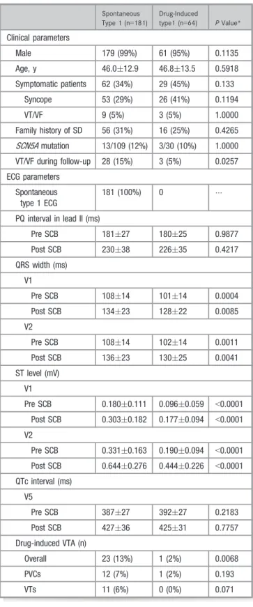
Table 4. Differences Between Patients With Spontaneous and Drug-Induced Type 1 ECG
Spontaneous Type 1 (n=181)
Drug-Induced
type1 (n=64) PValue*
Clinical parameters
Male 179 (99%) 61 (95%) 0.1135
Age, y 46.012.9 46.813.5 0.5918
Symptomatic patients 62 (34%) 29 (45%) 0.133
Syncope 53 (29%) 26 (41%) 0.1194
VT/VF 9 (5%) 3 (5%) 1.0000
Family history of SD 56 (31%) 16 (25%) 0.4265 SCN5Amutation 13/109 (12%) 3/30 (10%) 1.0000 VT/VF during follow-up 28 (15%) 3 (5%) 0.0257 ECG parameters
Spontaneous type 1 ECG
181 (100%) 0
PQ interval in lead II (ms)
Pre SCB 18127 18025 0.9877
Post SCB 23038 22635 0.4217
QRS width (ms) V1
Pre SCB 10814 10114 0.0004
Post SCB 13423 12822 0.0085
V2
Pre SCB 10814 10214 0.0011
Post SCB 13623 13025 0.0041
ST level (mV) V1
Pre SCB 0.1800.111 0.0960.059 <0.0001 Post SCB 0.3030.182 0.1770.094 <0.0001 V2
Pre SCB 0.3310.163 0.1900.094 <0.0001 Post SCB 0.6440.276 0.4440.226 <0.0001 QTc interval (ms)
V5
Pre SCB 38727 39227 0.2183
Post SCB 42736 42531 0.7757
Drug-induced VTA (n)
Overall 23 (13%) 1 (2%) 0.0068
PVCs 12 (7%) 1 (2%) 0.193
VTs 11 (6%) 0 (0%) 0.071
PVCs indicates premature ventricular contractions; SCB, sodium channel blocker; SD, sudden death; VTA, ventricular tachyarrhythmia; VT/VF, ventricular tachycardia/
ventricularfibrillation.
*Pvalue: comparison of ECG parameters in patients with and those without spontaneous type 1 ECG.
NALRESEARCH
Downloaded from http://ahajournals.org by on January 27, 2019
patients (2%). PVC/VT did not occur in all of the female patients. We did not avoid performing an SCB test for female patients. A high prevalence of males (>90% of the patients) with BrS was frequently observed in previous
Japanese studies,27,39 and it might be a racial characteristic of BrS.
Conclusion
VTAs and augmentation of ST-segment elevation after an SCB challenge test were associated with an increased risk of the development of VT/VF events in patients with BrS, especially in patients with spontaneous type 1 ECG. An SCB challenge test can serve as not only a diagnostic tool in patients without spontaneous type 1 ECG but also a risk stratification tool for patients with spontaneous type 1 ECG.
Sources of Funding
This study was supported by JSPS KAKENHI (15K09082 to Morita) and Tailor-made Medical Treatment Program with the BioBank Japan Project (BBJ) from Japan Agency for Medical Research and Development (AMED) (15km0305015h0101 to Morita).
Table 6. HR for Predicting VTA Events
HR 95% CI PValue
Clinical parameters
Male 0.62 0.13 to 10.98 0.6588
Age, y 0.99 0.96 to 1.02 0.5741
Symptomatic patients 4.35 2.10 to 9.67 <0.0001
Syncope 1.49 0.70 to 3.05 0.2851
VT/VF 13.81 5.97 to 29.39 <0.0001
Family history of SD 1.12 0.51 to 2.32 0.7657 SCN5Amutation 1.90 0.64 to 4.62 0.2253 ECG parameters
Spontaneous type 1 ECG 3.72 1.09 to 12.69 0.0279 PQ interval in lead II
Pre SCB 1.01 0.99 to 1.02 0.3054
Post SCB 1.01 1.00 to 1.02 0.0006
DPQ 1.02 1.01 to 1.03 0.0066
QRS width V1
Pre SCB 1.03 1.01 to 1.04 0.0109
Post SCB 1.01 1.00 to 1.02 0.0155
DQRS 1.01 0.99 to 1.02 0.2221
V2
Pre SCB 1.03 1.01 to 1.04 0.0059
Post SCB 1.01 1.00 to 1.02 0.0157
DQRS 1.01 0.99 to 1.02 0.2798
ST level V1
Pre SCB 7.69 0.33 to 118.40 0.1914
Post SCB 11.43 2.03 to 54.72 0.0069
DST 12.14 1.55 to 66.11 0.0087
V2
Pre SCB 0.78 0.07 to 6.84 0.8348
Post SCB 0.64 0.16 to to 2.34 0.512
DST 0.56 0.10 to 2.85 0.499
QTc interval V5
Pre SCB 1 0.99 to 1.01 0.8812
Post SCB 1.01 1.00 to 1.01 0.3075
DQT 1.01 0.99 to 1.02 0.1736
Continued
Table 6. Continued
HR 95% CI PValue
Drug-induced VTA
Overall 6.95 3.28 to 14.19 <0.0001
PVCs 6.36 2.53 to 14.05 0.0003
VTs 4.66 1.57 to 11.17 0.0082
HR of the ECG parameters represents risk increase/1 unit. CI indicates confidence interval; HR, hazard ratio for predicting VT/VF; PVCs, premature ventricular contractions;
SCB, sodium channel blocker; SD, sudden death; VTA, ventricular tachyarrhythmia; VT/
VF, ventricular tachycardia/ventricularfibrillation.
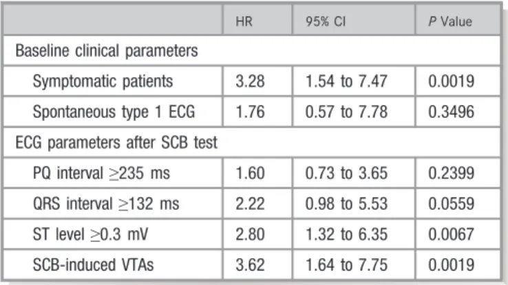
Table 7. Multivariable Analysis of Clinical and ECG Parameters for Predicting VTA Events
HR 95% CI PValue
Baseline clinical parameters
Symptomatic patients 3.28 1.54 to 7.47 0.0019 Spontaneous type 1 ECG 1.76 0.57 to 7.78 0.3496 ECG parameters after SCB test
PQ interval≥235 ms 1.60 0.73 to 3.65 0.2399 QRS interval≥132 ms 2.22 0.98 to 5.53 0.0559 ST level≥0.3 mV 2.80 1.32 to 6.35 0.0067 SCB-induced VTAs 3.62 1.64 to 7.75 0.0019
CI indicates confidence interval; HR, hazard ratio; SCB, sodium channel blocker; VTA, ventricular tachyarrhythmia.
NALRESEARCH
Downloaded from http://ahajournals.org by on January 27, 2019
Disclosures
Morita and Nishii are affiliated with the endowed department by Japan Medtronic Inc. The remaining authors have no disclosures to report.
References
1. Antzelevitch C, Brugada P, Borggrefe M, Brugada J, Brugada R, Corrado D, Gussak I, LeMarec H, Nademanee K, Perez Riera AR, Shimizu W, Schulze-Bahr E, Tan H, Wilde A. Brugada syndrome: report of the second consensus conference: endorsed by the Heart Rhythm Society and the European Heart Rhythm Association.Circulation. 2005;111:659–670.
2. Smits JP, Eckardt L, Probst V, Bezzina CR, Schott JJ, Remme CA, Haverkamp W, Breithardt G, Escande D, Schulze-Bahr E, LeMarec H, Wilde AA. Genotype- phenotype relationship in Brugada syndrome: electrocardiographic features differentiate SCN5A-related patients from non-SCN5A-related patients.J Am Coll Cardiol. 2002;40:350–356.
3. Meregalli PG, Tan HL, Probst V, Koopmann TT, Tanck MW, Bhuiyan ZA, Sacher F, Kyndt F, Schott JJ, Albuisson J, Mabo P, Bezzina CR, Le Marec H, Wilde AA.
Type of SCN5A mutation determines clinical severity and degree of conduction slowing in loss-of-function sodium channelopathies. Heart Rhythm.
2009;6:341–348.
4. Hong K, Brugada J, Oliva A, Berruezo-Sanchez A, Potenza D, Pollevick GD, Guerchicoff A, Matsuo K, Burashnikov E, Dumaine R, Towbin JA, Nesterenko V, Brugada P, Antzelevitch C, Brugada R. Value of electro- cardiographic parameters and ajmaline test in the diagnosis of Brugada syndrome caused by SCN5A mutations. Circulation. 2004;110:3023– 3027.
5. Dobbels B, De Cleen D, Ector J. Ventricular arrhythmia during ajmaline challenge for the Brugada syndrome.Europace. 2016;18:1501–1506.
6. Poli S, Toniolo M, Maiani M, Zanuttini D, Rebellato L, Vendramin I, Dametto E, Bernardi G, Bassi F, Napolitano C, Livi U, Proclemer A. Management of untreatable ventricular arrhythmias during pharmacologic challenges with sodium channel blockers for suspected Brugada syndrome. Europace.
2018;20:234–242.
7. Probst V, Veltmann C, Eckardt L, Meregalli PG, Gaita F, Tan HL, Babuty D, Sacher F, Giustetto C, Schulze-Bahr E, Borggrefe M, Haissaguerre M, Mabo P, Le Marec H, Wolpert C, Wilde AA. Long-term prognosis of patients diagnosed with Brugada syndrome: results from the FINGER Brugada Syndrome Registry.
Circulation. 2010;121:635–643.
8. Priori SG, Gasparini M, Napolitano C, Della Bella P, Ottonelli AG, Sassone B, Giordano U, Pappone C, Mascioli G, Rossetti G, De Nardis R, Colombo M. Risk stratification in Brugada syndrome: results of the PRELUDE (PRogrammed ELectrical stimUlation preDictive valuE) registry. J Am Coll Cardiol.
2012;59:37–45.
9. Sieira J, Ciconte G, Conte G, de Asmundis C, Chierchia GB, Baltogiannis G, Di Giovanni G, Saitoh Y, Casado-Arroyo R, Julia J, La Meir M, Wellens F, Wauters K, Pappaert G, Brugada P. Long-term prognosis of drug-induced Brugada syndrome.Heart Rhythm. 2017;14:1427–1433.
10. Brugada R, Brugada J, Antzelevitch C, Kirsch GE, Potenza D, Towbin JA, Brugada P. Sodium channel blockers identify risk for sudden death in patients with ST-segment elevation and right bundle branch block but structurally normal hearts.Circulation. 2000;101:510–515.
11. Brugada J, Pappone C, Berruezo A, Vicedomini G, Manguso F, Ciconte G, Giannelli L, Santinelli V. Brugada syndrome phenotype elimination by epicardial substrate ablation. Circ Arrhythm Electrophysiol. 2015;8:1373–
1381.
12. Priori SG, Wilde AA, Horie M, Cho Y, Behr ER, Berul C, Blom N, Brugada J, Chiang CE, Huikuri H, Kannankeril P, Krahn A, Leenhardt A, Moss A, Schwartz PJ, Shimizu W, Tomaselli G, Tracy C; Document R, Ackerman M, Belhassen B, Estes NA III, Fatkin D, Kalman J, Kaufman E, Kirchhof P, Schulze-Bahr E, Wolpert C, Vohra J, Refaat M, Etheridge SP, Campbell RM, Martin ET, Quek SC; Heart Rhythm S, European Heart Rhythm A and Asia Pacific Heart Rhythm S. Executive summary: HRS/EHRA/APHRS expert consensus statement on the diagnosis and management of patients with inherited primary arrhythmia syndromes. Europace. 2013;15:1389– 1406.
13. Morita H, Morita ST, Nagase S, Banba K, Nishii N, Tani Y, Watanabe A, Nakamura K, Kusano KF, Emori T, Matsubara H, Hina K, Kita T, Ohe T.
Ventricular arrhythmia induced by sodium channel blocker in patients with Brugada syndrome.J Am Coll Cardiol. 2003;42:1624–1631.
14. Tada T, Kusano KF, Nagase S, Banba K, Miura D, Nishii N, Watanabe A, Nakamura K, Morita H, Ohe T. Clinical significance of macroscopic T-wave
alternans after sodium channel blocker administration in patients with Brugada syndrome.J Cardiovasc Electrophysiol. 2008;19:56–61.
15. Therasse D, Sacher F, Petit B, Babuty D, Mabo P, Martins R, Jesel L, Maury P, Pasquie JL, Mansourati J, Dupuis JM, Kyndt F, Thollet A, Guyomarch B, Barc J, Schott JJ, Le Marec H, Redon R, Probst V, Gourraud JB. Sodium-channel blocker challenge in the familial screening of Brugada syndrome: safety and predictors of positivity.Heart Rhythm. 2017;14:1442–1448.
16. Ueyama T, Shimizu A, Yamagata T, Esato M, Ohmura M, Yoshiga Y, Kanemoto M, Kametani R, Sawa A, Suzuki S, Sugi N, Matsuzaki M. Different effect of the pure Na+channel-blocker pilsicainide on the ST-segment response in the right precordial leads in patients with normal left ventricular function. Circ J.
2007;71:57–62.
17. Hermida JS, Denjoy I, Jarry G, Jandaud S, Bertrand C, Delonca J. Electrocar- diographic predictors of Brugada type response during Na channel blockade challenge.Europace. 2005;7:447–453.
18. Batchvarov VN, Govindan M, Camm AJ, Behr ER. Significance of QRS prolongation during diagnostic ajmaline test in patients with suspected Brugada syndrome.Heart Rhythm. 2009;6:625–631.
19. Conte G, Sieira J, Sarkozy A, de Asmundis C, Di Giovanni G, Chierchia GB, Ciconte G, Levinstein M, Casado-Arroyo R, Baltogiannis G, Saenen J, Saitoh Y, Pappaert G, Brugada P. Life-threatening ventricular arrhythmias during ajmaline challenge in patients with Brugada syndrome: incidence, clinical features, and prognosis.Heart Rhythm. 2013;10:1869–1874.
20. Evain S, Briec F, Kyndt F, Schott JJ, Lande G, Albuisson J, Abbey S, Le Marec H, Probst V. Sodium channel blocker tests allow a clear distinction of electrophysiological characteristics and prognosis in patients with a type 2 or 3 Brugada electrocardiogram pattern. Heart Rhythm. 2008;5:1561– 1564.
21. Dubner S, Azocar D, Gallino S, Cerantonio AR, Muryan S, Medrano J, Bruno C.
Single oralflecainide dose to unmask type 1 Brugada syndrome electrocar- diographic pattern.Ann Noninvasive Electrocardiol. 2013;18:256–261.
22. Veltmann C, Wolpert C, Sacher F, Mabo P, Schimpf R, Streitner F, Brade J, Kyndt F, Kuschyk J, Le Marec H, Borggrefe M, Probst V. Response to intravenous ajmaline: a retrospective analysis of 677 ajmaline challenges.
Europace. 2009;11:1345–1352.
23. Rolf S. The ajmaline challenge in Brugada syndrome: diagnostic impact, safety, and recommended protocol.Eur Heart J. 2003;24:1104–1112.
24. Morita H, Takenaka-Morita S, Fukushima-Kusano K, Kobayashi M, Nagase S, Kakishita M, Nakamura K, Emori T, Matsubara H, Ohe T. Risk stratification for asymptomatic patients with Brugada syndrome. Circ J.
2003;67:312–316.
25. Kakihara J, Takagi M, Hayashi Y, Tatsumi H, Doi A, Yoshiyama M. Utility of 12- lead and signal-averaged Holter electrocardiograms after pilsicainide provo- cation for risk stratification in Brugada syndrome. Heart Vessels.
2017;32:1151–1159.
26. Shimizu W, Antzelevitch C, Suyama K, Kurita T, Taguchi A, Aihara N, Takaki H, Sunagawa K, Kamakura S. Effect of sodium channel blockers on ST segment, QRS duration, and corrected QT interval in patients with Brugada syndrome.J Cardiovasc Electrophysiol. 2000;11:1320–1329.
27. Chinushi M, Komura S, Izumi D, Furushima H, Tanabe Y, Washizuka T, Aizawa Y. Incidence and initial characteristics of pilsicainide-induced ventricular arrhythmias in patients with Brugada syndrome.Pacing Clin Electrophysiol.
2007;30:662–671.
28. Gasparini M, Priori SG, Mantica M, Napolitano C, Galimberti P, Ceriotti C, Simonini S. Flecainide test in Brugada syndrome: a reproducible but risky tool.
Pacing Clin Electrophysiol. 2003;26:338–341.
29. Wolpert C, Echternach C, Veltmann C, Antzelevitch C, Thomas GP, Spehl S, Streitner F, Kuschyk J, Schimpf R, Haase KK, Borggrefe M. Intravenous drug challenge usingflecainide and ajmaline in patients with Brugada syndrome.
Heart Rhythm. 2005;2:254–260.
30. Zorzi A, Migliore F, Marras E, Marinelli A, Baritussio A, Allocca G, Leoni L, Perazzolo Marra M, Basso C, Buja G, Thiene G, Iliceto S, Delise P, Corrado D.
Should all individuals with a nondiagnostic Brugada-electrocardiogram undergo sodium-channel blocker test?Heart Rhythm. 2012;9:909–916.
31. Gandjbakhch E, Fressart V, Duthoit G, Marquie C, Deharo JC, Pousset F, Hebert JL, Simon F, Himbert C, Klug D, Charron P, Hidden-Lucet F. Malignant response to ajmaline challenge in SCN5A mutation carriers: experience from a large familial study.Int J Cardiol. 2014;172:256–258.
32. McMillan MR, Day TG, Bartsota M, Mead-Regan S, Bryant R, Mangat J, Abrams D, Lowe M, Kaski JP. Feasibility and outcomes of ajmaline provocation testing for Brugada syndrome in children in a specialist paediatric inherited cardiovascular diseases centre.Open Heart. 2014;1:e000023.
33. Meregalli PG, Ruijter JM, Hofman N, Bezzina CR, Wilde AA, Tan HL. Diagnostic value offlecainide testing in unmasking SCN5A-related Brugada syndrome.J Cardiovasc Electrophysiol. 2006;17:857–864.
NALRESEARCH
Downloaded from http://ahajournals.org by on January 27, 2019
34. Conte G, Dewals W, Sieira J, de Asmundis C, Ciconte G, Chierchia GB, Di Giovanni G, Baltogiannis G, Saitoh Y, Levinstein M, La Meir M, Wellens F, Pappaert G, Brugada P. Drug-induced brugada syndrome in children: clinical features, device-based management, and long-term follow-up. J Am Coll Cardiol. 2014;63:2272–2279.
35. Mizusawa Y, Morita H, Adler A, Havakuk O, Thollet A, Maury P, Wang DW, Hong K, Gandjbakhch E, Sacher F, Hu D, Amin AS, Lahrouchi N, Tan HL, Antzelevitch C, Probst V, Viskin S, Wilde AA. Prognostic significance of fever-induced Brugada syndrome.Heart Rhythm. 2016;13:1515–1520.
36. Viskin S, Rosso R, Friedensohn L, Havakuk O, Wilde AA. Everybody has Brugada syndrome until proven otherwise?Heart Rhythm. 2015;12:1595– 1598.
37. Viskin S, Rosso R. Read my lips: a positive ajmaline test does not always mean you have Brugada syndrome.JACC Clin Electrophysiol. 2017;3:1409–1411.
38. Antzelevitch C, Yan GX, Ackerman MJ, Borggrefe M, Corrado D, Guo J, Gussak I, Hasdemir C, Horie M, Huikuri H, Ma C, Morita H, Nam GB, Sacher F, Shimizu W, Viskin S, Wilde AA. J-Wave syndromes expert consensus conference report:
emerging concepts and gaps in knowledge.Heart Rhythm. 2016;13:e295–e324.
39. Kamakura S, Ohe T, Nakazawa K, Aizawa Y, Shimizu A, Horie M, Ogawa S, Okumura K, Tsuchihashi K, Sugi K, Makita N, Hagiwara N, Inoue H, Atarashi H, Aihara N, Shimizu W, Kurita T, Suyama K, Noda T, Satomi K, Okamura H, Tomoike H; Brugada Syndrome Investigators in J. Long-term prognosis of probands with Brugada-pattern ST-elevation in leads V1–V3.Circ Arrhythm Electrophysiol. 2009;2:495–503.
NALRESEARCH
Downloaded from http://ahajournals.org by on January 27, 2019
SUPPLEMENTAL MATERIAL
Downloaded from http://ahajournals.org by on January 27, 2019
mutations.
SCN5A (+) n = 16
SCN5A (-)
n = 123 p value Clinical
parameters
Male (patients) 16 (100%) 120 (98%) 1.0000
Age (years) 40.9 ± 17.4 45.7 ± 11.6 0.3032
Symptomatic patients (patients) 10 (63%) 49 (40%) 0.0948 Syncope (patients) 8 (50%) 41 (33%)
VT/VF (patients) 2 (13%) 8 (7%) 0.2973 Family history of SD (patients) 7 (44%) 44 (36%) 0.5904
SCN5A mutation (patients) 16 (100%) 0 -
VT/VF during follow-up (patients) 5 (31%) 23 (19%) 0.3197 ECG
parameters
Spontaneous type 1 ECG (patients) 13 (81%) 96 (78%) 1.0000 PQ interval (ms) II Pre SCB 195 ± 29 180 ± 27 0.0247 Post SCB 259 ± 39 231 ± 36 0.0037 QRS width (ms) V1 Pre SCB 113 ± 24 107 ± 14 0.8274 Post SCB 163 ± 42 132 ± 20 0.0012 V2 Pre SCB 114 ± 23 108 ± 15 0.4853 Post SCB 167 ± 43 135 ± 20 0.0015 ST level (mV) V1 Pre SCB 0.182 ± 0.103 0.161 ± 0.104 0.7426 Post SCB 0.210 ± 0.131 0.287 ± 0.177 0.0905 V2 Pre SCB 0.291 ± 0.152 0.290 ± 0.177 0.7024 Post SCB 0.482 ± 0.290 0.593 ± 0.276 0.1359 QTc interval (ms) V5 Pre SCB 386 ± 30 392 ± 30 0.6927 Post SCB 451 ± 51 432 ± 34 0.1757 Drug-induced VA (n) overall 4 (25%) 15 (12%) 0.2386
PVCs 1 (6%) 9 (7%) 1.0000
VTs 3 (19%) 6 (5%) 0.0713
* p value: comparison of ECG parameters before and after the SCB test.
SCB: sodium channel blocker, SD: sudden death, VTA: ventricular
tachyarrhythmia, VT/VF: ventricular tachycardia/ventricular fibrillation.
Downloaded from http://ahajournals.org by on January 27, 2019
during follow-up. ROC curves of PQ interval (A), ST level in lead V1 (B) and QRS interval in lead V1 (C) after administration of pilsicainide.
Downloaded from http://ahajournals.org by on January 27, 2019