Kinematics Analysis of Different Types of Prosthesis in Total Knee Arthroplasty with a Navigation System
Hiroshi T AKAGI
*1,2), Soshi A SAI
1), Atsushi S ATO
1),
Humiyoshi K AWASHIMA
1), Koji K ANZAKI
1)and Katsunori I NAGAKI
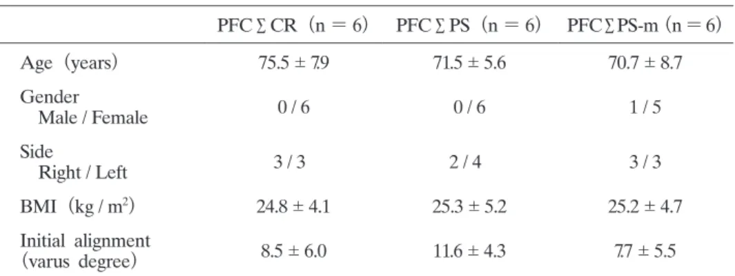
2)Abstract : Different total knee arthroplasty (TKA) designs showed different intra- operative kinematics when evaluated with a navigation system. This study com- pared the kinematics of three different prosthesis designs using navigation-based in vivo simulation. Studies were carried out on 18 osteoarthritic knees using a com- puted tomography (CT) -free navigation system. The TKA designs used for these patients were six each of cruciate-retaining (CR) type, posterior-stabilized (PS)
type, and PS mobile type. Intraoperative knee kinematics were measured during a passive range of motion from full extension to 130 degrees of knee flexion.
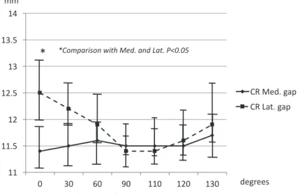
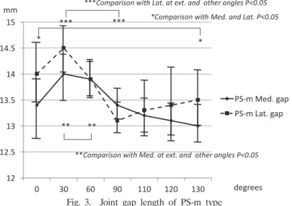
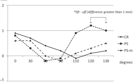
Three types of prosthesis were compared for the following factors : the presence of condylar lift-off (gap difference greater than 1 mm between medial gap and lateral gap) and anterior-posterior (AP) displacement of the center of femur relative to the tibia. The lift-off was found in patients with the PS type between 120 and 130 of flexion, but not in the other two types of implant. The pattern of AP displacement was similar among the three implants. The femur moved to the ante- rior side from extension to 50-55 of flexion, after that, the femur moved to the posterior side to full flexion. The function of PCL could prevent lift-off in the CR type of implants. In the PS-mobile implants, the mechanism of rotation platform might have contributed to lift-off prevention. The paradoxical anterior slide of the femoral condyles during mid flexion was observed in all types of implant.
Key words : navigation system, in vivo knee kinematics, total knee arthroplasty
Introduction
Recent studies of total knee arthroplasty (TKA) using a navigation system, have provided data on clinical results and alignment
1, 2)but also on the kinematics during surgery. Changes in the amount of pre and postoperative anterior-posterior (AP) displacement, rotation of tibia, and soft tissue balance were reported
3, 4), suggesting that varus / valgus stability could be acquired with TKA. Furthermore, in a study assessing intraoperative soft tissue balance using a navigation system with tensor, balance was found to be associated with postoperative outcome
5). Moreover, some reports have compared various systems based on the soft tissue balance and kinematics achieved
6). We previously compared two different TKA systems, the cruciate-retaining Original
1)
Department of Orthopaedic Surgery, Showa University Fujigaoka Hospital, 1-30 Fujigaoka, Aoba-ku, Yokohama 227- 8501, Japan.
2)
Department of Orthopaedic Surgery, Showa University School of Medicine.
*