Introduction
Patients with either partial or complete aplasia of the posterior arch of the atlas are generally asymptomatic. This anomaly is often detected incidentally when a patient has cervical X rays. However, some cases with this anomaly demonstrate neurological symptoms.1) 4) It is im- portant to predict the cases demonstrating this anomaly which may be accompanied by neurologi- cal symptoms. Based on both our clinical experi- ence and a review of the literature, we developed a new classification system of this condition for clini- cal use.
Materials and Methods
Between April 2001 and March 2005, 11,512 new patients were registered at the Department of Or- thopaedic Surgery at Chikushi Hospital, Fukuoka University. The cervical X rays examinations were performed for 4187 patients. Of these, three patients(0.071%)were encountered who had an anomalous posterior arch of the atlas. The diag- nosis was made based on the findings of lateral cer- vical X rays. Functional lateral cervical X rays were taken in all patients. To demonstrate the anomaly in more detail, threedimensional(3D)
computed tomography(CT)and magnetic reso- nance imaging(MRI)of the cervical spine were performed. A neurological examination was per-
Aplasia of the Posterior Arch of the Atlas:
A Proposal for a New Clinical Classification
Teruaki I
ZAKI1), Etsuji S
HIOTA2)and Jingfan Z
HANG2)1)Department of Orthopaedic Surgery, Faculty of Medicine, Fukuoka University
2)Department of Orthopaedic Surgery, Chikushi Hospital, Fukuoka University
Abstract:Partial or complete aplasia of the posterior arch of the atlas is often detected inciden- tally when a patient has cervical Xrays. Patients with this anomaly are generally asymptomatic. However, some cases demonstrate neurological symptoms. It is important to predict cases of this anomaly that may be accompanied with neurological symptoms. Between April 2001 and March 2005, three patients were encountered who had an anomalous posterior arch of the atlas. The anomalies encountered in the three patients were midline cleft(two pa- tients)and an absence of the posterior arch with persistent posterior tubercle(one patient). Based on our clinical experience and a review of the literature, we developed a new clinical classification system for this condition. From a clinical point of view, aplasia of the pos- terior arch of the atlas can be classified into three types. Type Ⅰ is complete aplasia. Type Ⅱ is partial aplasia with the isolated posterior fragment. Type Ⅱ is divided into subtypes depend- ing on the inward mobility of the isolated posterior fragment(Type Ⅱa without and Type Ⅱ b with mobility). Type Ⅲ is partial aplasia without the isolated posterior fragment. Our new
classification is simple and unambiguous, and it also provides a clear direction for developing ap- propriate diagnostic and treatment strategies.
Key words:Cervical spine, Atlas, Arch, Anomaly, Classification
Correspondence to:Teruaki IZAKI
Department of Orthopaedic Surgery, Faculty of Medicine, Fukuoka University, 7 45 1 Nanakuma, Jonanku, Fukuoka City, 8140180, Japan
Tel: +81 92 801 1011 Fax:+81 92 864 9055 E mail address:izaki@fukuoka u.ac.jp
formed at the initial and the last followup visit.
Results
The subjects included one male and two females with an average age of 30.7 years(range, 2052 years). The cervical Xrays were obtained be- cause of nonspecific neck pain or headache, tired- ness in the upper extremities(Table 1). The anomalies encountered in the three patients were midline cleft(two patients)and an absence of the posterior arch with persistent posterior tubercle
(one patient)(Table 1). No atlantoaxial instabil- ity or anterior displacement of the bony fragment during extension of the cervical spine was found on functional lateral cervical Xrays in all patients.
An isolated posterior tubercle and a precise posi- tion of the defect were confirmed by CT and 3DCT
(Figures 1, 2, Table 2). In all patients, MR im- ages did not show a narrowing of the spinal canal nor any signal alternation of the spinal cord on T2 weighted images at the level from C1 to C7. In all patients, a neurological examination, including the mental status, gait and posture, cranial nerve re- flexes, postural reactions, spinal reflexes and pain perception, was normal at the initial and the last followup visit. The symptoms of the patients subsided quickly after conservative treatments
(Table 1).
Discussion
Partial or complete aplasia of the posterior arch of the atlas is an uncommon anomaly. In a study of 1,613 dissections by Geipel, a cleft of the poste- rior arch of the atlas was found in 4% of all
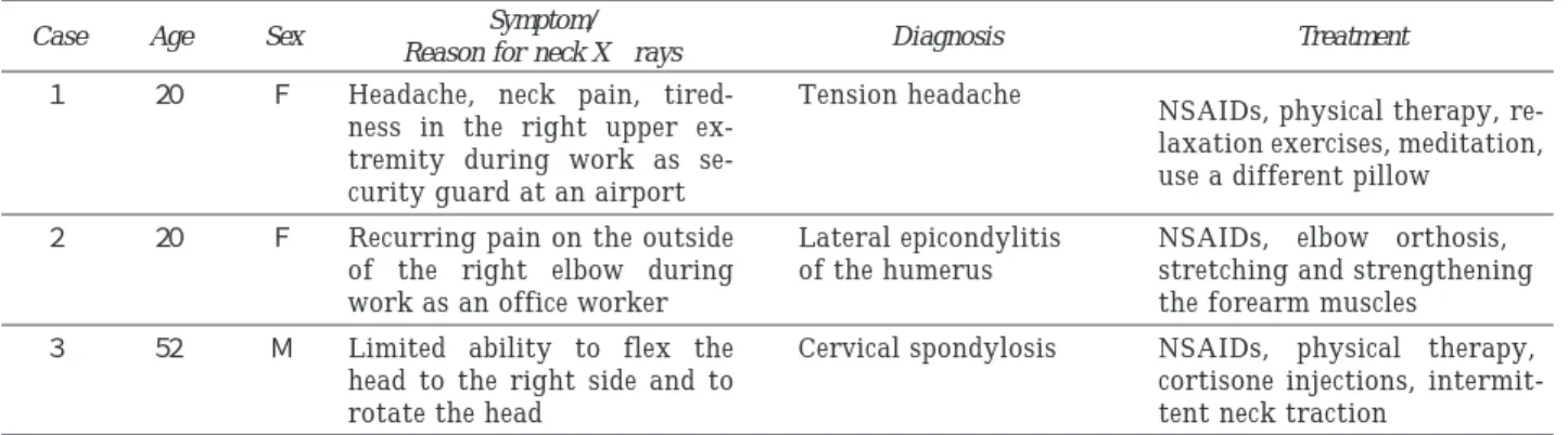
Table 1. Clinical features in the three patients
Treatment Diagnosis
Symptom/
Reason for neck Xrays Sex
Age Case
NSAIDs, physical therapy, re- laxation exercises, meditation, use a different pillow
Tension headache Headache, neck pain, tired-
ness in the right upper ex- tremity during work as se- curity guard at an airport F
20 1
NSAIDs, elbow orthosis, stretching and strengthening the forearm muscles
Lateral epicondylitis of the humerus Recurring pain on the outside
of the right elbow during work as an office worker F
20 2
NSAIDs, physical therapy, cortisone injections, intermit- tent neck traction
Cervical spondylosis Limited ability to flex the
head to the right side and to rotate the head
M 52 3
NSAIDs:nonsteroidal antiinflammatory drugs
Table 2. Radiological findings in the three patients
MRI Inward mobility of the CT
posterior fragment Atlantoaxial
instability Plain Xray
Case
spinal cord compression(−)
signal alteration(−)
Partial aplasia of the posterior arch of the atlas with an isolated posterior tubercle(Fig. 1b)
(−)
(−)
Absence of the poste- rior arch of the atlas except for the poste- rior tubercle(Fig. 1a)
1
spinal cord compression(−)
signal alteration(−)
Posterior arch of the atlas became gradually thin Spinal canal re- mains wide at the C1 level(Fig. 2b)
N/A
(−)
ADI:2 mm, SAC:12 mm S/O congenital stenosis at the C1 level(Fig. 2a)
2
spinal cord compression(−)
signal alteration(−)
A bony defect is present in the mid- line posteriorly N/A
(−)
Double bulbous ends of the hemiarches Absence of a spino laminar line(Fig. 3)
3
ADI:atlantodental interval SAC:space available for the cord S/O:suspected of N/A:not applicable
cases.5) It appears to be more common in women and an autosomal dominant inheritance has been suggested.6) Autopsies and surgical explorations have shown that the bony gap in the posterior arch tends to be bridged by loose3) 7) or dense5) connec- tive tissue rather than cartilage. Furthermore, based on the shape and developmental pattern
of the structures of the cartilaginous human craniovertebral junction, some authors have sug- gested that certain pathologies most likely origi- nated during the chondrification phase of devel- opment.8) 10) These observations support the no- tion that these anomalies result from a defective de- velopment of the cartilage rather than a distur- Figure 1. a Case 1:A lateral radiograph of the cervical spine, showing the
absence of the posterior arch of the atlas except for the poste- rior tubercle.
b Case 1:A lateral view of a threedimensional computed tomo- graphy of the atlas, showing aplasia of the posterior arch of the atlas with an isolated posterior tubercle.
Figure 2. a Case 2:A lateral radiograph of the cervical spine, showing hy- poplasia of the atlas; the anterior atlantodens interval was 2 mm(black arrowheads), whereas the space available for the cord was 12 mm(white arrowheads).
Figure 2. b Case 2:An axial view of computed tomography at the C1 level. The posterior arch of the atlas had gradually become thin;as a result, the space available for the cord was remained wide(white arrow).
a b
a
b
bance of ossification by itself.
The diagnosis of this anomaly can be made either incidentally on plain films in cases of trauma to the neck,1 ) 2 ) 11 ) nonspecific neck or shoulder pain,1) 2) 4) 12) 13) an evaluation of the tonsil size or epiglottitis,1) and in an examination of edema in the upper extremities.13) In some patients with partial aplasia of the posterior arch of the atlas with an isolated posterior tubercle, the isolated posterior fragment moves anteriorly into the spi- nal canal during extension of the cervical spine.4) 12) 14) This inward mobility of the isolated posterior fragment may compress the cord during the extension of the cervical spine. Therefore, the clinical manifestations, neck pain or neurologi cal deficits, may be caused by this malforma- tion.1)4) 11)15) The TorklusGehle classification16)
and the Currarino classification1 for aplasia of the posterior arch of the atlas have all been accepted
worldwide. The existence of an isolated posterior tubercle was noted in these previous classifications based on the morphological criteria, but its clinical significance was not described. These morphologi- cal classifications were not suitable for evaluating the clinical features because of its complexity and a lack of information about the movement of an iso- lated posterior tubercle. From a clinical point of view, the existence of an isolated posterior tubercle and its movement during cervical motion thus seem to be most important factors for predicting the development of neurological symptoms. There- fore, a new classification should be based not only on the morphological findings but also on func- tional factors.
Our classification for aplasia of the posterior arch of the atlas can be achieved by Xrays and CT scans(Figure 4). Functional lateral cervical X rays provide better information about atlantoaxial Figure 3. Case 3:A posterior median cleft of the arch of the atlas
could be recognized in the double bulbous ends of the hemi arches and the absence of a spinolaminar line in a lateral view of Xrays which indicates the absence of the posterior wall of the spinal canal. A posterior cleft also is shown par- ticularly clear on an oblique view of Xrays.
instability and/or inward mobility of the isolated posterior fragment. Aplasia of the posterior arch of the atlas can be classified into three basic types;
Type Ⅰ to Ⅲ, with Type Ⅱ having two subtypes.
Type Ⅰ is complete aplasia. Type Ⅱ is partial aplasia with the isolated posterior fragment. Type
Ⅱ is divided into two subtypes depending on the inward mobility of the isolated posterior frag- ment(Type Ⅱ a without and Type Ⅱ b with mobility). Type Ⅲ is partial aplasia without the isolated posterior fragment. According to our new classification, case 1 corresponded to Type Ⅱ a, while cases 2 and 3 corresponded to Type Ⅲ. If atlantoaxial instability or inward mobility of the isolated posterior fragment(Type Ⅱ b)is suspect- ed based on functional lateral cervical Xrays, then MRI is required. MRI is also required to evaluate the spinal cord in Type Ⅰ, because the filamentous fibrous band replacing the absent bony arch may act on the unprotected cord in a manner similar to that of a guillotine in traumatized cases. Weisz re- ported a case of trauma to the cervical spine with partial aplasia of the posterior arch of the atlas with an isolated posterior tubercle.15) This case corresponds to our Type Ⅱa, because of the lack
of the inward mobility of the isolated posterior fragment. The injury was severe enough to pro- duce a vertebral body compression fracture, but it did not affect the stable occipitoatlantoaxial mechanism or the posteriror fragment position, thus resulting in no neurological deficit.15) All re- ported patients with neurological symptoms, who had to undergo operative treatment, thus corre- spond to our Type Ⅱb.3) 17)19) If atlantoaxial in- stability is not present, then good results have only been reported after an excision of an isolated posterior tubercle.
Our new classification for aplasia of the poste- rior arch of the atlas is simple, unambiguous and functional while also providing a clear direction for developing appropriate diagnostic and treat- ment strategies.
One limitation of the present study is the lack of experience with Type Ⅰ and Type Ⅱ b cases.
However, we believe that our review of the litera- ture was extensive enough to allow us to develop a new classification system.
Figure 4. A new clinical classification for aplasia of the posterior arch of the atlas. TypeⅠ:complete aplasia. TypeⅡ:partial aplasia with an isolated posterior fragment. Type Ⅱa;
without inward mobility of the isolated posterior frag- ment. Type Ⅱb;with inward mobility of the isolated poste- rior fragment. Type Ⅲ is partial aplasia without an iso- lated posterior fragment.
Conclusion
Aplasia of the posterior arch of the atlas is often detected incidentally when a patient has cervical X rays. Patients with this anomaly are generally asymptomatic. However, in some cases, clinical manifestations may be caused by this malfor- mation. It is therefore clinically important, to predict cases demonstrating this anomaly that may be accompanied by neurological symptoms.
We believe that our new classification described in this report may thus be a useful tool for predicting the clinical manifestations while also providing a clear direction for developing appropriate diagnos- tic and treatment strategies in the clinical manage- ment of aplasia of the posterior arch of the atlas.
Acknowledgments
The authors thank Professor Masatoshi Naito, Department of Orthopaedic Surgery, Fukuoka Uni- versity, Fukuoka, Japan, for critical comments and his review of this manuscript.
References
1)Currarino G, Rollins N, Diehl JT. Congenital de- fects of the posterior arch of the atlas:a report of seven cases including an affected mother and son. Am J Neuroradiol 1994;15:249 254.
2)Lipson SJ, Hammerschlag SB. Atlantoaxial ar- throdesis in the presence of posterior spondyloschisis
(bifid arch) of the atlas. Spine 1984;9:65 69.
3)Richardson EG, Boone SC, Reid RL. Intermittent quadriparesis associated with a congenital anomaly of the posterior arch of the atlas. Case report. J Bone Joint Surg Am 1975;57:853 854.
4)Sharma A, Gaikwad SB, Deol PS, Mishra NK, Kale SS. Partial aplasia of the posterior arch of the atlas with an isolated posterior arch remnant:findings in three cases. Am J Neuroradiol 2000;21:1167 1171.
5)Geipel P. Zur Kenntnis der Spaltbildungen des At- las und Epistropeus. Teil Ⅳ. Zentralbl Allg Pathol 1955;94:19 84.
6)Saltzman, CL; Hensinger, RN;Blane, CE;Phillips, WA. Familial cervical dysplasia. J Bone Joint Surg Am 1991;73:163 171.
7)Logan WW, Stuard ID. Absent posterior arch of the atlas. Am J Roentgenol Radium Ther Nucl Med 1973;118:431 434.
8)David KM, McLachlan JC, Aiton JF, Whiten SC, Smart SD, Thorogood PV, et al. Cartilaginous de- velopment of the human craniovertebral junction as visualised by a new threedimensional computer re- construction technique. J Anat 1998;192:269 277.
9)Kimura T. Studies of the early development of the vertebral column in human embryos. J Nippon Med Sch, 1956;23:333 352(in Japanese).
10)Tompsett, A. C., S. W. Donaldson. The anterior tu- bercle of the first cervical vertebra and the hyoid bone. Their occurence in newborn infants. Am J Roentgenol 1951;65:582 584.
11)Duong DH, Chadduck WM. Reconstruction of the hypoplastic posterior arch of the atlas with calvarial bone grafts for posterior atlantoaxial fusion:techni- cal report. Neurosurgery 1994;35:1168 1170.
12)Chigira M, Kaneko K, Mashio K, Watanabe H.
Congenital hypoplasia of the arch of the atlas with abnormal segmentation of the cervical spine. Arch Orthop Trauma Surg 1994; 113:110 112.
13)Schulze PJ, Buurman R. Absence of the posterior arch of the atlas. Am J Roentgenol 1980;134:178 180.
14)Motateanu M, Gudinchet F, Sarraj H, Schnyder P. Case report 665. Congenital absence of poste- rior arch of atlas. Skeletal Radiol 1991;20:231 232.
15)Weisz GM. Trauma to anomalous cervical spine.
Spine 1983;8:2257.
16)Torklus DV, Gehle W. The Upper Cervical Spine.
London:Georg Thieme Verlag, Stuttgart;1972.
pp.14 44.
17)Torreman M, Verhagen IT, Sluzewski M, Kok AJ, van Rooij WJ. Recurrent transient quadriparesis af- ter minor cervical trauma associated with bilateral partial agenesis of the posterior arch of the atlas. Case report. Rev Hosp Clin Fac Med Sao Paulo;2002;57:73 76.
18)Klimo P Jr, Blumenthal DT, Couldwell WT.
Congenital partial aplasia of the posterior arch of the atlas causing myelopathy:case report and review of the literature. Spine;2003;28:E224 228.
19)Sagiuchi T, Tachibana S, Sato K, Shimizu S, Ko- bayashi I, Oka H, Fujii K, Kan S. Lhermitte sign during yawning associated with congenital partial aplasia of the posterior arch of the atlas. Am J Neu- roradiol;2006;27:258 260.
(Received on October 10, 2008, Accepted on December 11, 2008)