Hirosaki Med. 1.
56: 45-54, 2005ORIGINAL ARTICLE
CHARACTERIZATION OF DECORIN OF THE HUMAN YELLOW LIGAMENT
Kazunari Takeuchi", Seiko Harata
U,Keiichi Takagaki", and Masahiko Endo
2)Abstract Decorin, a component of the low-molecular-weight proteoglycans (PG) of yellow Iigaments was purified, and the structures of the core protein and the glycosaminoglycan (GAG) chains were analyzed. The crude PG fraction extracted under dissociative conditions was subjected to DEAE-Sephacel ion-exchange chromatography, gel-filtration on Sepharose CL-4B, and Octyl-Sepharose CL-4B chromatography, and decorin was obtained. The amino-terminal amino acid sequence of decorin was found to be identical to those of human bone PG-II and human fetal membrane PG-II. The analyses of the GAG chains using two-dimensional electrophoresis on cellulose acetate membranes, affinity high-performance liquid chromatography (HPLC) with a hydroxyapatite column and gel-filtration HPLC after chondroitinase digestion, showed the presence of dermatan sulfate (DS) chains and chondroitin sulfate (CS). Further, it was suggested that CS located in the reducing termini and DS in the non-reducing termini in the hybrid chain.
Hirosaki Med. J. 56: 45-54, 2005 Key
words: decorin; proteoglycan: glycosaminoglycan: yellow ligament, dermatan sulfate.
fO EB
~~ g~~~0~~~0h~~m~~~~~0~~.~.*~GM~G, ~.~0~~~57~-~M~G~~, ~0~
7 -3l~J\~:BetU!fI)~-!t-2 J ~"0fJ ~ (GAG)mt~ji0t~m~~~fJTGt.::. ~7 -3l~)\ ~0N*jJiM7-2 J~~2:71J~J:e:l-~PG-II:B
etUe: ~ij~ij~PG-II0~hC:-3&Gll;)t.::. GAGM~ji0-t)vD-;Z7-t~-~ij~=jzjI;~%r*J}], /\-1 ~0:::f:~7/\-3l-1 ~fJ :;; b.~ffll;)t.::~fD'11~Jm¥&{*~0~l-~57
-r -
(HPLC) :BetU~.>
~0-1TT-t'1~{~13f:07)v6J&IHPLC~::et~~fJT~~J:, GAGM~ji~J:JijI;*j/iM{WH::~ ~ ~0 -1T ~1Jj1t~ (CS), ~FJijI;*j/iM{J!lH:: ~)v~-3l~~1rE~ (DS) i6~~tt9 ~/\-1
:1'
I)'Y ~ ~M~ji~~·:::d.::.
iJlxMl2f¥
56: 45-54, 2005Introduction
Decorin, which is one of the low-molecular-weight p r o t c o g l
yc a n s (LMWPG), is a complex macromolecule
1)Department of Orthopaedic Surgery, Hirosaki University School of Medicine
2) First Department of Biochemistry, Hirosaki University School of Medicine
Correspondence: K. Takeuchi
Received for publication, August 10, 2004 Accepted for publication November 1,2004
composed of a g lycosaminog lycan (GAG) chain bound covalently to a core protein",'
Itis distributed in extracellular matrices (ECM) of various connective tissues. In recent years, the functions of decorin in ECM have been
1) ~b*tk~~~ff~~*H1.*4~~fiJ~
2) %iW*~~~ff~~-j::1t;~~fiJ~
)JIJijlJ~~*)t :
Ifrl*JfoJJ.X:
SjIpt161:F 8J=j10
B §t11
SjIpt161:F11J=j 1
B
§t~reported. For example, decorin can interact with transforming growth factor-
(32)and epidermal growth factor". Decorin can also bind to type I, II, III, VI and XIV collagens
4-6),and it has been found that the PGs as decorin were involved in the regulation, stabilization and assembly of collagen fibers. Therefore, decorin would be considered to have various functions specific to the tissue.
The human yellow ligaments are a series of short ligaments that pass between the vertebrae along the length of the spine.
Endochondral ossification may occur with aging in the ligaments", and patients with this disease present neurological symptoms such as myelopathy and r adicu lopath
y,and easily sustain spinal cord injuries. The pathogenesis of ossification might involve many factors, such as genes, cv tokiries.
hormones, and circumference, but the details of the ossification process remain uncertain.
From past studies, we found that GAG chains change with aging from dermatan sulfate (DS) chains to chondroitin 6-sulfate near the protein-carbohydrate linkage region, and in the composition of GAG chains, /). Di-6S instead of /). Di-4S increases with aging and ossified ligaments". In addition, we found that decorin accounted for the major part of PGs from yellow ligaments"
By the present methods, it is impossible to sequence a GAG chain like a protein, or DNA.
In this paper, we report the structure of GAG chains of decorin contained in human yellow ligament analyzed by using two-dimensional electrophoresis and the specific action of chondroitinase ABC· and B.
Material and Methods
Materials
Chondroitin 6-sulfate (Ch6S, from shark cartilage), chondroitin 4-sulfate (Ch4S, from whale cartilage), dermatan sulfate (DS,
from pig skin), heparin (Hep, from bovine lung), chondroitinase ABC (from Proteus vulgaris) and chondroitinase B (from Flavobacterium aurescens) were purchased from Seikagaku Kogyo Co., Tokyo, Japan.
Hyaluronic acid (HA, from human umbilical cord) and endo-
(3-xylosidase were prepared by the methods of Nakamura et al.
I O)and Takagaki et al.:", respectively. Actinase E was purchased from Kaken Kag ak u Co., Tokyo, Japan, and the immunoblotting ABC POD (R) Kit and 2-aminopyridine were purchased from Wako Pure Chemical Co., Osaka, Japan. DEAE-Sephacel, Sepharose CL-4B, Octyl-Sepharose CL-4B and the electrophoresis calibration kit were purchased from Pharmacia LKB Biotechnology AB, Uppsala, Sweden. Cellulose acetate membranes and 5-20% acrvlarnide gradient slab gels were purchased from J ookoo Co., Tokyo and A tto Co., Tokyo, Japan, respectively and anti-human decorin antibody (PG-40) was purchased from Gibco, Grand Island, New York, U.S.A. All the other reagents used were obtained from commercial sources.
Extraction and separation of PGs from human yellow ligaments
Normal human yellow ligaments (268g) were taken from 305 patients from teenagers to those in their eighties, at the time of operation for intervertebral disc herniation or lumber spinal canal stenosis. This study was approved by The Committee of Medical Ethics of Hirosaki University School of Medicine, Hirosaki, Japan. All of the patients gave their informed consent.
The extraction of PGs from human yellow
ligaments was carried out in 5 volumes of
4 M guanidine-HCl!50 mM sodium acetate
buffer (pH 5.6) containing protease inhibitors
at 4°C for 24 h, as described by Saidera
and Hascall
12).The extracts were collected
Decorin of the Human Yellow Ligament 47
and designated the crude PG fraction. This fraction was dialyzed against 7 M urea/50 Mm Tris-HCI buffer (pH 7.2) at 4°C for 48 h.
After that, the dialyzed inner solution was applied to a DEAE-Sephacel column 0.0 x 15.0 ern) equilibrated with 7 M urea/50 mM Tris-HCI (pH 7.0), which was eluted with a linear concentration-gradient of NaCI (0-1.0 M) at a flow rate of 60 ml/h and 20-ml fractions were collected. Each fraction was assayed for uronic acid by the carbazole reaction'", and for protein by the protein-dye binding method'", The uronic acid-containing fractions were pooled. The precipitate by 80% ethanol was collected by centrifugation, and then soluted in 4 M guanidine-HCl/50 mM sodium acetate buffer (pH 5.6). These fractions were subjected to gel-filtration chromatography on a Sepharose CL-4B column (3.3 x 175 ern) at a flow rate of 60 ml/h, 20-ml fractions were collected, and the LMWPG fractions were separated.
For further purification, chromatography on an Octyl-Sepharose CL-4B column (3,4 x 29 em) was carried out under the conditions described by Choi et al.
1 5).The uronic acid-containing (LMWPG) fractions were dialyzed against 2 M guanidine-HCl!50 mM sodium acetate buffer (pH 5.6) at 4°C for 24 h. After that, the fractions were applied to the column, which was equi libr a ted with 2 M guanidine-HCl/50 mM sodium acetate buffer (pH 5.6), and eluted with a linear concentration-gradient of 2-6 M guanidine-HCl!50 mM sodium acetate buffer (pH 5.6) at a flow rate of 60 ml/h and 20-ml fractions were collected, which were designated the LMWPG fractions.
Enzymic digestions
To obtain the GAG chain, first, the core protein was digested with Actinase E (0.1 M Tris-HCI buffer, pH 8.0, containing 10 mM CaCI
2 )which was one of the proteases.
Secondly, the residual core protein was eliminated from the GAG chains with endo-{3 -xylosidase (0.1 M sodium acetate buffer, pH 4.0Y6) in the reducing termini. This enzyme catalyzes the eliminative cleavage of Xylose of the GAG chain linkages to Serine residue of the core protein. The GAG chains were digested with chondroitinase ABC (0.1 M Tris-HCI buffer, pH 8.0)17) which can be used for selective removal of the chondroitin sulfate or dermatan sulfate side chains, and chondroitinase B (0.1 M Tris-HCI buffer, pH 8.0)18) which can be used for selective removal of the dermatan sulfate side chains.
Electrophoresis
Sodium dodecyl sulfate-polyacrylamide gel electrophoresis (SDS-PAGE) was carried out, as described by Rosenberg et al.
I 9).SDS-PAGE was carried out on 5-20% acrylamide gradient slab gels at 4°C at 200 V for 4 h. The gels were stained successively with Coomassie brilliant blue R-250 followed by Alcian blue, as described by Fisher et al.""
Two-dimensional electrophoresis was carried out on cellulose acetate membranes (lOx 10 em), as described by Yokoyama et al.". The first dimension was obtained in 0.1 M pyridine/Oxl? M formic acid buffer (pH 3.0) at 1 mA/cm for 1 h, and the second with 0.1 M barium acetate (pH 8.0) at 1 mA/cm for 4 h. The membranes were stained with 0.1 % Alcian blue in 70% ethanol.
Immunoassay
Western blotting immunoassay for PGs using anti-human decorin antibody was carried out, as described by Towbin et al.
22).Anti-human decorin antibody was from polyclonal antiserum of rabbits immunized with a peptide representing the carboxyl terminus of human decorin.
Ithas specific activity against only decorin"
Fluoro-Iabeling
The prepared GAG chains were
0.2 r - - - . 8
4 6
2
120 o
80 100 60
40
o 20
I
o
0.1
Fraction number
Figure 1 Separation of the low-molecular-weight proteoglycans by Octyl-Sepharose CL-4B.
In order to separate the low-molecular-weight proteoglycans, chromatography was carried out on an Octyl-Sepharose CL-4B column (3.4 x 29 em). The column was eluted with a linear concentration-gradient of 2-6 M guanidine-HCl/50 mM sodium acetate buffer (pH 5.6) at a flow rate of 60 ml/h and 20-ml fractions were collected. Each fraction was monitored for uronic acid (530 nm),
pyridylaminated at their reducing termini by the method of Kon et a1.
24).The GAG chains were coupled with 2-aminopyridine with reductive amination to give pyridylaminated GAG chains (PA-GAG chains).
Analysis of the amino acid compositions and sequence
The amino acid compositions of the core protein were analyzed. The samples were hydrolyzed with 6 N HCl at 135°C for 3 h.
This hydrolyzed solution was evaporated and soluted in distilled water again. This solution was analyzed with a Hitachi L-8500 amino acid automatic analyzing meter (Hitachi Co., Tokyo, Japan).
The amino-terminal sequence, and the inner sequence of PG-II core proteins followed by cyanogeris bromide (BrCN) cleavage"?
were analyzed using a Hewlett Packard
G1OOA protein sequencer'".
High-performance liquid chromatography (HPLC)
Affinity HPLC was carried out with a KB-OO 12 column (7.8 x 100 mm, Koken Co., Tokyo, Japan). The column was eluted with a linear gradient of increasing molarity (10-100 mM) of sodium phosphate buffer (pH 6.8) for 10-40 min at flow rate of 1.0 ml/min at 30°C
27)
Size-fractionation HPLC was carried out with a Shodex OHpak SB-803 HQ column (8 x 300 mm, Shoko Co., Tokyo, Japan) using 0.2 M NaCl. The flow rate was 0.5 ml/min and the column temperature was 30°C.
For detection, an excitation wavelength of
320 nm and an emission wavelength of 400
nm were used (Hitachi F-I050).
A 220kDa-+ :
1
Decorin of the Human Yellow Ligament
+-94kDa +-67 kDa
2
B
1 2
49
Figure2 Analysis of the low-molecular-weight proteoglycans by SDS-PAGE and immunoassay.
The proteoglycan fraction eluted from an Octyl-Sepharose CL-4:B column (peak I and II in Figure 1) were collected and subjected to SDS-PAGE on 5 to 20% acrylamide gradient slab gels at 4°C at 200 V for 4 h, after which the gels were stained with Coomassie brilliant blue R-250 followed by Alcian blue (A). Western blotting immunoassay using anti-human decorin was carried out (B).
Lane 1,peak I shown in Figure 1; lane 2, peak II shown in Figure 1.
Results
Separation of the LMWPGs
The crude PG fraction extracted with 4 M guanidine-HCl/50 mM sodium acetate buffer (pH 5.6) was applied to a DEAE-Sephacel column, and the eluted uronic acid-containing fractions using a NaCl concentration were collected and subjected to Sepharose CL-4B gel-filtration chromatography. The two peaks, which were reacted with the carbazole reaction, were found. They were pooled as the LMWPG fractions (figures are not shown).
For further separation, the LMWPG fractions were applied to an Octyl-Sepharose CL-4B column (Figure 1). The column was eluted with a linear-concentration gradient of 2-6 M guanidine-HCl/50 mM sodium acetate buffer (pH 5.6), and the uronic acid-containing
fractions were eluted at guanidine-HCl concentrations of about 3.35 M (peak I) and 5.0 M (peak II). The total amount of uronic acid which was contained in peak I was about 27 mg.
Itwas suggested that more than two types of the LMWPGs which possess different hydrophobities of core protein are contained in human yellow ligaments.
Analysis by SDS-PAGE and immunoassay
The PGs contained in the fractions of
each peak eluted from an Octvl-Sepharose
CL-4B column were analyzed by SDS-PAGE
(Figure 2-A). A single band appeared for the
PG of peak I fractions at molecular weight of
70,000-95,000, while two bands appeared for
those in peak II fractions at molecular weights
of 70,000-95,000 and 180,000-200,000, and
were designated PG-II, and PG-III and PG-I,
respectively. These numbers of PG-I and
PG-II were the same as those of human bone?" and fetal membrane'?" PG-III was the characteristic number of the present study.
Western blotting immunoassay for PG-I, PG-II and PG-III was carried out (Figure 2-B).
Anti-human decorin reacted with PG-II, but not with PG-I and PG-III.
Itwas suggested that PG-II was decorin, while PG-I and PG-III possess different core proteins from that of PG-II.
Analysis of the amino acid compositions and sequence of the core protein of PG-II
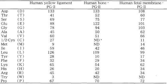
The amino acid compositions of PG-II are shown in Table 1. The compositions of human yellow ligaments PG-II were not identical but were similar to those of human bone PG-II
28)and fetal membrane PG-II
29).The core protein contained 329 residues and its molecular weight was 36,400.
The amino-terminal sequence of PG-II is shown in Table 2. The sequence of human
yellow ligament PG-II was identical to those of human bone PG-II
2 8)and human fetal membrane '".
Itwas suggested that this LMWPG is possessed of the same core protein as the decorins previously reported.
Analysis of the GAG chains of PG-II
In order to identify the GAG chains of PG-II, GAG chains were first prepared by digesting PG- II with Actinase E and endo-
{3-xylosidase and then analyzed by two-dimensional electrophoresis on cellulose acetate membranes (Figure 3).
Itwas found that DS, and Ch6S extended from DS were contained in the GAG chains of PG-II.
Secondly, after the GAG chains were fluoro-labclcd at their reducing termini with 2-aminopyridine, they were analyzed by HPLC on a hydroxyapatite column (Figure 4). The P A -G AG chains were eluted as bimodal distribution with a linear gradient of increasing molarity (10-100 mM) of sodium
Table 1 Amino acid analysis residues/1000
Asp (D)
Thr (T)
Ser ( S)
Glu (E)
Gly (G)
Ala (A)
Val (V)
l/2-Cys (C)
Met (M)
Ile ( I )
Leu (L)
Tyr (Y)
Phe (F)
Lys (K)
His (H)
Arg (R)
Try (W)
Pro (P)
Human yellow ligament PG-II
133 41 69 88 78 45 60 27 9 59 126 28 32 65 26 45 3 67
Human bone"
PG-II 133
52 75 122 94 50 51 ND"
ND 42 109 10 29 54 20 42 ND 70
Human fetal membranec:
PG-II 108
60 77 101 103 62 66 11 14 46 99 17 34 62 34 34 ND 72
" Ref. No. [28].
" ND, not determined.
cRef. No. [29].
Table 2 Amino acid sequences
X, no assigned amino acid; ( ), tentative amino acid assignment
1 6 11 16
Human yellow ligament PG-II NH2 DEASG IGPEV PDDRD
Human bone PG-II" NH2 (D) EAXG I (A) PEV PDD (R) (P) FEPS Human fetal membrane PG-IIc NH2 DEAXG IGPEV PDDR
aRef. No. [28].
" Ref. No. [29].
Ch6S , DS Rep
2nd
Decorin of the Human Yellow Ligament
RA DS Ch6S
51
1st
Figure 3 Two-dimensional electrophoresis on cellulose acetate membranes of the glycosaminoglycanchains of PG-II.
The glycosaminoglycan chains from PG-II obtained by Actinase E and endo-{3-xylosidase digestions were subjected to electrophoresis on cellulose acetate membranes (10 x 10 em) in the first dimension with 0.1 M pyridine/0.47 M formic acid buffer (pH 3.0) at 1 mA/cm for 1 h, and in the second with 0.1 M barium acetate (pH 8.0) at 1 mA/cm for 4 h. The membranes were stained with 0.1% Alcian blue in 70% ethanol. DS. dermatan sulfate; Ch6S, chondroitin 6-sulfate; Hep, heparin; HA, hyaluronic acid.
phosphate buffer (pH 6.8). The analysis of the GAG chains showed that those of decorin were distributed from DS to CS compared with standards.
Itwas suggested that the GAG chains contained in this decorin are composed of DS and CS.
Next, in order to examine the existing form of DS and CS in the GAG chains, the chondroitinase ABC and chondroitinase B digests of the PA-GAG c h a in s were analyzed by HPLC on a Shodex SB 803 HQ gel-filtration column (8 x 300 mm) (Figure 5). After chondroitinase ABC treatment, the GAG chains were completely digested to PA-tetrasaccharides and PA-hexasaccharides, however, after chondroitinase B treatment, some were only partially digested. That is to say, the GAG chains, which possess the parts of CS and resist the chondroitinase B
digestion, were present.
It
was suggested that two types of GAG chains exist in decorin of yellow ligaments, one of which is a major component, a DS chain, and the other a hybrid chain of DS and CS. Further, it was suggested that part of CS is located in the reducing terminal side of the part of DS in the hybrid chains.
Discussion
Three types of LMWPGs could be separated from human yellow ligaments in the current study. PG-II was identified as decorin, because the core protein of PG-II reacted with anti-human decorin antibody.
The authors analyzed the structure of decorin,
which is considered to be a major component
in the connective tissue of human yellow
ligaments. N-Terminal amino acid sequence
a
o 10 20 30
Retention time (min)
a
o 10 20 30 40
Retention time (min)
(1)
o
(1)s:::::
o
(1)
b
s:::::o
(1)r/'J
o
(1) r/'J
$-..i (1)
0 $-..i
~ 0
....-I ~
~ ....-I~
C
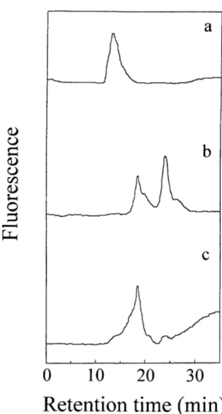
Figure 4 Affinity HPLC of the PA-glycosaminoglycan chains of PG-II on a hydroxyapatite column.
The PA-glycosaminoglycan chains were examined with HPLC using a KB-O 012 hydroxyapatite column (7.8 x 100 mm), The column was eluted with a linear gradient of increasing molarity (l0-1 00 mM) of sodium phosphate buffer (pH 6.8) for 10-40 min at a flow rate of 1.0 ml/min at 30°C. For detection, an excitation wavelength of 320 nm and an emission wavelength of 400 nm were used. a, PA-dermatan sulfate; b. PA-chondroitin 6-sulfate;
c, PA-glycosaminoglycan chains from PG-II.
Figure 5 Size-fractionation HPLC of the
PA-glycosaminoglycan chains of PG-II before and after chondroitinase digestion.
The P A -glycosaminoglyc a n chains after digestion without or with chondroitnase ABC and chondroitnase B were analyzed by HPLC using a Shodex SB803 HQ column (8 x 300 mm), The column was eluted with 0.2 M NaCI at a flow rate of 0.5 ml/min at 30°C. For detection, an excitation wavelength of 320 nm and an emission wavelength of 400 nm were used. a, PA-glycosaminoglycan chains before digestion;
b. after chondroitinase ABC digestion; c. after chondroitinase B digestion.
of the core protein of decorin was identical to those from human fetal membrane and human bone. These results were also identical to the results reported by Yukawa et aL
3 0>.The authors analyzed the composition of the GAG chain from decorin by the methods of two-dimensional electrophoresis and HPLC using P A -GAG chains. From these results, it was demonstrated that the GAG chain of decorin was composed of two kinds of GAG, which were DS and CS, and were found to be a single molecular size. Further,
from observation of digestion patterns of
PA-GAG, the locations of CS and DS in GAG
chains were speculated. PA-GAGs of decorin
were digested with chondroitinase ABC, and
two peaks were found indicating that GAG
chains were completely digested to tetra- and
hexasaccharides. While, after chondroitinase
B digestion, the peak was shifted to the
location of hexasaccharides, PA-GAGs were
not completely digested and mor e than
hexasaccharides were contained. This finding
indicated that the GAG chains were hybrid, in
which CS was located in the reducing terminal
Decorin of the Human Yellow Ligament 53
site and DS was located in the non-reducing terminal site because chondroitinase B is an exo type enzyme. Recently, Yukawa et al. reported on the structures of the GAG chains and core proteins of LMWPGs:
30).They analyzed decorin by HPLC analysis after chondroitinase digestion and demonstrated that part of CS is located in the
l