Difference in the Susceptibility to Brugia pahangi Infection between Male and Female BALB/c
Mice: Differences of Effector Cell Responses between Sex
Hirokum NAKANISHI
Department of Medical Zoology, Nagasaki University School of Medicine, 12‑4 Sakamoto‑machi, Nagasaki 852,
Abstract: Difference in the susceptibility to Brugia pahangi infection between male and female BALB/c mice was examined. In a primary infection, male mice showed greater susceptibility than did females as defined by counting the number of worms recovered from the peritoneal cavity. Sex difference appeared at the early phase of primary infec‑
tion (5‑10 days) and continued for at least 7 weeks of infection. Inoculation with the 3‑
rd stage larvae was effective in inducing sex difference, whereas inoculation with the 4‑
th stage larvae had no effect. Therefore, sex difference in susceptibility seems to be in‑
duced by the 3rd stage larvae (possibly molting stage larvae). Since sex difference was only observed in a primary infection but not in a challenge infection, the expression of sex difference might be dependent upon some differences of development of early phase
of immune response to the larval worm of B. pahangi between the sex. Thus, in the present study, the difference in immunological response of host to filarial worm was ex‑
amined. Significantly great number of effector cell infiltrations (macrophages and eosinophils were predominant) were observed in the peritoneal cavities of female mice 10 days after infection. Whereas no significant difference in the levels of anti‑B. pahangi IgG, IgM and IgE were observed at this time of infection, the cellular reaction that occured in female mice 10 days postinfection may be one of the factors which reflects the dif‑
ferential sex susceptibility of host in B. pahangi‑mouse model.
Key words: sex difference, susceptibility, Brugia puhangi, BALB/c mouse, effector cells
INTRODUCTION
It is a common knowledge that male hosts show higher susceptibility to certain species of parasites than do female hosts (Solomon, 1969; Goble and Konopka, 1973)I The sex difference in susceptibility of the host to filarial worm is frequently reported in the
epidemiological studies on filarial infection (Jakowski et at. cited in Ash, 1971; Beye and Gurian, 1960; Dondero and Menon, 1972; Murray, 1948; Nagatomo, 1960; Napier, 1944;
Received for Publication, July, 20, 1987.
Contribution No. 313 from the Department of Medical Zoology, Nagasaki University
School of Medicine.
Nelson et al., 1962; Omori et al, 1962; Rosen, 1955; Sasa et al.t 1970) and in expenmen‑
tal filariasis by using the rodents model (Ash and Riley, 1970; Ash, 1971; 1973; Suchant and MacDonald, 1972)・ Although some possible explanation are given to differential sex susceptibility, Wesley (1973) concluded that androgen is one of the factors which regulate the susceptibility of the jird to Bγug由♪uhangi infection.
Recently Abe et al. (1985) reported that differential sex susceptibility of mouse to stron甜Ioides ratti is due to the difference in strength of host defence mechanism at a given stage of infection・ Their studies encourage to examine whether preferential suscep・
■
tibility of the male host to filarial worms depends on the difference in host immune response at any specified stage of infection・
so far the jird‑5. ♪vhangi model has been commonly used for the experimental ro‑
dent filariasis, because it is easy to handle model, however, is less useful for the im‑
munological studies, because the inbred jirds are not commercially available. Recently
・
BALEIc mice were reported to show less susceptibility, but allow some larvae to develope to the adult worm, when the intraperitoneal route of infection is used (Macken‑
zie et al・ 1985). The BALEIc mice, therefore, were used in the present study as the ex‑
perimental animal host・
MATERIALS AND METHODS Animals
lnbred male and female BALEIc mice were raised in our laboratory・ The parental stocks were kindly given by Dr. S. Matsuo, Institute of Tropical Medicine, Nagasaki
%
●
university. Mice, over 12 weeks old, were used in all the experiments.
Preparation of antigen
B. pahangi adult worms, obtained from peritoneal cavities of jirds which have been intraperitoneally inoculated with 300‑400 infective 3rd stage larvae (L3) 9‑10 weeks previously, were washed extensively in phosphate‑buffered saline (PBS, pH 7.2). Worms were homogenized in a glass homogenizer and extracted in PBS by continuous stirring
・
overnight at 4 C. The extract was centrifuged at 12,000 r.p・m・ for 1 hr. After centnfuga…
tion, the protein concentration of the supernatant was measured by the method of Lowry et α/. (1951).
Preparation of sera
Male and female BALB/c mice intraperitoneally inoculated with 50 B・ pahangi L3 were bled on day 0, 5, 10 and 15 of infection.
Inoculation of ♪arasttes
lnfective stage larvae of B・ pahangi were obtained from mosquitoes (Aedes aegyμil which had been fed on microfilaremic Wistar rats 2 weeks previously・ Mice were in・
collated intraperitoneally with 50 L3 suspended in 0.5ml of Hanks'balanced salt solution
(HBSS)・
The 4th stage larvae (L4) were obtained from BALE/c mice which had been in‑
oculated mtraperitoneally with 400‑500 L3 of B・ pahangi 20 days previously・ Mice were inoculated with 40 L4 in 0.5ml of HBSS through a 18‑gauge needle into the peritoneal cavity・
Immunization and challenge infection
BALB/c mice, 7 to 8 weeks old, were immunized by a subcutaneous injection of 100 naive B. pahangi L3 in the left groin. Five weeks later, immunized mice were challenged in‑
trapentoneally with 50 homologous L3.
Recovery of worms from peritoneal cavity
The peritoneal cavity was examined for recovery of worms at various time points postmoculation (PI). To avoid coagulation of peritoneal cells, 0.5ml of heparinized HBSS (4
u/ml) was injected into peritoneal cavity of each animal… Then, they were killed by ether,
the peritoneal cavity was flushed with 4・5ml of HBSS. Worms were collected in a petri dish and counted immediately with a stereo microscope at 6 x magnification. Then the mouse was soaked in HBSS in a 50ml tube for 2 hours, after that live worms at the hot‑
torn of the tube were collected and counted under a stereo microscope. The summation of live worm counts from a petri dish and a bottom of the tube was expressed as the total number of recovered worms for each mouse.
Differential count of peritoneal exudate cells
Peritoneal exudate cells (PEC) were collected from peritoneal cavities of male and female mice by washing with 5ml of heparinized HBSS・ Total number of PEC was deter‑
mined using an improved Neubauer's hemocytometer. Differential count was made by counting 200 cells in a smear stained with Giemsa.
Passive cutaneous anaphylaxis (PCA) reaction
PCA reaction was carried out according to the method of Ovary et al (1975) with a slight modification・ Test sera were serially diluted with PBS and injected intradermally into normal indicator rats in 0.05ml volume. Four hours later, these rats received lmg of
B. pahangi antigen intravenously together with lml of a l% Evans blue solution. Thirty
minutes later, the animals were sacrificed and their skin was reversed to determine the
bluing reactions… Bluings m′ore than 5mm in diameter were considered as a PCA positive.
Enzyme ‑ linked immunosorbent assay (ELISA)
ELISA for detecting specific lgG or lgM levels of B. pahangi antigen were carried out according to the method of Tanaka et al (1983) using crude B. pahangi adult worm antigen (2・5 μg/ml)・ Peroxidase‑conjugated rabbit antトmouse lgG and lgM were commer‑
cially obtained・
Statistical analysis
Statistical significance of differences in mean values was assessed using Student's
トtest or Welch'sトtest・ Statistical significance was determined using P=0・05 criterion.
RESULTS Susceptibility to a primary infection
Adult male and female mice (more than 12 weeks old) were injected mtrapentoneal‑
1y with 50 L3・ At 7 weeks PI, some live worms were recovered from the peritoneal cavi‑
ty. And there was considerable difference in the number of worms recovered from male
and female mice (Fig… 1)… This preliminary experiment showed that male mice were more
susceptible to B・ ♪ahangi infection than females (P<0.01)… To know the time when sex
= =
difference became apparent, adult male and female mice were injected intrapentoneally with 50 L3 and their peritoneal cavities were subsequently examined at a given interval.
No significant difference was observed on day 5 PI (Fig・ 2)I Significant difference in
10
IK 也 i(
<u I
。 U
<u L
2 5
,I
。 き
a
0
ト
Mal e Female
Fig… 1・ Recovery rates of worms from the
peritoneal cavities of male (open bar) and female (dotted bar) BALE/c mice at 7 weeks after a primary infection with B. pahangi L3. Ten male mice and eleven females were inoculated in‑
traperitoneally with 50 B. ♪ahangi L3 and the live worms were collected from their peritoneal cavities at 7 weeks postinoculation・ Vertical bar in‑
dicates the standard deviation of the mean. *Recovery rate of worms from female mice was significantly less than that from males (P<0.01).
100
てコ 0) r13
<u td 0) U む
」50
Ul
=
叩■
0 き
㌔
。
■……■
・■■…■●■■
=コ==
■■・・
■・…■
/・■■
■■,I
■■●・
・■■…・■・・
●・…■
I,■・
・■t・
==8=
■■・,,
■・・■・
■●,…■
■■・・●
=⊂%∵8
■■■■,
●●・■
,■・,
●■■●
・■・■
==∴=
■■■…
■・●・
====,■■■
,■■■
===∵
:I:・
・■■■
Hd;/
tT・・
::::
:・:・
■・,■
;;;:
∵==::
・…・・
●■■●
;:≡:
■●,I
≡:=1
=8==
:・:I
■■…■
…■■…
■●■・
:・:・
‑:,:
●■■■
■…・●
*
*
10 15 Days after inlecti。n
Fig・ 2… Recovery rates of worms from the
peritoneal cavities of male (open bars) and female (dotted bars) BALE/c mice at various times after an in‑
traperitoneal inoculation of 50 L3.
From left to right each bar represents the mean from eleven, ten, eleven,
eleven, ten and seven mice, respective…
1y… Vertical bars indicate the st弧dard
deviation of the mean. *As shown in
Fig. 1 (10 days PI, P<0.005; 15 days
PI, P<0・001).
the susceptibility between sex to B. pahangi infection was observed on day 10 and 15 PI (P<0.005 and P<0.001, respectively). The percent of worms recovered from male mice on day 10 and day 15 PI were as high as those on day 5 PI・ However, those from females were significantly reduced on days 10 and 15 PI・
=
Difference in survi・val of L3 and L4 in peritoneal cavities of male and female mice To clarify which stage of worms to be important for the tar酢t that induces host sex difference in the susceptibility to B. pahangi infection, L3 and L4 were inoculated in‑
to the peritoneal cavities of male or female mice・ As shown in Fig. 3, statistically signifi‑
cant (P<0.001) sex difference (the female mice showed less susceptibility than the males)
was observed only when L3 were inoculated into叫ce. The recovery rate of L4 was
rather higher in female mice than that in male mice, but it was not statistically signifi‑
●
cant.
Serological data
Kinetics of specific serum lgG and lgM antibody levels of B・ pahangi was examin‑
ed by using ELISA・ As shown in Table 1, both classes of antibody levels increased
=
gradually but remained low during the observation period of 15 days PI and no dif‑
ference in levels of these antibodies between sex were observed except lgM which
100
T3 也
<b Lg I 。 fe U
%50 ,∩ ∈
。 し?
Q o* o
。 …享…重量董…
*
L3 k
Fig. 3・ Recovery rates of worms at 15 days after inoculation of 50 L3 and 40 L4 from male (open bars) and female (dotted bars) mice. From left to right each bar represents the mean from seven, seven, five and five mice, respec‑
tively. Vertical bars indicate the standard deviation of the mean. ・As shown
in Fig. 1 (P<0.001).
Table 1. Anti‑B. pahangi antibodies in male and female mice infected intraperitoneally with 50 L3 。f且♪ahangi
Days IgG IgM
postinfection M le Female Male Female
0 5 10 15
0.000±0.000 …000±0・OnO 0.080±0・072 0.149±0・051 0.212±0・117 0.224±0.129
0…195±0… 137 0.162±0.0930・000±0,000 ・000±0・000 0・000±o.ooo ・042±0…079 0.082±0.093 0. 108±0.062 0・062±0…093 0.223±0・097*
Values are the mean absorbance (experimental … control) at 490nm and standard deviation
from five mice・ Antibodies to crude B. pahangi adult worm antigen were measured using an ELISA.
*Significantly higher than males (P〈O. 05 ・
′■■■ヽ
qJ l/l
̲ul岩 石E
距B王
コ X o>
8E/
q)
⊂
。
■・一
・ ̲ .
L5 4J Q.
5
t/1
ド?
<L JD IS コ
≡
40
2。
0
10
0 10
0 。 10 15 Days 。fte「 infection
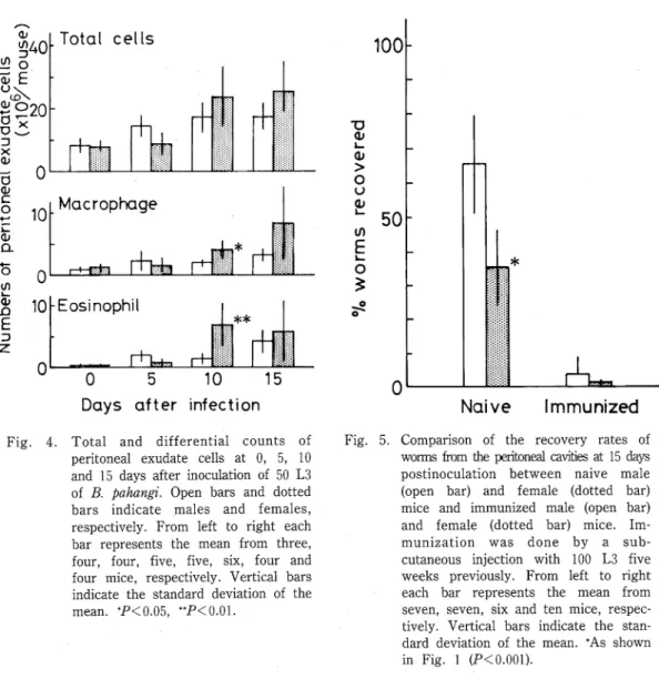
Fig. 4. Total and differential counts of peritoneal exudate cells at 0, 5, 10 and 15 days after inoculation of 50 L3 of B. pahangi. Open bars and dotted bars indicate males and females, respectively・ From left to right each bar represents the mean from three, four, four, five, five, six, four and four mice, respectively・ Vertical bars indicate the standard deviation of the mean. *P<0.05, **P<0.01.
100
■ロ qノ ドll Ol
>
。 U 巴50 1/1 ∈
。 L き
・o o*
0
*
「「.zz凹
Naive Immunized
Fig・ 5. Comparison of the recovery rates of worms from the pentoil由1 cavities at 15 da野 postm。culation between naive male (open bar) and female (dotted bar) mice and immunized male (Open bar) and female (dotted bar) mice. Im‑
munizati。n was done by a sub…
cutaneous injection with 100 L3 five weeks previously・ From left to right each bar represents the mean from seven, seven, six and ten mice, respec‑
tively. Vertical bars indicate the stan‑
dard deviation of the mean. *As shown
in Fig. 1 (P<0.001).
was higher in females than in males at 15 days PI・ Specific lgE antibody against B.
♪uhangi worm antigen was measured by PCA reaction・ No significant level of lgE an‑
tibody was detected during 15 days of observation…
■
Cytological data
The responsiveness of peritoneal cells against B・ ♪vhangi larvae was examined (Fig・ 4). However, no difference in the total number of PEC and differential counts of macrophages and eosinophils between sex was observed until 5 days PI・ On days 10 and 15 PI, total PEC, macrophages and eosinophils showed a tendency to increase in number‑ in females rather than in males. Statistically significant increase in numbers were observed especially in macrophages and eosinophils on day 10 PI (P<0.05, P<0.01 respectively).
Susceptibility to a challenge infection
As shown in Fig・ 5, the recovery rate of worms in the challenge infection was significantly lower than that in the primary infection (P<0.001). No significant difference in the recovery rate of worms was observed between male and female mice in the challenge infection・
DISCUSSION
The present paper clearly discribed that BALE/c mice show differential sex suscep‑
tibility to B・ pahangi infection. Adult male BALB/c mice showed higher susceptibility than female animals as defined by percent recovery of worms from the peritoneal cavity.
This difference became significant at very early phase of infection (10 days PI) and remained unchanged for 7 weeks PI, the time when worms developed into young adult stage in mice (Sakamoto et al・ 1982). Sex difference seems to appear around the time of the 3rd molt, because the molt from the 3rd to the 4th stage occurs at about 7‑10 days after infection in the mouse‑Brugia system (Sakamoto et at., 1982). Then, the experi‑
merit was designed to investigate which stage of larvae (3rd or 4th stage larvae) can in‑
duce this difference between sex… Sex difference (expressed as a higher susceptibility in
males than in females) was observed only when the 3rd stage larvae were inoculated・
When the 4th stage larvae were inoculated, there were not significant difference of recovery rates between sex, though slightly less recovery rates were observed in males than in females. These results suggest that difference in the susceptibility to a primary
=
B. pahangi infection between sex was induced by the 3rd stage larvae and/or the molting stage larvae・
In general, the causes of the failure of the parasite to mature in the undefinitive
host were complicated・ Mouse has been considered to be an undefinitive host to …
phatic dwelling filarial worm. However, in the mouse‑Brugia system an immune response, rather than some innate physiological or biochemical insufficiency, played an
=
important role in the failure of B・ ♪ahangi to develope in normal mice, since the sue…
cessful development of the same parasite was seen in athymic (nude) mice (Vincent et al., 1982) and T‑celトdeprived mice (Suswillo et at., 1981). So present paper examined
whether host immune response to filarial infection differs between male and female BALE/c mice・
so far antibody…dependent cell adherence to parasite and their叫otoxic activity against worms have been identified as one of the major effector mechanisms to parasite
worms (Butterworth et al, 1977; Mackenzie et at・, 1977; Perrudet…Badoux et aL 1978;
weiss and Tanner, 1979; Butterworth, 1984). Thus, serum levels of antibodies and effec‑
tor cells infiltrated in the peritoneal cavities of mice during the early phase of infection
●
(15 days PI) were examined. The present results did not show any difference of antibody (IgG and lgM) levels between sex except only one data of lgM obtained on day 15 PI・ And increased level of lgE was not detected in both sex of mice. In contrast to humoral immune response, the number of periton占al exudate cells was likely to increase in female mice than in males 10 days PI, and the number of macrophages and eosinophils (P<0.05, P< 0.01, respectively) significantly increased in the peritoneal cavities of female mice (Fig. 4)・ The time of appearance of differential response in macrophages and eosinophils between the sex corresponded with the time when sex difference in susceptibility to B. ♪αhangi infection was observed. These observations suggest that the high resistance to B. ♪ahangi infection in female mice could be attributed to the high cellular responsiveness (macrophages and eosinophils) in females, because macrophages and eosinophils have been identified as important effector cells for filarial worm (Higashi and Chowdhury, 1970; Mehta et al, 1981; Haque et at., 1982; Chandrashekar et at., 1985;
1986)I Therefore the differences of these cellular responses between sex seem to cor‑
relate with the expression of the differential sex susceptibility to B・ pahangi infection in the BALE/c mouse. Antibodies seem to have a minor role on the expression of sex dif‑
ference in susceptibility to B. ♪ahangi infection. The complement‑dependent an‑
tibody‑independent cell‑mediated killing of B. pahangi and B. malayi larvae have been
reported (Chandrashekar et al., 1985; 1986)…
In contrast to a primary infection, sex difference was not observed in a challenge
infection (Fig… 5)… This result clearly indicates that sex difference in the susceptibility to
B. pahangi infection in mice was induced only in a primary infection.
Recently, it has been reported that in Strongyloides ratti infection in C57BL/6 mice, sex difference in susceptibility (expressed as a higher susceptibility in males than in females, similar to B, ♪ahangi infection in mice) was observed at the early tissue migratory phase of a primary infection (Kiyota, 1984; Kiyota et al., 1984). More recently, Abe et al. (1985) reported that mononuclear phagocyte system played an important role on the expression of the resistance of female C57BL/6 mice to infection with S. ratti.
chandrashekar et al (1986) reported that eosinophil enhances macrophage functions in the killing of B. malayi larvae. These reports suggest that different degree of effector cells not only in population kinetics but also in functions may alter the susceptibility to B・
%
♪ahangi infection in BALE/c mice. It may cause a differential sex susceptibility as a
result… Therefore, the sex difference in functions of effector cells such as macrophages or
eosmophils should be further clarified.
AcKNOWLEDGEMENTS
The author would like to express his gratitude to Prof・ K… Fuuta, Department of
Medical Zoology, Nagasaki University School of Medicine, and Prof・ Y. Aoki, Depart‑
ment of Parasitology, Institute of Tropical Medicine, Nagasaki University, for encourage‑
=ment and critical advices throughout the course of this study. The author is grateful to Dr. Y. Horii and Dr・ K・ Terashima without their help this work would have been im‑
possible. Grateful thanks are also due to Prof・ Y. Nawa and Dr. T・ Abe, Department of Parasitology, Miyazaki Medical College, for their valuable discussions・ The technical
assistance of Mr. M. Ueda and Mr・ K… Kurokawa is also acknowledged.
REFERENCE S
1 ) Abe, T., Kiyota, M. & Nawa, Y. (1985): Strongyloides ratti: Increase in susceptibility to infection following blockade of the mononuclear phagocyte system in female mice・ Aust. J・ Exp・ Biol Med. Sci., 63, 651‑653.
: ) Ash, L. R. (1971): Preferential susceptibility of male jirds (Meriones unguiculatus) to infection
・,with Brugia pahangi, J. ParasitoL, 57, 777‑780.
3 ) Ash, L. R. (1973): Chronic Brugia pahangi and Brugia ma吻′i infections in Meriones unguiculatus.
J. ParasitoL, 59, 442‑447.
4〕 Ash, L. R・ & Riley, J. M. (1970): Development of Brugia pahangi in the jird. Meriones ungmculatus, with notes on infections in other rodents. J. Parasitol., 56, 962‑968.
5 ) Beye, H. K. & Gurian, J. (1960): The epidemiology and dynamics of transmission of Wuchereria bancwfti and Bmgia malayi. Ind. J. MalarioL, 14, 415‑440.
6 〕 Butterworth, A. E. (1984): Cell‑mediated damage to helminths. Adv. Parasitol., 23, 143‑235.
7) Butterworth, A・ E., David, T・ R., Franks, D…. Mahmoud. A・ A・ F…, David, P… H・, Sturrock, R・
F. & Houba, V. (1977): Antibody‑dependent eosinophil‑mediated damage to 5iCr‑labeled schistosomula of Schistosoma mansoni: Damage by purified eosinophils. J. Exp. Med., 145, 136‑
150.
8) Chandrashekar, R., Rao, U. R., Parab, P. B. & Subrahmanyam, D. (1986): Brugia malayi: Rat cell interactions with infective larvae mediated by complement・ Exp. Parasitol., 62, 362‑369, 9 ) Chandrashekar, R., Rao, U・ R・ & Subrahmanyam, D. (1985): Serum dependent cell‑mediated im‑
mune reactions to Bmgia pahangi infective larvae. Parasite lmmunol・, 7, 633‑641.
10) Dondero, T. J. & Menon, V. V. V. (1972): Clinical epidemiology of filariasis due to Brugia malayi on a rubber estate in West Malaysia. Southeast Asian J. Trop. Med. Pub. Hlth., 3, 355
‑365.
ll) Gobie, F・ C. & Konopka, E. A. (1973): Sex as a factor in infectious disease. Trans.′NY. Acad・
Sci., 35, 325…346・
12) Haque, A., Ouaissi, AリSantoro, F…. des Moutis, I. & Capron, A. (1982): Complement‑mediated
leukocyte adherence to infective larvae of Dipetalonema m・teae (Filarioidea): Requirement for eosinophils or eosinophil products in effecting macrophage adherence・ J・ ImmunoL, 129, 2219‑
2225・
13) Higashi, G・ Ⅰ・ & Chowdhury, A・ B. (1970): In vitro adhesion of eosinophils to infective larvae of Wuchereria bancrofti. Immunology., 19, 65‑83.
14〕 Kiyota, M・ (1984): Regulatory role of testosterone on the natural defence mechanism against in‑
faction with Strongyloides ratti in C57BL/6 mice. Jpn・ J. Parasitol, 33, 49‑54・
15) Kiyota, M., Korenaga, M., Nawa, Y. & Kotani, M. (1984): Effect of androgen on the expression of the sex difference in susceptibility to infection with Stron甜Ioides ratti in C57BL/6 mice. Aust・
J・ Exp・ Biol・ Med・ ScL, 62, 607‑618.
16) Lowry, 0・ HリRosebrough, N. J= Farr, A. L・ & Randall, R. J. (1951): Protein measurement
with the Folin phenol reagent. J・ Biol・ Chem., 193, 265‑275.
17) Mackenzie, C. D., Oxenham, S. L., Liron, D. A., Grennan, D. & Denham, D. A. (1985): The in‑
duction of functional mononuclear and multinuclear macrophages in murine brugian filanasis:
Morphological and immunological properties・ Trop. Med. Parasitol・, 36, 163‑170…
18) Mackenzie, C・ D., Ramalho‑Pinto, F. JリMcLaren, D・ J… & Smithers, S・ R・ (1977): An‑
tibody‑mediated adherence of rat eosinophils to schistosomula of Schistosoma mansoni in vitro.
Clin・ Exp. ImmunoL, 30, 97‑104・
19) Mehta. K., Subrahmanyam, D.∴Hopper, KリNelson, D・ S. & Rao, C・ K・ (1981): IgG dependent human eosinophil‑mediated adhesion and cytotoxicity of Litomosoides carinii larvae. Ind. J.
Med. Res., 74, 226‑230.
20) Murray, W. D・ (194帥Filariasis studies in American Samoa. US. Nav. Med. Bullリ48, 327‑341・
21) Nagatomo, I・ (1960): Epidemiology and control of bancroftian filariasis in some villages of
Nagasaki prefecture・ 1… Incidence of filariasis and natural infection rate of mosquitoes in
Nanatugama and Taira villages. Endem. Dis・ Bull. Nagasaki Univ・, 2, 296…306.
22) Napier, L. E・ (1944): Filariasis due to Wuchereria bancrofti. Medicine, 23, 149‑179,
23) Nelson, G・ S., Heisch, R… B. & Furlong, M. (1962): Studies in filariasis in East Africa… ⅠⅠ・
Filarial infections in man, animals and mosquitoes on the Kenya coast. Trans… Roy. Soc. Trop・
Med・ Hyg・, 56, 202‑217・
24) Omori, N., Suenaga, 0. & Nakachi, K・ (1962): Epidemiology of bancroftian filariasis in Hisamat‑
su village, Miyako island, the R叩kyus・ 1. Results of a survey made in October, 1961・ Endem.
Dis. Bull. Nagasaki Univ・, 4, 194‑205・
25) Ovary, Z., Caiazza, S・ S・ & Kojima, S・ (1975): PCA reaction with mouse antibodies in mice and rats. Int. Archs. Allergy Appl・ Immunol・,略16…21・
26) Perrude卜Badoux, A・f Anteunis, A・, Dumitrescu, S. M・ & Binaghi, R… A. (1978): Ultrastructural study of the immune interaction between peritoneal cells and larvae of Trichinella spirahs. J.
Reticuloendothel. Soc., 24, 311‑314・
27) Rosen, L・ (1955): Observations on the epidemiology of human filariasis in French Ocea血Am・
J. Hyg., 61, 219‑248.
28) Sakamoto, M., Aoki, Y・ & Nakajima, Y・ (1982): Experimental transmission of Brugia pahangi in‑
to mice. Trop・ Med., 24, 17‑25. (In Japanese with English summary)
29〕 Sasa, M・, Kanda, TリMitsui, GリShirasaka, A., Ishii, A・ & Chinzei, H・ (1970): The filanasis con・
trol programs in Japan and their evaluation by means of epidemiological analysis of microfilana
survey data, pp 3‑72. In M, Sasa (ed・). Recent Advances in Researches on Filariasis and
Schistosomiasis in Japan… University of Tokyo Press, Tokyo.
30) Solomon, G・ B・ (1969): Host hormones and parasitic infection. Rev. Trop. Med・, 8, 101‑158・
31) Sucharit, S. & MacDonald, W. W. (1972): Brugia pahangi in small laboratory animals: The screening of infection rate. Southeast Asian J・ Trop. Med・ Pub・ Hlth・, 3, 347‑354.
32) Tanaka, HリMatsuda, HリBias, B… LリNosenas, J・ S., Hayashi, M., luchi, M・. Nakao, M… 良
Santos, A. T., Jr. (1983): Evaluation of micro‑ELISA for schistosomiasis iaponica using crude
egg antigen・ Jap・ J. Exp. Med., 53, 147‑154…