Acta Med. Nagasaki 33: 189-192
Histochemical Nature of Eosinophilic Globules in Pheochromocytoma of Adrenal Medulla
Kioko KAWAI, Masachika SENBA * * , Kazuto SHIGEMATSU, Junji IRIE, Kuniko YOSHIDA, Akira NAKATANI, Takihiro KAMIO, Hirosi KANETAKE*,
Yutaka SAITO * and Hideo TSUCHIYAMA
Depatment of Pathology, Department of Urology*, Nagasaki University School of Medicine, Nagasaki 852, Japan
**
Department of Pathology, Institute of Tropical Medicine, Nagasaki University, Nagasaki 852, Japan
Received for publication, June 21, 1988
SUMMARY : Eosinophilic globules were observed in 7 out of 11 cases of pheo- chromocytoma of the adrenal medulla. All of these globules were present in the cytoplasm, and were round and eosinophilic, measuring 3 µm to 30 µm in diameter.
These globules were periodic acid Schiff (PAS) -positive with and without diastase predigestion, phosphotungstic acid hematoxylin (PTAH) positive, acid fuchsin posi- tive, and autofluorescent under ultraviolet illumination. These findings were very similar to the eosinophilic globules of yolk sac tumor, hepatocellular carcinoma, Kaposi's sarcoma, and alpha-l-antitrypsin deficiency in light microscopy and histochemistry. They were not stained with Grimelius's method for argyrophil reac- tion, and Fontana-Masson's method for argentaffin reaction. It might be suggested that eosinophilic globules in pheochromocytoma of the adrenal medulla were not related to the chromaffin secretory granules and these globules were glycoprotein.
INTRODUCTION
Eosinophilic globules have been reported in
the adrenal medulla, 2 )5) lung carcinoma, )6) hepatocellular carcinoma, 3)8)11) breast car- cinoma, 3) yolk sac tumor,9)15)16) renal cell car- cinoma,') Kaposi's sarcoma, 13) and alpha-l-anti- trypsin deficiency. 3)10) In the adrenal medulla, these globules were stained with periodic acid Schiff (PAS) with and without diastase predi- gestion, phosphotungstic acid hematoxylin (P TAH ), and autofluorescent with ultraviolet illumination. 2 )5) Such globules have been de- tected rarely in the pheochromocytoma of the adrenal medulla.) Their nature and genesis in pheochromocytoma have not been reported.
The purpose of this report is to present the histochemical characteristics of eosinophilic globules in pheochromocytoma of the adrenal medulla.
MATERIALS AND METHODS
Eleven cases of pheochromocytoma of the adrenal medulla were studied by light micro- scope. The materials of seven cases were de- rived from surgical operations in the Nagasaki University Hospital and four other cases were from other hospitals in Nagasaki City.
For light microscopic study. the tissues from the surgical specimen were fixed in 10% forma- lin, routinely embedded in paraffin. Paraffin- embedded sections were stained with hematoxy-
lin-eosin stain (HE ), periodic acid schiff (PAS) stain, PAS with and without diastase predigestion method, phosphotungstic acid hematoxylin (PTAH) method, Mallory's method for collagen fiber, Prussian blue reaction for iron, trichrom method for connective tissue, Mayer's method for mucin, silver im- pregnation method for reticulum fiber, alcian blue (pH 2.5 ) stain for acid mucopoly- saccharide, Grimelius's method for argyrophil reaction, Fontana-Masson's method for argen- taffin reaction, and acid fuchsin method for easy detection of eosinophilic globules.") Addi- tionally, unstained and HE stained sections of paraffin-embedded tissues were examined with ultraviolet illumination using fluorescence microscope (Zeiss).
RESULTS
Eosinophilic globules were recognized in seven (63.6 % ) of 11 cases of pheochromo- cytoma in the adrenal medulla. In HE sections, all of the eosinophilic globules were present in the cytoplasm, and were round and eosino- philic, measuring about 3 ,u m to 30 ,u m in di- ameter (Fig. 1). They usually occurred singular- ly within cells, but occasionally in groups with- nut+ fiicinn
not stained with Grimelius's and Fontana- Masson's methods. They were easily distin- guished from red blood cells by using acid fuch- sin method (Fig. 2 ). These globules were autofluorescent intensely in unstained and HE stained sections with ultraviolet illumination by the transmitted fluorescence microscope (Zeiss) (Fig. 3).
Fig. 2. Differently sized round eosinophilic glob- ules are seen in the cells of pheochro-
mnrvtmmq (Acid f,ichcin mnt.hnrl X40(1)
Fig. 1. A cluster of many globules are ob- served in the cells of pheochromocytoma.
(Hematoxylin-eosin, X400)
The globules were stained with PAS with and without diastase predigestion, phospho- tungstic acid hematoxylin (PTAH ), and trich- rom stain. Mayer's method for mucin, alcian blue (pH 2.5 ), Prussian blue reaction for iron, and silver impregnation method for reticulum fiber did not stain the globules. They were also
Fig. 3. Eosinophilic globules are autofluorescent
under ultraviolet illumination with BP 450- 490 and FT 510 filters. (X400)
COMMENT
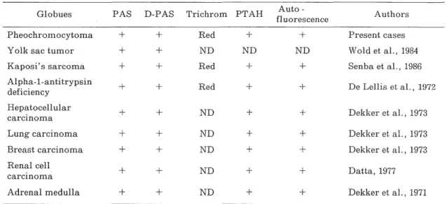
The eosinophilic globules were recognized in seven of 11 (63.6%) pheochromocytoma of the adrenal medulla. This incidence is higher than in other neoplasms. 13 ) Light microscopically, these globules were similar to the globular bodies found in yolk sac tumor9~15)16) Kaposi's sarcoma, l3) alpha-1-antitrypsin deficiency, 3)9) hepatocellular carcinoma, l l) lung carcinoma,
3)6) breast carcinoma
, renal cell carcinoma, l and adrenal medulla 2)5) They also have similar histochemical properties, such as PAS, with and without diastase predigestion, phospho-
tungstic acid hematoxylin (PTAH ), and auto- fluorescence positive (Table 1). Some of these globules were stained positively by immuno- histochemical techniques for alpha- 1-antitry- psin, alpha-fetoprotein, and beta-subunit of human gonadotropin, or human albumin 9) 10)15) 16) Hart described that eosinophilic globules in the adrenal medulla had a possible relation to infections and certain neurologic disease such as multiple sclerosis and Parkinson's disease 5 ) Dekker suggested that hyaline globules in the adrenal medulla were similar to lipof uscin pig- ments and had a relationship to products of lipid peroxidation.3) But we could not confirm these findings in our cases of pheochro- mocytoma. Eosinophilic globules are certainly different from lipofuscin pigments in the light microscopic and histochemical properties.
Lipofuscin pigments are brown, PTAH negative and yellow-orange autofluorescent under ultra-
violet illumination. These globules in pheochro- mocytoma of the adrenal medulla were not stained with argyrophil and argentaffin reac- tions. These histochemical findings suggested to us that eosinophilic globules of pheochro- mocytoma of the adrenal medulla were not re- lated to the chromaffin granules and were glycoprotein.
REFERENCES
1) D vrTA,M. N.: Hyaline intracytoplasmic glob- ules in renal carcinoma. Arch. Pathol. Lab.
Med., 101:391, 1977.
2) DEKKrR, A. and OPIu LE, J. S.: Hyaline glob- ules of the adrenal medulla of man. Arch.
Pathol., 91:353-364, 1971.
3) Diouci R, A. and KRAusE, J.R.: Hyaline glob- ules in human neoplasms. Arch. Pathol., 95:
178-181, 1973.
4) Dr Li.r.r,rs, R. A. and MI:Rrc, F. B.: Distinctive
hepatic cell globules in adult alpha- 1-anti try- psin deficiency. Arch. Pathol., 94:308-316,
1972.
5) HART, M. N. and CYrzus, A.: Hyaline globules of the adrenal medulla. Am. J. Clin. Pathol.,
49:387-391, 1968.
6) HART, M. N.: Hyaline globules. Arch. Pa- thol., 96:144, 1973.
7) LEDDER, L. D., Rrcr-rrrR, H. J.: Pheochrmocy-
toma, in Lohre, LederL-D (eds ): Renal and adrenal tumors. Berlin Heidelberg,
Springer-Verlag, 1987, pp 54-58.
8) Norzru:, S. A. and Campagna-Pinto, D.:
Cytoplasmic hyaline inclusions in hepatoma:
Histochemical study. Arch. Pathol., 86:25- 32,1968.
Table 1. Characterization of eosinophilic globulues
Globues PAS D-PAS Trichrom PTAH flAuto - Authors uorescence
Pheochromocytoma + + Red + + Present cases
Yolk sac tumor + + ND ND ND Wold et al., 1984
Kaposi's sarcoma + + Red + + Senba et al., 1986
Alpha-l-antitrypsin + + R
ed + + De Lellis et al., 1972 d
eficiency
Hepatocellular + + ND + + D
ekker et al., 1973 carcinoma
Lung carcinoma + + ND + + Dekker et al., 1973
Breast carcinoma + + ND + + Dekker et al., 1973
Renal cell + + ND + + D
atta, 1977 carcinoma
Adrenal medulla + + ND + + Dekker et al., 1971
PAS : periodic acid Schiff, D-PAS : periodic acid Schiff reagent after diastase predigestion, PTAH : phosphotungstic acid hematoxylin, ND: not done, +: positive.
9) PALMER, P. E. SAFAII, H. and WOLr1:, II. J.:
Alpha- l-antitrypsin and alpha-fetoprotein:P
rotein markers in endodermal sinus (yolk sac) tumors. Am. J. Clin. Pathol., 65:575-
582, 1976.
10) PALMER. P. E. and WOLFE, H. J.: Immunocyto-
chemical localization of oncodevelopment
proteins in human germ cell and hepatic tumor. J. Histochem. Cytochem., 26:523-531,
1978.
11) PAI.MER, P. E., Uccr, A. A. and WoI.FI;, H. J.:
Expression of protein markers in malignant
hepatoma: Evidence for genetic epigenetic mechanisms. Cancer, 45:1424-1431, 1980.
12) PrEI1nIt, U. and BnN.Tnsc1I, P.: Zum Problem- der " hyalinen Eiwei /3 tropfen " im Cyto-
plasma der Leberparenchymzellen: Licht-
und elektronenmikroskopische Untersuch-
ungen nach 3 /4-Hepatektomie. Virchows Arch. B Zell-pathologie, 1:365-388, 1968.
13) S1: n3n, M., IT!\I uRA, H., YA,4ASIuT/, H. and Huiwc, H. J.: Eosinophilic globules in Ka-
posi's sarcoma: A histochemical, immuno-
histochemical, and ultrastructural study.
Acta Pathol. Jpn., 36:1327-1333, 1986.
14) SE\-o:1, M. and ITAIcCRA, H.: A staining method for easy detection of eosinophilic globules in
Kaposi's sarcoma. Tohoku J. Exp. Med.,
152:209-210, 1987.
15) SIURAI, T., ITOi-i, T., Yosl-rixr, T., Nono, T., Tomf\o, Y. and H\YnsAK\, T.: Immunofluor-
escent demonstration of alpha-fetoprotein
and other plasma proteins in yolk sac tumor.
Cancer, 38:1661-1667, 1976.
16) WoLo, L, E., KRA ER, S. A. and FAIu ow, G.
M.; Testicular yolk sac tumor and embry-
onal carcinoma in pediatric patients: Com-
parative immunohistochemical and clinic-
opathologic study. Am. J. Clin. Pathol., 84:
427-435, 1984.