Introduction
Metal implants are frequently used in orthopaedic surgery
and have substantially contributed to improving surgical outcomes. Implant-related infection (IRI) is a serious complication that might require repeated debridement or implant removal.
One reason that IRI can become difficult to control is biofilm formation on the surface of implants. Once adhered, the bacteria proliferate and produce extracellular polysaccharides that can form a biofilm
1. It has been reported that the biofilm
prevents antibiotics and immune system components from reaching bacteria
2,3.
Staphylococci are the most common causal bacteria of
IRI, accounting for approximately 50% of cases
4,5. Among members of this genus, coagulase-negative staphylococci have especially high ability to adhere to implants and form biofilms
6. Biofilms have been reported to exhibit resistance even toward vancomycin (VCM)
7, which is effective against most Gram-positive bacteria including methicillin-resistant
Staphylococcus aureus8-10. Antibiotic concentrations greater
MS#AMN 07219
Effect of Vancomycin and Glucose on Biofilm Formation by Coagulase-negative Staphylococci: Investigation of Standard Strains and Clinical Strains isolated from Implant-related Infection
Shinji A
dAchi1, Akihiko Y
onekurA1, Toshiyuki S
AkimurA2, Shiro k
AjiYAmA1, Masato T
omiTA1, Toshiyuki T
SurumoTo3, Makoto o
SAki11 Department of Orthopaedic Surgery, Nagasaki University Graduate School of Biomedical Sciences
2 Department of Orthopaedic Surgery, National Hospital Nagasaki Medical Center
3 Department of Macroscopic Anatomy, Nagasaki University Graduate School of Biomedical Sciences
[Objective] The objective of this study was to investigate the suppressive effect of antibiotics and presence or absence of glucose on biofilm formation on metal surfaces by coagulase-negative Staphylococci.
[Materials and Methods] Bacterial strains included a standard biofilm-forming strain of Staphylococcus epidermidis, non- biofilm-forming strain of Staphylococcus hominis, and two clinical strains of Staphylococcus epidermidis isolated from implant- related infection (IRI). Vancomycin (VCM) was used as the antibiotic. Trypticase soy broth (TSB) and TSB without dextrose (TSB w/o D) were used as growth media. Each strain was adhered to stainless steel washers and cultured in different growth media with various VCM concentrations for different lengths of time. The degree of biofilm formation was measured as the biofilm coverage rate (BCR).
[Results] BCR decreased with increasing VCM concentration and shorter incubation time; however, the degree of suppression varied by strain. BCR was lower for strains cultured in TSB w/o D than for those in TSB. BCR for 8-h incubation group was significantly reduced at lower VCM concentrations in TSB w/o D than in TSB.
[Conclusions] Biofilm formation was suppressed in absence of glucose environments, suggesting the importance of controlling blood glucose levels during the perioperative period to prevent IRI.
ACTA MEDICA NAGASAKIENSIA 61: 159−166, 2018 Key words: coagulase-negative staphylococci, biofilm, biofilm coverage rate (BCR), vancomycin, glucose
Address correspondence: Akihiko Yonekura, M.D., Ph.D., Department of Orthopaedic Surgery, Nagasaki University Graduate School of Biomedical Sciences, 1-7-1 Sakamoto, Nagasaki 852-8501 Japan
Tel: +81-95-819-7321, Fax: +81-95-849-7325, E-mail: [email protected] Received December 12, 2017; Accepted January 9, 2018
than minimum inhibitory concentrations (MIC) are needed to treat IRI
11,12. Treatment of IRI is further complicated by the fact that optimal antibiotic dose varies depending on the bacterial species and strain.
Diabetes mellitus (DM) is one of the primary host factor
complicating IRI treatment. Persistent hyperglycemia due to DM reduces the ability of neutrophils to migrate, adhere, phagocytize bacteria, and kill bacteria, thereby increasing the risk of infection
13,14. Treatment of IRI in DM patients become difficult by the fact that the immune systems are prevented from reaching bacteria within biofilms
3. No study has investigated about the importance of blood glucose level to biofilm formation on metal surfaces.
The objective of this study was to clarify the effect of
glucose and the suppressive effect of antibiotics on biofilm formation. We investigated the effect of different concentrations of VCM and the presence or absence of glucose in growth media on biofilm formation using not only standard strains but also clinical strains isolated from IRI cases.
Materials and Methods
The study was conducted following the method used in a
previous study by Sakimura et al.
10. Bacterial strains consisted of RP62A (ATCC 35984), a standard biofilm- forming strain of Staphylococcus epidermidis, SP2 (ATCC 35982), a non-biofilm-forming strain of S. hominis, and two clinical strains of
S. epidermidis (Clinical strain 1 and 2)from IRI cases treated at our institution (Table 1).
Vancomycin hydrochloride (VCM, Wako, Osaka, Japan)
was used as the antibiotic. The MIC of VCM was 1 μg/mL for all bacterial strains. Trypticase soy broth (TSB; Becton- Dickinson, Sparks, MD, USA) and TSB without dextrose (TSB w/o D; Becton-Dickinson) were used as liquid media with different glucose concentrations. Glucose concentration of TSB was 250 mg/dL. Stainless steel washers (diameter:
6.0 mm, thickness 0.5 mm; UW-0306-05; Wilico, Tokyo, Japan) sterilized by ultrasonic cleaning followed by autoclaving were used as the metal substrate for adherence of the bacterial
strains.
After preculturing overnight in liquid medium (TSB or
TSB w/o D), bacterial suspensions of each strain in exponential phase were prepared by creating 10-fold dilutions of each preculture using fresh liquid medium. These suspensions were incubated by shaking at 37°C until an OD
600of 0.2 (2.0
× 107
colony forming unit/mL). The bacterial suspensions (1 mL) were then added to 24-well polystyrene microplates (Iwaki, Funabashi, Japan) containing steel washers in each well. After allowing bacteria to adhere to the washers for 5 min, the wells were rinsed twice with phosphate-buffered saline (PBS).
The washers adhered with bacteria were placed in 1 mL
of fresh liquid medium and incubated for 2, 4, or 8 h to allow biofilm formation on washer surfaces. The resulting samples were divided into three incubation groups (2-h, 4-h, and 8-h). After biofilm formation, washers in each of the three incubation groups were again rinsed twice with PBS. The washers were then transferred to 1 mL of liquid medium with VCM concentrations ranging from 0 to 1,024
μg/mL and incubated for 20 h at 37°C.The quantity of biofilm formation on washers was evaluated
as biofilm coverage rate (BCR) following the method by Kajiyama et al.
15. After fixing the washers with 95% ethanol for 1 min, the washers were dried, stained with 0.5% crystal violet for 5 min, and rinsed with distilled water. After drying the washers, digital images were captured at eight arbitrarily- selected locations on each washer at 450
× magnificationusing a digital microscope (VHX-1000; Keyence, Osaka, Japan). BCR was calculated from the captured images using image analysis software (ImageJ; National Institutes of Health, Bethesda, MD, USA). The experiment was repeated
five times.BCR at each VCM concentration was compared with BCR at
the VCM concentration of 0 μg/mL for each strain, incubation time, and presence or absence of glucose. We performed two-way analysis of variance and multiple comparison tests using the Bonferroni method on BCR results, with p < 0.05 considered as significant. SPSS version 22.0 for Windows (IBM, North Castle, NY, USA) was used for statistical analyses.
Results
As representatives of typical macro images, the photographs
of the stainless steel washers of 2-h incubation group are shown in Figure 1. The washers of RP62A (Standard biofilm- forming strain) cultured in TSB were stained in blue (Figure
Table 1. The bacteria used in present experiment;IRI: implant-related infection
Strain Bacterial Species Characteristics RP62A (ATCC 35984) S. epidermidis Standard biofilm-forming strain
SP2 (ATCC 35982) S. hominis Non-biofilm-forming strain Clinical strain 1 S. epidermidis Isolated from IRI patient Clinical strain 2 S. epidermidis Isolated from IRI patient
1a, 1b). BCR was 100% at VCM concentration 0 μg/mL and was 79% at 8 μg/mL, indicating suppressive effect by VCM against biofilm formation. The washers of RP62A cultured in TSB w/o D were stained in blue (Figure 1c, 1d). BCR was 88% at VCM concentration 0 μg/mL, indicating suppressive effect by absence of glucose against biofilm formation (Figure 1c). BCR was 69% at VCM concentration 8 μg/mL, indicating suppressive effect by both VCM and absence of glucose against biofilm formation (Figure 1d). The washers of SP2 (Non-biofilm-forming strain) cultured in TSB were almost not stained in blue despite at VCM concentration 0
μg/mL (Figure 1e, 1f).BCR at different VCM concentrations of RP62A incubated
in TSB or TSB w/o D for 2, 4, or 8 h were examined (Figure 2). In both TSB and TSB w/o D, BCR tended to decrease with increasing VCM concentration. The reduction of BCR was especially evident for shorter incubation times. BCR of TSB w/o D was lower than that of TSB at most VCM concentrations.
The same experiment was examined using Clinical strain
1 (Figure 3). As with RP62A, BCR tended to decrease with increasing VCM concentration in both TSB and TSB w/o D.
The reduction of BCR was especially evident for shorter incubation times. BCR of TSB w/o D was lower than that of TSB at most VCM concentrations. In 8-h incubation group at high VCM concentration, BCRs in TSB w/o D of Clinical
strain 1 were especially lower than those of RP62A (Figure 3c).
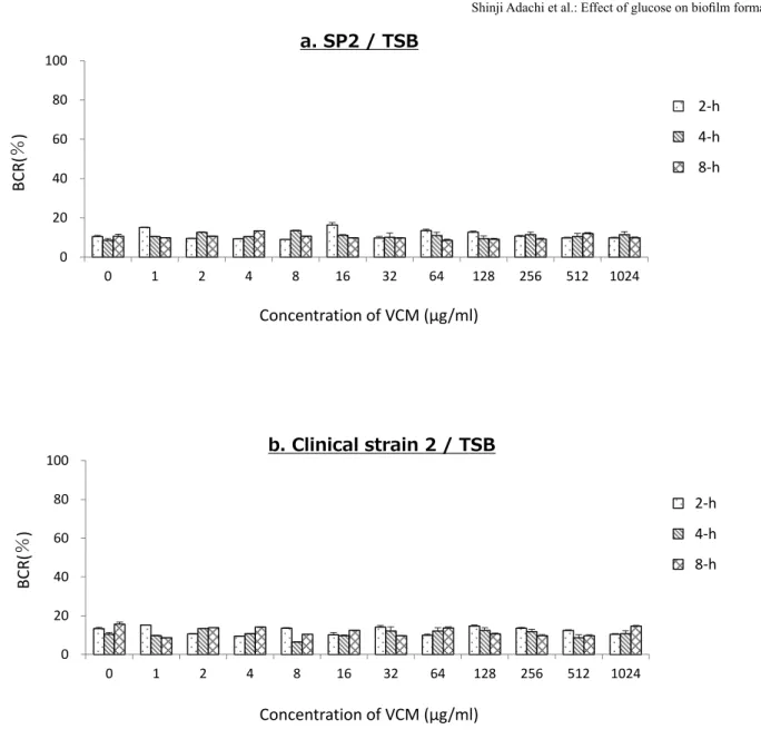
BCR at different VCM concentrations using SP2 (Figure
4a) and Clinical strain 2 (Figure 4b) incubated in TSB for 2, 4, or 8 h were examined. The BCRs of these two bacterial strains were as low as about 15% or less at all VCM concentrations, indicating that Clinical strain 2 was considered a non-biofilm- forming strain.
Discussion
In this study, we demonstrated for the first time that biofilm
formation on metal surfaces is suppressed in absence of glucose environments. In addition, we clarified this suppressive effect using not only standard strains but also clinical strain isolated from IRI. Our results indicated importance of perioperative control of blood glucose levels for preventing IRI.
It is believed that antibiotic-resistance mechanisms ofbacteria involve the expression of various genes and proteins
16,17. The development of antibiotic resistance following exposure
18and differences in resistance among bacterial species and phenotypes
19have also been reported. In many cases, biofilm formation plays a role in this antibiotic resistance
17-19. Numerous studies have investigated the development of antibiotic resistance of bacteria adhered to a surface and in biofilms.
Nishimura et al.
9reported that the antibiotic resistance of bacteria in biofilms is 1,024-fold or greater than that of bacteria in suspension using Staphylococcus strains isolated from infected total hip arthroplasty cases. Sakimura et al.
10used BCR and the viable cell count method to investigate the onset timing of antibiotic resistance accompanying biofilm development by RP62A adhered to stainless steel washers. They found that VCM resistance develops immediately after bacterial adherence and nearly plateaus at 8 h. The MIC of vancomycin of RP62A and Clinical strain 1 were 1
μg/mL, however BCR was suppressed when the vancomycinconcentration was 4 μg/mL or more after formation of biofilm.
This results were consistent with the previous report by Sakimura et al
10.
Regarding the regulation mechanism of biofilm formation
by glucose, You et al. reported the existence of GbaAB (glucose induced biofilm accessory gene), a novel gene of
Staphylococcus aureus20. GbaAB regulates the formation of PIA (polysaccharide intercellular adhesin) which is a component of the biofilm through the ica operon in the presence of glucose. They suggested that the GbaAB regulated biofilm formation at the multicellular aggregation stage rather than
Shinji Adachi Figure 1VCM
0 μg/ml
8 μg/ml
RP62A SP2
TSB TSB w/o D TSB
a b
c d
e f
100 88 10
79 69 9
(%)
Figure 1. The photographs of the stainless steel washers of 2-h incubation group as representatives of typical macro images are shown. They indicated the effects of the absence of glucose (c, d) and administration of VCM (b, d, f) on biofilm formation by RP62A (a, b, c, d) and SP2 (e, f). The numbers on the images represent BCR (%).VCM: vancomycin, BCR: biofilm coverage rate, TSB: trypticase soy broth, TSB w/o D: trypticase soy broth without dextrose.
Shinji Adachi Figure 2
b. RP62A / 4-h incubation
c. RP62A / 8-h incubation a. RP62A / 2-h incubation
0 20 40 60 80 100 120
0 1 2 4 8 16 32 64 128 256 512 1024
TSB w/o D
Concentration of VCM (μg/ml)
BCR(%)
0 20 40 60 80 100 120
0 1 2 4 8 16 32 64 128 256 512 1024
TSB w/o D
Concentration of VCM (μg/ml)
BCR(%)
0 20 40 60 80 100 120
0 1 2 4 8 16 32 64 128 256 512 1024
TSB w/o D
Concentration of VCM (μg/ml)
BCR(%)
*
*
**
*
*
** * * * * * * * * * * * *
* *
*
*
*
*
*
*
* *
*
*
*
*
*
*
*
*
*
*
TSB w/o D TSB
TSB w/o D TSB TSB w/o D TSB
Figure 2. BCR of RP62A treated with different concentrations of VCM. The culture media used were TSB (black) and TSB w/o D (white). Measurements were conducted after incubating samples for 2 h (a), 4 h (b), and 8 h (c). Values represent mean and error bars indicate SD (n=80). * p < 0.001 versus BCR at the VCM concentration of 0 μg/mL. BCR:
biofilm coverage rate, VCM: vancomycin, TSB: trypticase soy broth, TSB w/o D: trypticase soy broth without dextrose.
Shinji Adachi Figure 3
0 20 40 60 80 100 120
0 1 2 4 8 16 32 64 128 256 512 1024
TSB w/o D
Concentration of VCM (μg/ml)
BCR(%)
0 20 40 60 80 100 120
0 1 2 4 8 16 32 64 128 256 512 1024
TSB w/o D
Concentration of VCM (μg/ml)
BCR(%)
0 20 40 60 80 100 120
0 1 2 4 8 16 32 64 128 256 512 1024
TSB w/o D
Concentration of VCM (μg/ml)
BCR(%) **
**
* * * * * * * * * * * * ** * *
*
*
*
*
*
*
*
*
** ** ** ** ** **
*
*
*
*
* *
*
TSB w/o D TSB
TSB w/o D TSB TSB w/o D TSB
b. Clinical strain 1 / 4-h incubation
c. Clinical strain 1 / 8-h incubation a. Clinical strain 1 / 2-h incubation
Figure 3. BCR of Clinical strain 1 treated with different concentrations of VCM. The culture media used were TSB (black) and TSB w/o D (white). Measurements were conducted after incubating samples for 2 h (a), 4 h (b), and 8 h (c). Values represent mean and error bars indicate SD (n=80). * p < 0.001 versus BCR at the VCM concentration of 0 μg/mL. BCR:
biofilm coverage rate, VCM: vancomycin, TSB: trypticase soy broth, TSB w/o D: trypticase soy broth without dextrose.
initial attachment stage. In our study, the VCM concentration at which BCR were suppressed, were the same for TSB and TSB w/o D in the 2-h and 4-h incubation groups. On the other hand, BCR of TSB w/o D were suppressed at a lower VCM concentration than that of TSB in the 8-h incubation group. This result was consistent with the regulation mechanism of biofilm formation via GbaAB reported by You et al. In the 4-h incubation group of RP62A and the 8-h incubation group of Clinical strain 1, the BCR of TSB w/o D was markedly lower than that of TSB at high VCM concentration.
This indicates that the presence or absence of glucose in the culture medium markedly regulated BCR in the long time incubation group. The fact that the incubation time and degree of BCR suppression differs between the two strains
may be due to the timing and level of expression of GbaAB.
Further investigation of GbaAB or genes with similar actions in various bacterial species may lead to clarify the regulation mechanism of biofilm formation by glucose.
Compromised immune function that is caused by DM is
the primary host factor complicating IRI treatment. DM causes microangiopathy and alters cytokinin signaling networks, increasing the risk of infection
21. DM is also reported to reduce the ability of neutrophils to migrate, adhere, phagocytize bacteria, and kill bacteria
13,14. In a clinical study involving 2,316 spinal surgery cases, Olsen et al.
22found DM to be a risk factor for surgical site infection (odds ratio of 3.5). Mraovic et al.
23investigated 1,948 cases of total hip arthroplasty and total knee arthroplasty and
Shinji Adachi Figure 4
0 20 40 60 80 100
0 1 2 4 8 16 32 64 128 256 512 1024
2hr 4hr 8hr
0 20 40 60 80 100
0 1 2 4 8 16 32 64 128 256 512 1024
2hr 4hr 8hr
Concentration of VCM (μg/ml) Concentration of VCM (μg/ml) BC R( % ) BC R( % )
a. SP2 / TSB
b. Clinical strain 2 / TSB
4‐h 2‐h 8‐h
4‐h 2‐h 8‐h
Figure 4. BCR of SP2 (a) and Clinical strain 2 (b) treated with different concentrations of VCM. The culture media used was TSB. Measurements were conducted after incubating samples for 2 h, 4 h, and 8 h. BCR: biofilm coverage rate, VCM: vancomycin, TSB: trypticase soy broth.
found that the risk of post-operative infection doubled when blood glucose was 200 mg/dL or greater. Efforts are currently being made to advocate perioperative control of blood glucose for preventing IRI. The Guideline for the Prevention of Surgical Site Infection 2017
24issued by the United States Centers for Disease Control and Prevention strongly advises that blood glucose be maintained under 200 mg/dL in the perioperative period. Our study demonstrated that biofilm formation was suppressed in absence of glucose environments. This suppression was already evident at short incubation times and persisted even after longer incubation times when the suppressive effect of antibiotics was reduced.
It suggests the importance to control blood glucose during the perioperative period for preventing IRI.
The ability to form biofilms also differed among strains
within the same
S. epidermidis species. Although the twoclinical strains used in this study were both S. epidermidis, Clinical strain 1 formed biofilms while Clinical strain 2 had the exact opposite characteristic and did not form biofilms.
Even among biofilm-forming strains, the pattern of suppression of biofilm formation by VCM differed between RP62A and Clinical strain 1. The suppressive effect in the absence of glucose environment also differed between these two strains.
In particular, one characteristic of Clinical strain 1 was the very evident suppression of biofilm formation after 8-h incubation in TSB w/o D at high VCM concentration. When IRI occurs, it is recommended that identification of the causal bacteria be prioritized over administration of antibiotics.
In our study, the antibiotic dose at which antibiotic resistance occurs differed between bacterial strains within the same species. It suggests that assessing the ability to form biofilms also important in the treatment of IRI. It will be useful for decision making to preserve or remove infected implants.
The first limitation of this study is the small number of
strains tested. Only two biofilm-forming strains were tested, leaving open the possibility of the existence of strains whose ability to form biofilms is not reduced in absence of glucose environments. The second limitation is that we did not investigate the effect of different glucose concentrations.
The glucose concentration of TSB used in this study was 250 mg/dL, which reasonably reproduces high-glucose levels in the clinical environment. In contrast, the glucose concentration of TSB w/o D is 0, representing a profoundly low-blood glucose environment. In this study, we focused on biofilm formation and development over time. Investigating the dependence of biofilm development on glucose concentration may enable us to determine target blood glucose levels for the perioperative period. The third limitation is that we did not investigate the effect of antibiotics other than VCM.
Raad et al. examined the inhibitory effect of VCM, Linezolid, and Daptomycin on biofilm formation of catheter-related infection. Antibiotics were exposed after adherence of MRSA to silicone disks, followed by counting the number of bacteria detached from the biofilm formed on the silicone disks. As a result, Daptomycin suppressed biofilm formation significantly more than VCM and Linezolid
8. Although there is a difference between silicone disk and metal surface of stainless steel, different results might be obtained when using Daptomycin or Linezolid. The fourth limitation is that the number of viable cell count (VCC) in biofilm was not measured in this study. Sakimura et al. reported the VCC of RP62A at VCM concentration of 32
μg/mL or more in the 2-h incubationgroup was almost zero. BCR before addition of VCM in the 2-h incubation group was reported to be 9.5%
10. In this study, BCR was lower than 9.5% when the VCM concentration was 32
μg/mL or more. It was considered that BCR wasdecreased by the decreasing of VCC in 2-h incubation group of RP62A. Due to the different incubation time and bacteria strains, it was unclear whether the decrease in BCR was due to the suppression of biofilm formation or the decrease in the number of viable bacteria in this study. The
fifthlimitation has to do with the use of stainless steel as the only metal substrate. Biofilm formation and suppression of biofilm formation may differ depending on the type of metal substrate. Given that various metals such as titanium and cobalt chrome are currently being used for implants in clinical settings, further investigation is expected.
Conclusion
This study demonstrated that biofilm formation on metal
surfaces is suppressed in absence of glucose environments.
The results of this study indicated importance of perioperative control of blood glucose levels for preventing IRI.
Acknowledgements
The authors declare that there are no conflicts of interest
regarding the publication of this paper.
References
1. Gristina AG, Costerton JW. Bacterial adherence to biomaterials and tissue: The significance of its role in clinical sepsis. J Bone Jt Surg Am 67: 264-273, 1985
2. Costerton JW, Stewart PS, Greenberg EP. Bacterial Biofilms : A Common Cause of Persistent Infections. Science 284: 1318-1322, 1999 3. Stewart PS, Costerton JW. Antibiotic resistance of bacteria in biofilms.
Lancet 358: 135-138, 2001
4. Ammon P, Stockley I. Allograft bone in two-stage revision of the hip for infection; Is it safe? J Bone Jt Surg Br 86: 962-965, 2004
5. Weinstein MA, McCabe JP, Cammisa FP. Postoperative spinal wound infection: a review of 2,391 consecutive index procedures. J Spinal Disord 13: 422-426, 2000
6. Mack D, Davies AP, Harris LG, Rohde H, Horstkotte MA, Knobloch JK-M. Microbial interactions in Staphylococcus epidermidis biofilms.
Anal Bioanal Chem 387: 399-408, 2007
7. Barna C, Williams DH. The structure and mode of action of glycopeptide antibiotics of the vancomycin group. Annu Rev Microbiol 38: 339-357, 8. Raad I, Hanna H, Jiang Y, et al. Comparative Activities of Daptomycin, 1984 Linezolid, and Tigecycline against Catheter-Related Methicillin-Resistant Staphylococcus Bacteremic Isolates Embedded in Biofilm. Antimicrob Agents Chemother 51: 1656-1660, 2007
9. Nishimura S, Tsurumoto T, Yonekura A, Adachi K, Shindo H. Antimicrobial susceptibility of Staphylococcus aureus and Staphylococcus epidermidis biofilms isolated from infected total hip arthroplasty cases. J Orthop Sci 11: 46-50, 2006
10. Sakimura T, Kajiyama S, Adachi S, et al. Biofilm-forming Staphylococcus epidermidis expressing vancomycin resistance early after adhesion to a metal surface. Biomed Res Int. 2015. http://dx.doi.org/10.1155/2015/943056.
11. Ceri H, Olson ME, Stremick C, Read RR, Morck D, Buret A. The Calgary Biofilm Device : New Technology for Rapid Determination of Antibiotic Susceptibilities of Bacterial Biofilms. J Clin Microbiol 37: 1771-1776, 12. Pettit RK, Weber CA, Kean MJ, et al. Microplate Alamar Blue Assay for 1999 Staphylococcus epidermidis Biofilm Susceptibility Testing. Antimicrob Agents Chemother 49: 2612-2617, 2005
13. Delamaire M, Maugendre D, Moreno M, Le Goff MC, Allannic H, Genetet B. Impaired Leucocyte Functions in Diabetic Patients. Diabet Med 14:
29-34, 1997
14. Alexiewicz JM, Kumar D, Smogorzewski M, Klin M, Massry SG.
Polymorphonuclear leukocytes in non-insulin-dependent diabetes mellitus:
abnormalities in metabolism and function. Ann Intern Med 123: 919- 924, 1995
15. Kajiyama S, Tsurumoto T, Osaki M, Yanagihara K, Shindo H. Quantitative analysis of Staphylococcus epidermidis biofilm on the surface of biomaterial.
J Orthop Sci 14: 769-775, 2009
16. Taniguchi K, Ono T, Murakami K, et al. Novel Pseudomonas aeruginosa Gene That Suppresses Tolerance to Carbapenems. Antimicrob Agents Chemother 47: 2997-3001, 2003
17. Parkins MD, Ceri H, Storey DG. Pseudomonas aeruginosa GacA, a factor in multihost virulence, is also essential for biofilm formation. Mol Microbiol 40: 1215-1226, 2001
18. Hoffman LR, D'Argenio DA, Maccoss MJ, Zhang Z, Jones RA, Miller SI. Aminoglycoside antibiotics induce bacterial biofilm formation. Nature 436: 1171-1175, 2005
19. Drenkard E, Ausubel FM. Pseudomonas biofilm formation and antibiotic resistance are linked to phenotypic variation. Nature 416: 740-743, 2002 20. You Y, Xue T, Cao L, Zhao L, Sun H, Sun B. Staphylococcus aureus
glucose-induced biofilm accessory proteins, GbaAB, influence biofilm formation in a PIA-dependent manner. Int J Med Microbiol 304: 603- 612, 2014
21. Turina M, Fry DE, Polk HC. Acute hyperglycemia and the innate immune system: Clinical, cellular, and molecular aspects. Crit Care Med 33:
1624-1633, 2005
22. Olsen MA, Nepple JJ, Riew KD, et al. Risk Factors for Surgical Site Infection Following Orthopaedic Spinal Operations. J Bone Jt Surg Am 90: 62-69, 2008
23. Mraovic B, Suh D, Jacovides C, Parvizi J. Perioperative Hyperglycemia and Postoperative Infection after Lower Limb Arthroplasty. J Diabetes Sci Technol 5: 412-418, 2011
24. Berríos-Torres SI, Umscheid CA, Bratzler DW, et al. Centers for Disease Control and Prevention Guideline for the Prevention of Surgical Site Infection, 2017. JAMA Surg 152: 784-791, 2017